




 Inglês (pdf)
 Artigo em XML
 Referências do artigo
 Tradução automática
 Enviar este artigo por email
 Citado por SciELO
  Similares em
SciELO
Similares em
SciELO 
 Permalink

Introduction
Syphilis is a sexually transmitted disease caused by Treponema pallidum,1 a helical anaerobic bacterial species.2,3 It represents a significant public health issue worldwide, with more than 6 million estimated new cases of syphilis each year and severe health consequences if left untreated.4,5,6,7 Brazil is experiencing a significant increase in syphilis cases compared to other Latin American countries, following a trend contrary to the eradication goal.8,9 This increase may be related to the compulsory notification throughout the country in force since 2010,10 and the reduction in condom use.11 This is a significant risk to public health and underscores the importance of prioritizing investments in prevention and promotion that target Health Care Networks, especially for improving the quality of care provided to populations engaging in risky behaviors.9
Syphilis can have various clinical manifestations and progresses through four different stages with varying levels of activity and infectivity: primary, secondary, latent, and tertiary syphilis.5 The typical lesion of primary syphilis consists of a frequently single, painless, and non-pruritic cutaneous ulceration called a “chancre,” which appears at the site of inoculation approximately three weeks after exposure and may be associated with regional lymphadenopathy, both of which resolve spontaneously.10 The secondary stage results from hematogenous and lymphatic dissemination of treponemes, usually causing mucocutaneous and systemic manifestations six to eight weeks after the initial infection.3,12 In secondary syphilis, the manifestations are more heterogeneous and nonspecific.13 Primary and secondary lesions can resolve spontaneously, and the infection enters a latent stage that can persist for years. Then, the disease may progress to the tertiary stage, in which a range of late manifestations might lead to destructive neurological and cardiovascular complications, severe skin or visceral lesions (gummas), or bone involvement.4,5
Syphilis can exhibit pathognomonic signs in the oral cavity in different stages.14 However, multiple oral lesions are more commonly seen in secondary syphilis. Among its clinical oral manifestations, the most common are described as “mucous patches” or “macules” and may appear as slightly elevated, usually oval-shaped plaques, occasionally ulcerated, covered by a grayish or whitish fibrinous pseudomembrane.2,3,15,16
Another manifestation consists of multiple mucous patches that can coalesce, giving rise to serpiginous lesions described as “slug track ulcers.”3,17,18 Syphilitic lesions of the oral mucosa are often associated with systemic and cutaneous manifestations, and exclusive oral involvement is quite uncommon in secondary syphilis.15,17,18
This study aimed to report a clinical case of an isolated oral manifestation of secondary syphilis diagnosed in a public oral health service, confirmed by treponemal and non-treponemal serological tests.
Case Report
The patient provided written consent for the publication of her clinical case in accordance with the Declaration of Helsinki by signing an informed consent form prior to the drafting of this study.
A Caucasian 30-year-old female patient sought medical attention at a public health service in Southern Brazil, complaining of symptomatic oral ulcerative lesions that did not show spontaneous regression. After clinical evaluation, treatment with triamcinolone acetonide 1 mg/g for 7 days was prescribed due to suspicion of aphthous ulcers. The patient returned five days later seeking dental services, reporting the lack of lesion regression and persistent painful symptoms. During the dental consultation, the clinical history of the lesions was thoroughly investigated for differential diagnosis.
The patient reported that the oral manifestations had first presented at least 30 days previously and were initially asymptomatic before becoming symptomatic. She reported no other skin or genital lesions and no systemic manifestations such as fever, sore throat, or lymphadenopathy.
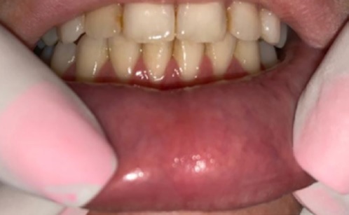
Intraoral clinical examination diagnosed a larger lesion in the central region of the hard palate with confluent mucous plaques, giving rise to “slug-shaped ulcers” surrounded by erythema (Figure 1). Smaller mucous patches with irregularly shaped, whitish fibrinous pseudomembranes were identified in the region of the lower vestibular sulcus near teeth 41, 42, 33, and 34 (Figure 2). Another larger mucous patch with na irregular appearance and coalescence, slightly elevated with a whitish fibrinous pseudomembrane and surrounded by erythema, was detected in the central region of the lower labial mucosa (Figure 3).
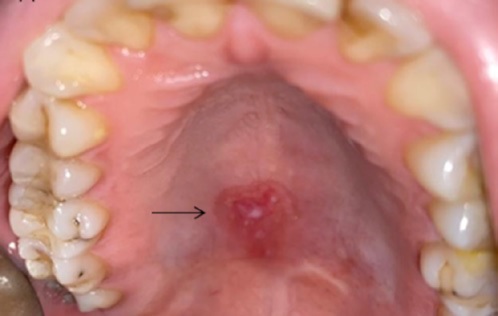
Figure 1 Larger lesion in the central region of the hard palate with confluent mucous plaques, giving rise to “slug-shaped ulcers” surrounded by erythema
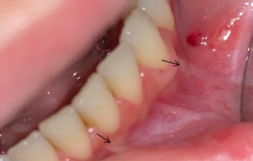
Figure 2 Smaller mucous patches with irregularly shaped white fibrinous pseudomembranes in the lower vestibular sulcus near teeth 41 and 42 and teeth 33 and 34
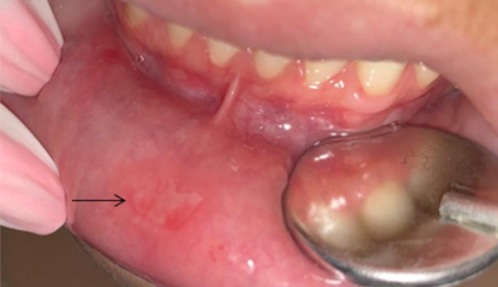
Figure 3 Larger mucous patch with an irregular appearance and coalescence, slightly elevated with white fibrinous pseudomembrane and surrounded by erythema in the central region of the lower labial mucosa
Upon suspicion of syphilitic oral lesions and for differential diagnosis, rapid tests were requested and performed by the nursing service at the healthcare unit. The results showed a reactive test for syphilis (treponemal serological test VDRL) and non-reactive results for other rapid screening tests (HIV, hepatitis C, and hepatitis B). After confirming the diagnosis, the patient was referred to the medical service for treatment with Penicillin G Benzathine 2.4 million IU, administered via intramuscular injection once a week for three weeks. The diagnostic VDRL test showed a titer of 1:128.
Dental follow-up was conducted to observe the regression of oral manifestations during the treatment period (Figures 4 and 5) and 15 days after the last dose of antibiotic therapy, when complete regression of oral manifestations was observed (Figures 6 and 7). After completing the treatment with Penicillin G Benzathine, a new VDRL test showed a titer of 1:16. A drop in titer from 1:128 to 1:16 after the treatment period indicates treatment success since a four-fold or higher change in titer, equivalent to a change of at least two dilutions, is considered a significant difference between two sequential tests.19
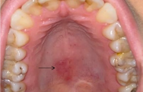
Figure 4 Partial regression of the larger lesion in the central region of the hard palate 14 days after starting treatment with Penicillin G Benzathine
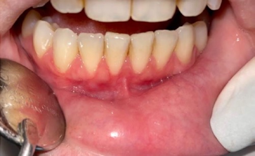
Figure 5 Complete regression of the larger mucous patch in the central region of the lower labial mucosa 14 days after starting treatment with Penicillin G Benzathine
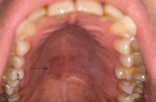
Figure 6 Complete regression of the larger lesion in the central region of the hard palate 30 days after starting treatment with Penicillin G Benzathine
Discussion and Conclusions
This case report emphasizes the importance of paying attention to oral manifestations of syphilis, especially to the clinical differential diagnosis of this disease, which may initially have an uncertain etiology. Moreover, the possibility of isolated oral manifestations should be considered in the absence of other systemic signs or symptoms. Delayed diagnosis of syphilis can lead to disease progression to the latent and tertiary stages, potentially causing more severe and sometimes fatal consequences.20 It is important to consider that syphilitic lesions can be confused with other diseases as they can mimic various conditions and have significantly variable manifestations. The differential diagnosis of syphilis includes, for example, lichen planus, eosinophilic ulcers, traumatic ulcers, leukoplakia, HIV, gonorrhea, necrotizing sialometaplasia, and squamous cell carcinoma.21
In the present case, the suspicion of a syphilitic lesion arose from the lesions’ clinical appearance, consistent with what is normally seen in these cases, namely, prolonged (over four weeks) erythematous macules and plaques in lips, tongue, gums, or tonsils. Although the patient did not report other skin or genital lesions or systemic manifestations such as fever, sore throat, or lymphadenopathy, the VDRL test was conducted. This test is available in the public health network in Brazil and is always recommended when syphilis is suspected, such as in the case of a small painless wound, swelling in the lymph nodes near the wound, or skin rashes that do not cause itching.
Before the discovery of penicillin, syphilis was a public health problem controlled for decades with the use of antibiotics.7,22 However, considering the current increasing incidence of syphilis,23 it is of utmost importance to discuss the dentist’s knowledge and experience for the differential diagnosis of lesions affecting the oral cavity. Awareness is especially important when the only manifestation is oral ulcers, as in the present case, making the differential diagnosis and the possible etiological origin of these lesions more challenging.
The oral manifestations of syphilis can occur in all three stages of the disease, as described earlier. In secondary syphilis, the lesions are usually multiple and more diverse,11 but a single lesion can often be the only manifestation of the disease,21 more commonly presenting as mucous patches, papules, or ulcerations with erosive areas.3,16,21 Such reports corroborate the findings of the present case, where multiple ulcerated lesions with diverse characteristics were observed.
The Unified Health System, a Brazilian public health policy, advocates for the prevention of Treponema pallidum contamination and provides free diagnosis and treatment to the population.24 Treponemal tests, such as the rapid syphilis test, and non-treponemal tests, such as VDRL, are available through the Unified Health System and, due to being quick and low-cost, are widely used for screening and disease detection. The rapid test is easier to perform since it is available in Unified Health System Primary Care for sexually transmitted disease screening, and it is reactive in the secondary and latent stages, although it is less sensitive in primary syphilis.25 Serosurvey tests, in conjunction with a well-conducted clinical examination and diagnosis, play a crucial role in diagnosing syphilis.26 Accordingly, in this case, after conducting a differential clinical diagnosis during the dental consultation, serological testing for syphilis was immediately performed, confirming the diagnosis and expediting treatment onset.
This case report emphasizes the need for effective integration and interdisciplinary collaboration within healthcare teams, particularly within public health. The relationship between oral and systemic health is well-documented in the literature, highlighting that the exchange of diagnoses between dental and medical professionals plays a crucial role in the early identification of health issues.
Additionally, it is important to underscore that the rising number of new syphilis cases relates to the social and behavioral aspects of society,21 namely, risky sexual behavior such as reduced safe sexual practices, which is the primary reason for this resurgence.25 This situation highlights the urgent need for public health education measures or campaigns, especially in the affected countries.
This case report emphasizes the importance of integrating and incorporating oral health into the public health context. Moreover, it reflects the importance of updating and continuously training health professionals, as many systemic diseases have oral manifestations that can be diagnosed and monitored by dentists. Additionally, the multidisciplinary approach of public health teams is essential for comprehensive healthcare coverage for patients.

