Serviços Personalizados
Journal
Artigo
Indicadores
-
 Citado por SciELO
 -
 Acessos
Links relacionados
-
 Similares em
SciELO
Compartilhar
Acta Obstétrica e Ginecológica Portuguesa
versão impressa ISSN 1646-5830
Acta Obstet Ginecol Port vol.14 no.2 Coimbra jun. 2020
ISSUE IMAGE/IMAGEM DO TRIMESTRE
Primary syphilis - an atypical presentation
Sífilis primária - um caso de apresentação clínica atípica
Elisa Soares1, Pedro Brandão2, Ana Torgal3
Centro Hospitalar do Tâmega e Sousa, EPE
1 Ginecologia e Obstetrícia, Interna de Formação Específica, Centro Hospitalar Tâmega e Sousa
2 Ginecologia e Obstetrícia, Assistente Hospitalar, Instituto Valenciano de Infertilidad (IVI) - Valência, Espanha
3 Ginecologia e Obstetrícia, Assistente Hospitalar Graduada, Centro Hospitalar Tâmega e Sousa
Endereço para correspondência | Dirección para correspondencia | Correspondence
ABSTRACT
Syphilis is a sexually transmitted disease caused by the spirochete Treponema pallidum. The clinical manifestations of this disease vary widely, according to its stage. Primary syphilis typically presents as a single painless genital ulcer.
The authors report a case of an atypical presentation of primary syphilis: multiple vulvar, perianal and between the buttocks ulcerated and very painful papules and plaques, with seropurulent exudate.
Keywords: Primary Syphilis, Vulvar ulcers, Treponema pallidum.
Syphilis is a sexually transmitted disease that can represent a great diagnostic challenge. This disease is also known as "the great imitator", due to its variety of possible clinical presentations.
Although primary syphilis typically presents as a single painless genital ulcer, it can become painful if secondarily infected1. In addition, the presence of multiple lesions may occur, particularly in immunocompromised patients2.
Aetiology determination of vulvar ulcers is often hampered, either by the broad spectrum of possible clinical manifestations or by frequent of overlapping infections3.
In these cases, it is particularly important to carry out a detailed anamnesis, with thorough evaluation of the sexual history, physical examination, and request of laboratory tests, allowing the establishment of a differential diagnosis among the different causes of vulvar ulcers.
The authors report a case of 43-year-old women, 3G3P, without relevant medical history, and history of penicillin allergy, who went to the general emergency department with multiple painful vesicular vulvar lesions. Due to clinical suspiction of genital herpes, she was medicated with acyclovir.
A month later, due to the worsening of the lesions, she returned to the emergency department and was observed by a gynaecologist.
A thorough anamnesis revealed non-compliance with previously prescribed therapeutic plan and sexual risk behaviors.
Physical examination revealed multiple vulvar, perianal and between the buttocks ulcerated, and very painful plaques and papules, with seropurulent exudate (which was collected for cultural exam), and one umbilical papular lesion (Figure 1). There were no vaginal lesions or inguinal adenopathies.

(clique para ampliar ! click to enlarge)
Cultural exam of lesion exudate was positive for Staphylococcus aureus and Methicillin-sensitive Streptococcus agalactiae. PCR for HSV 2 in the lesions swab came positive, and for HSV 1 came negative.
Serological results were as follows: positive anti-HCV antibody, negative HIV 1 and 2 antibodies and positive VDRL (1:84).
Given these clinical manifestations and analytic findings, syphilis was the main hypothesis of diagnosis. However, FTA-abs test was negative. Despite this, the presence of T. pallidum in the exudate of the lesions was verified at darkfield microscopy.
As a first approach, treatment with 500 mg/day intravenous erythromycin was started empirically. Seven days after, given the presence of T. pallidum in the lesions and the history of penicillin allergy (standard therapy for primary syphilis4) the patient was treated with doxycycline 100 mg/day per os during 14 days.
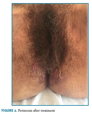
At the time of discharge, significant clinical improvement was observed with erythematous plaques, without exudate, ulceration or pain (Figure 2).
One month after discharge, FTA-abs came positive, supporting the diagnosis of syphilis.
REFERÊNCIAS
1. Janier M , Hegyi V, Dupin N, Unemo M , Tiplica GS , Potočnik M , French P , Patel R. 2014 European Guideline on the Management of Syphilis. [ Links ]
2. French P. Syphilis. BMJ. 2007;334(7585):143. [ Links ]
3. Rosen T, Brown TJ. Genital ulcers. Evaluation and treatment. Dermatol Clin. 1998;16(4):673. [ Links ]
4. WHO GUIDELINES FOR THE Treatment of Treponema pallidum (syphilis). World Health Organization; 2016. [ Links ]
Endereço para correspondência | Dirección para correspondencia | Correspondence
Elisa Sofia Oliveira Soares
Centro Hospitalar Tâmega e Sousa, Penafiel, Porto
E-Mail: elisa.s.o.soares@gmail.com
Recebido em: 09/08/2019. Aceite para publicação: 18/05/2020


