Introduction
Cervical insufficiency is the incapacity of the uterine cervix to sustain a pregnancy in the second trimester in the absence of contractions1. Its pathophysiology is still poorly understood. Although structural cervical weakness may be the cause of many recurrent second-trimester abortions, the majority of cases are probably caused by other disorders such as decidual infection, placental bleeding or uterine overdistension, as is usually the case in multiple pregnancies2.
In Portugal, in 2018, multiple births accounted for 1,6 percent of total births with 62,6 percent of these births being preterm3. Twin pregnancy is associated with higher rates of several potential complications of pregnancy of which the most serious is preterm delivery. Besides neonatal death, complications of prematurity include respiratory distress syndrome, intraventricular hemorrhage, necrotizing enterocolitis, retinopathy of prematurity, among others.
Several interventions, including vaginal progesterone, pessary or placement of a cervical cerclage, have been proposed to reduce the risk of preterm birth in twin gestations but, to date, none has clearly proven its effectiveness2,4. In a 2017 meta-analysis by Jarde et al.4 that assessed the effectiveness of cerclage or pessary for preventing preterm birth the authors concluded that no intervention was beneficial for this purpose. Despite the routine use of these procedures in the management of twin pregnancies may be ineffective, the judicious utilization of some interventions in selected clinical circumstances may be of inestimable value, such as physical examination-indicated cerclage placement.
The authors describe a case of cervical insufficiency in a twin pregnancy identified in the 21st week of gestation in which the performance of a physical examination-indicated cerclage resulted in good maternal-fetal outcomes. For the elaboration of this article, an informed consent was obtained from the patient.
Case report
A 25-year-old patient, primigravida with no relevant medical/gynecological history, was referred for surveillance due to a spontaneous dichorionic twin pregnancy. In the first trimester ultrasound, no structural anomalies were detected and the screening for fetal chromosomal abnormalities was negative for both fetuses.
In the 17th week, although the patient was asymptomatic, the cervical length was 25 mm. We decided to initiate vaginal progesterone to reduce the risk of preterm birth. There were no signs of urinary tract infection as demonstrated by negative urine culture.
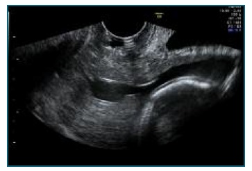
The 2nd trimester ultrasound, at 21 weeks and 2 days of gestation, showed normal morphology in both fetuses and amniotic fluid superior to normal, yet not reaching the threshold for diagnosing polyhydramnios. The transvaginal ultrasound (Figure 1) demonstrated a cervical length of 6 mm with pronounced funneling and in the speculum examination the cervix was dilated with exposed membranes. The patient maintained no symptoms of clinical contractions.

Figure 1 Transvaginal ultrasound at 21 weeks and 2 days: cervix with a length of 6 mm and pronounced funneling.
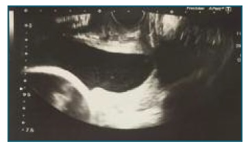
After discussion with the couple about the prognosis and potential interventions to decrease the risk of preterm labor, the decision was to perform a cervical cerclage. The procedure was executed at 21 weeks and 5 days of pregnancy using McDonald’s technique (Figure 2). Indomethacin 25 mg every 6 hours was prescribed for 24 hours post-procedure.

Figure 2 Cervical length of 4-6 mm at 21 weeks and 5 days (transvaginal ultrasound before the placement of cerclage).
In the 22nd week, although the pregnant woman remained asymptomatic, the speculum examination raised suspicion of the presence of amniotic fluid in the posterior pouch of vagina. There was no leakage of amniotic fluid through the external cervical os using the Valsalva maneuver. The cervical length was 4-6 mm and the suture was visible on the distal third of its extent. The patient was treated with ampicillin. During hospitalization, she remained asymptomatic and there were not any other signs of amniotic fluid loss. The patient was discharged after 7 days of surveillance.
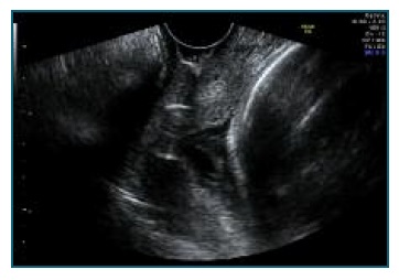
On the 3rd trimester ultrasound, performed in the 28th week, the cervical length evaluation (Figure 3) was similar to previous measurements (4-6 mm) and there were no clinical signs of uterine contractions.

Figure 3 Transvaginal ultrasound in the 28th week showing cerclage suture in the cervix and a cervical length of 4-6 mm.
In an ultrasound performed at 35 weeks and 5 days, the fetus 1 had an estimated fetal weight in the percentile 4 and normal Doppler evaluation and the fetus 2 had an estimated fetal weight in the percentile 0,5 with altered cerebroplacental ratio. We decided to terminate pregnancy and a cesarean section was performed due to breech presentation of fetus 2. Fetus 1 weighted 2310 grams at birth and fetus 2 weighted 1810 grams. Both had an Apgar score of 9/10/10 (at 1, 5 and 10 minutes post-birth respectively). After the procedure the cerclage was removed.
Discussion
The authors report a case of successful prenatal management with cerclage placement of a twin pregnancy that presented with cervical dilation in the 21st week.
In the present case, the first warning sign was a short cervix detected in the 17th week. In our practice we screen for short cervical length in multiple gestation since it is a good predictor of preterm birth in these pregnancies5,6 and initiate supplementation with vaginal progesterone when the cervix is short (≤ 25 mm). In a 2017 meta-analysis from six randomized trials of asymptomatic women with twin gestations and a cervical length ≤ 25 mm, vaginal progesterone reduced the risk of preterm birth <33 weeks of gestation by 31% and neonatal death by 47%7. The relative risks of neonatal death, respiratory distress syndrome and birth weight <1500g were also reduced significantly, without any demonstrable deleterious effects on childhood neurodevelopment.
In the 21st week, the patient presented with cervical dilation in speculum examination. Cervical dilation in the mid-trimester is associated with a more than 90% rate of spontaneous preterm birth and a poorer perinatal prognosis8,9. Although there are potentially beneficial interventions for prevention of preterm birth, the treatment options in the presence of cervical dilation are limited to physical examination-indicated cerclage (also known as emergency or rescue cerclage). After informing the couple about the unfavorable prognosis and discussing the available evidence about managing options, the decision was to place a rescue cervical cerclage.
The only randomized controlled trial to date that determined if physical exam indicated cerclage reduces the incidence of preterm birth in women with twin gestations and cervical dilation (1-5 cm) found a significant decrease in preterm birth at all evaluated gestational ages (including a 50% decrease in preterm birth <28 weeks) and a 78% decrease in perinatal mortality10. In this study, all women who underwent cerclage received prophylactic antibiotics and indomethacin. This was a small sample size study that was terminated early because of the significant decrease in perinatal mortality in the cerclage group.
In a 2019 meta-analysis by Li et al.11, which included 1211 women, the authors concluded that cerclage placement is beneficial for the reduction of preterm birth and the prolongation of pregnancy in twin pre-gnancies with a cervical length <15 mm or dilated cervix >10 mm. The benefit was not demonstrated for history-indicated or twin alone-indicated cerclage. In women with cervical dilation, cerclage placement was associated with a significant prolongation of pregnancy, reduction of preterm birth rate and improvement in perinatal outcomes.
Earlier meta-analyses showed no benefit of cerclage for women with twin pregnancies2,4,12. A 2014 Cochrane review4 that included five randomized controlled trials (two of obstetric history indicated-cerclage and three of cerclage indicated by transvaginal ultrasound) reported no differences in perinatal deaths, serious neonatal morbidity or preterm birth rates when cerclage was compared with no cerclage in women with twin gestations. Saccone et al. (12 evaluated the efficacy of cerclage in twin pregnancies with a short cervical length and found no differences in preterm birth <34 weeks and perinatal deaths. Very low birthweight and respiratory distress syndrome were more frequent in the cerclage group. Since these meta-analyses included few and small clinical studies, their conclusions are of limited value and need to be interpreted with caution.
Recently, several studies demonstrated a potential benefit of physical examination-indicated cerclage in reducing preterm birth rate in twin gestations13-16. Roman et al. (13 reported that an association of cerclage, indomethacin and antibiotics in twin pregnancies with cervical dilation ≥ 1 cm was associated with a significant prolongation of gestation, decreased incidence of preterm birth and improved perinatal outcome, compared with expectant management. In a 2018 retrospective cohort study of twin pregnancies14, cerclage placement significantly reduced the incidence of preterm birth <32 weeks compared with expectant management only in the subgroup of rescue cerclage. Abbasi et al. (15 compared the perinatal outcomes in twin pregnancies with cervical dilation treated with cerclage or expectant management and observed lower rates of preterm birth, neonatal intensive care unit admission and neonatal respiratory morbidity in the cerclage group. Importantly, in this study, all women managed expectantly gave birth prior to 28 weeks.
Other authors compared the outcomes of emergency cerclage in twin versus singleton gestations and concluded that the beneficial obstetric outcomes are similar between the two groups17-20.
In conclusion, preterm birth is one of the main challenges in the management of multiple pregnancies. Whilst it is the major source of perinatal morbidity and mortality in twin gestations, there is currently no proven effective intervention for the prevention of this complication. Despite the need for more high-quality trials of appropriate size and duration, physical examination-indicated cerclage may be an option for preventing preterm birth in twin pregnancies.


