SciELO - Scientific Electronic Library Online

 
vol.16 número1Anemia de Fanconi e gravidez: uma nova realidadeMigração de Essure® para a cavidade endometrial: um achado ecográfico incomum índice de autoresíndice de assuntosPesquisa de artigos
Home Pagelista alfabética de periódicos  

Serviços Personalizados

Journal

Artigo

Indicadores

Links relacionados

  • Não possue artigos similaresSimilares em SciELO

Compartilhar


Acta Obstétrica e Ginecológica Portuguesa

versão impressa ISSN 1646-5830

Acta Obstet Ginecol Port vol.16 no.1 Algés mar. 2022  Epub 31-Mar-2022


Imagem do trimestre/ Issue image

Uterine fibroid: size does matter

Mioma uterino: porque o tamanho importa

Mariana Crespo Marques1 

Vanessa Santos1 

José Silva Pereira2 

1. Hospital Professor Doutor Fernando Fonseca EPE. Portugal.

2. Director do Serviço de Ginecologia, Hospital Prof. Doutor Fernando Fonseca. Portugal.


Abstract

Leiomyomas are the most common benign tumors in reproductive age women. The incidence is higher in African descent women. Development and growth of fibroids is estrogen and progesterone dependent. Besides excessive menstrual bleeding, pelvic pain is also a common symptom. Doppler ultrasound (dUS) or magnetic resonance imaging (MRI) are useful for preoperative differentiation between benign and the more uncommon malignant forms. Treatment of leiomyomas depends on size, location, severity of symptoms and patient desire to preserve fertility. The authors present a case of a 16 cm subserous cervical myoma that occupied the pelvic cavity. Laparotomy was performed for tumor excision.

Keywords: Leiomyoma; Abnormal uterine bleeding; Fibroids treatment

Leiomyomas are benign monoclonal neoplasm that originate from the smooth muscle cells and fibroblasts of the myometrium. They represent the most common benign tumors in premenopausal women. (1 Incidence increases with age during pre-menopausal years, with a lifetime risk > 80% in black women.2 Development and growth of fibroids is estrogen and progesterone dependent. Aromatase in fibroids tissue leads to the endogenous production of estradiol and overexpression of hormonal receptors that facilitate the expanding of these tumors.1 Therefore, situations inducing prolonged exposure to high levels of estrogens and/or progesterone, such as obesity, polycystic ovarian syndrome, early menarche, or late pregnancy increase the risk of uterine fibroids.2,3 The rate of growth varies widely, even for a single individual. (4

Most commonly, patients present with abnormal uterine bleeding. Other symptoms include abdominal distention, pelvic pain or symptoms related with pressure on adjacent organs, like bowel dysfunction or urinary symptoms. Symptoms are related to the size, location, and number of tumors. (1 Based on the International Federation of Gynecology and Obstetrics, leiomyomas may be classified in eight subtypes: 0-2, submucosal; 3-4, intersticial; 5-7, subserosal; and 8, extrauterine. (4

Pathological evaluation is necessary to distinguish between benign and malignant forms. Risk factors for malignant forms include increasing age, radiation of the pelvis and use of tamoxifen. (1 Assessment of tumor vascularization by dUS or MRI is useful for preoperative differential diagnosis. (3

Treatment options should be individualized, considering not only the size and location of the fibroid, but also patient’s age, pregnancy desire, symptoms, and medical experience. (1

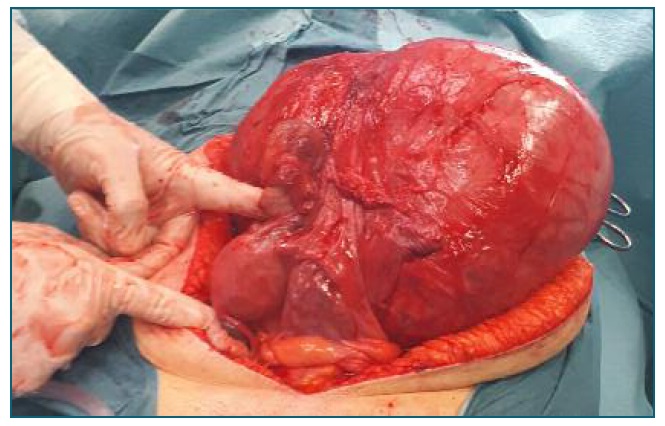
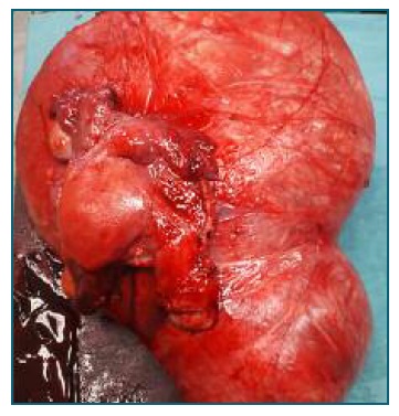
The authors present a case illustrating a huge subserous leiomyoma originating from the uterine cervix. The patient was a caucasian, 50 years-old, premenopausal, with no gynecological surveillance for the past 6 years. She presented with abdominal distention, pelvic pain, and dysmenorrhea for the past year. Clinical evaluation revealed a mass that occupied the entire pelvic cavity and extended into the abdominal cavity. MRI showed normal vascularization, suggesting a benign etiology. Due to the fibroid size and patient desire, an abdominal hysterectomy with bilateral adnexectomy was performed. Histopathological analysis revealed a benign leiomyoma with 16 cm and 855g of weight.

Figure 1 Laparotomy. Normal uterus and adnexa anteriorly pointed by the surgeon. 

Figure 2 Giant subserous myoma, originating from the cervix, adherent to the uterus and adnexa. 

Cervical myomas are extremely rare, with a reported incidence <1%5. Hysterectomy remains the first line treatment in postmenopausal women as it eliminates the risk of recurrence. Management of premenopausal patients is more challenging if there is a desire for fertility preservation. Laparotomic/laparoscopic myomectomy and radical trachelectomy5 have been described to treat giant cervical myomas and may be an option for younger patients.

Patient informed consent was obtained for publication.

Conflicts of interest

The authors declare that they have no conflict of interest.

References

1. De la Cruz MS, Buchanan E.M. Uterine Fibroids: Diagnosis and Treatment. Am Fam Physician, 2017, 95(2):100-107 [ Links ]

2. Pavone D, Clemenza S, Sorbi F, Fambrini M, Petraglia F. Epidemiology and Risk Factors of Uterine Fibroids, Best Pract Res Clin Obstet Gynaecol 2018, 46:3-11 [ Links ]

3. Chabbert-Buffet N, Esber N, Bouchard P. Fibroid growth and medical options for treatment, Fertil. Steril; 2014, 102:630-639 [ Links ]

4. Munro M, et al. FIGO classification system (PALM-COEIN) for causes of abnormal uterine bleeding in non-gravid women of reproductive age. Int J Gynecol Obstet, 2011, 113:3-13 [ Links ]

5. Wong, Jolene et al. Novel management of a giant cervical myoma in a premenopausal patient. BMJ case reports 2017 DOI: https://doi.org/10.1136/bcr-2017-221408 [ Links ]

Received: May 09, 2021; Accepted: September 05, 2021

Endereço para correspondência Mariana Crespo Marques E-mail: mcsmarques@gmail.com

Creative Commons License This is an open-access article distributed under the terms of the Creative Commons Attribution License