Introduction
Decidualization is a structural and functional modification of the endometrium caused by progesterone-mediated stimulation during pregnancy1,2. Although this intrauterine alteration is vital for the success of pregnancy, ectopic decidua (or deciduosis) can occur in other locations, such as the cervix, vagina, abdominopelvic cavity and even more rarely the lungs and skin1,3,4. This entity is most frequently described in pregnant women, but some cases of non-pregnant and even postmenopausal women have been reported5-7. The etiopathogeny is not fully understood, although there is some association to preexisting endometriosis8. It is usually an incidental finding during c-section, surgical management of ectopic pregnancy, postpartum tubal ligation or other surgical abdominal interventions during pregnancy in previously asymptomatic women9. Mild symptoms may occur, such as abdominal pain or irritable bowel syndrome10,11. However, due to the highly vascularized and friable lesions, it can also present as potentially life-threatening maternal and/or foetal situations, mostly due to haemorrhagic conditions: genital bleeding, hemoperitoneum (before, during or after labour) and even as lower gastrointestinal bleeding4,5,12-15.
In some cases, due to the exuberant macroscopic appearance, only a biopsy can establish the diagnosis and exclude the presence of malignancy or peritoneal tuberculosis7,15. Nonetheless, high suspicion and familiarity with this entity can spare a potentially highly haemorrhagic iatrogenic lesion, since deciduosis spontaneously regresses in four to six weeks postpartum, as well as in the physiologic process of decidualization5,6,9,16,17. If ectopic decidualization is highly likely, minimal manipulation and the most conservative measures should be performed to avoid adverse permanent morbidity, such as hysterectomy5,6.
Case report
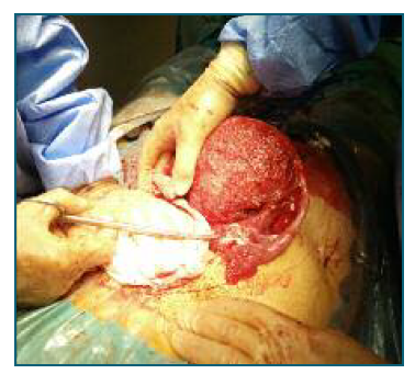
A primigravida in her early 30s, previously healthy, with an uneventful pregnancy except for the diagnosis of posterior placenta previa, for which she had no known risk factors, was admitted to the Emergency Department at 36 weeks and 1 day due to severe abdominal pain and regular uterine contractions. Gentle vaginal evaluation showed no vaginal bleeding or cervical dilation. Obstetric ultrasound showed a foetus in cephalic presentation with normal amniotic fluid and normal umbilical artery doppler velocimetry and a posterior placenta previa with no evidence of abruption. Cardiotocography showed no signs of foetal distress but regular uterine contractions. Persistence of intense complaints despite analgesic medication increased the suspicion of early-stage preterm labour. Due to the subsequent risk of placental haemorrhage, an immediate c-section was decided. When the abdominal cavity was reached and before the uterine incision, moderate volume hemoperitoneum was observed, mostly limited to the pelvic cavity and lower abdomen. The foetus was immediately extracted, with an Apgar Score of 9, 10 at the 1st and 5th minutes, respectively, and a weigh of 2790 g. The placenta was easily removed, showing no signs of previous detachment. Following the uterine incision closure, further abdominal cavity inspection revealed the only additional haemorrhagic source as an extensive lesion of friable and superficially vascularized tissue covering the posterior uterine wall, both adnexal regions and anterior wall of the rectum (Figure 1). Due to the macroscopic appearance and typical location, in spite of the extensive distribution, deciduosis was suspected, for which no biopsy was performed. Haemostasis was achieved with electrocoagulation and a reabsorbable oxidized cellulose meshwork. A drain was placed on the Douglas Pouch for blood loss surveillance. The postoperative hemoglobulin value was 9,2 g/dl (12 g/dl preoperative) and the patient was managed conservatively with iron supplementation. After an initial difficult pain management and constipation, she had a favourable evolution and was discharged on the 9th postoperative day wi-thout further complications. Puerperal evaluation at 4 weeks postpartum had no relevant findings, namely no lesions visualized on gynaecological ultrasound or fur-ther episodes of abdominal pain suggestive of intraabdominal bleeding. She recovered completely and had a second pregnancy 3 years later, also complicated by placenta previa. During the second c-section no deciduosis was described, but extensive intraabdominal adhesions were found.
Discussion
Ectopic decidualization is mostly an incidental finding during surgery, but may be the cause of severe hemoperitoneum and other haemorrhagic complications. The differential diagnosis of these lesions is malignancy (such as peritoneal mesothelioma) or tuberculosis. Due to the macroscopic resemblance of these diseases in some cases, biopsy should be done to stablish the diagnosis18. Whenever suspicion of this benign entity and surgical risk of biopsy are high, conservative haemostasis should be employed and unnecessary manipulation avoided due to the good prognosis and spontaneous regression of the lesions within four to six weeks postpartum5,6.
Authors’ contribution
DM and FS were directly responsible for the initial approach to the patient on the emergency department and surgical approach; DM, ISG, ML were closely involved in the follow up of the patient. All authors were major contributors in writing the manuscript. All authors read and approved the final manuscript.


