Conjoined twins are a complication of monozygotic twin pregnancies resulting from an aberrant splitting of the primitive node and streak, when that division occurs after the 14th post-conception day. Its incidence is 1:50 000 to 1:100 000 and no risk factors are identifiable1.
These twins are classified according to the anatomical site of union adding the suffix “pagus”: cephalopagus (head to umbilicus), thoracopagus (chest), omphalopagus (abdominal wall), ischiopagus (lower abdomen and pelvis), parapagus (chest, abdomen and pelvis), rachipagus (spines) and pyropagus (sacrum) 2.
The most common form of conjoined twins is fusion of the anterior or lateral facet of the thorax and/or abdomen, which constitutes 70% of conjoined twins2.
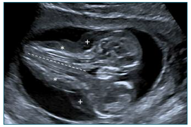
We present an image issue of a 33-year-old nulliparous woman, with unremarkable previous medical history and a spontaneous pregnancy. She presented to the Fetal Medicine Department for the first-trimester-scan and was diagnosed with an ongoing conjoined twin pregnancy. It was classified as dicephalus-parapagus since the ultrasound documented two alive foetuses fused by the lateral thorax, abdomen and pelvis in the caudal axis. They shared a common heart, diaphragm, gastrointestinal system and had four (two upper and two lower) limbs, it was also observed subcutaneous oedema. These conjoined twins are dicephalus because they had separated heads and faces. - Figure 1.

Figure 1 12th week ultrasound of the dichephalus-parapagus twins (coronal view). *Spines of both fetuses; + Subcutaneous oedema; --- lateral fusion of the chest, abdomen and pelvis in the caudal axis.
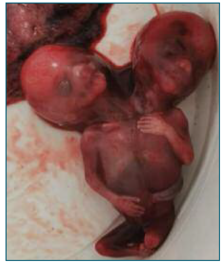
Chorionic villous sampling was performed and the results were normal (quantitative-fluorescence polymerase-chain-reaction and microarray). Regarding the poor prognosis and the unfeasibility of post-natal surgical separation, termination of pregnancy was offered upon parent`s request, after multidisciplinary team counselling (fetal medicine, pediatric surgery and neonatology) - Figure 2.
Conjoined twins are a rare phenomenon and a challenging medical situation. An anatomical scan must be performed in order to consider the post-natal surgical separation. The parapagus twins separation is not viable because they share a complex fused heart and is not possible to reconstruct one single functional organ2.
The first-trimester ultrasound scan of monochorionic monoamniotic twin pregnancy is extremely important: early diagnosis and classification of the type of conjoined twins is mandatory to enable proper counselling. On ultrasound, there are some clues that lead to the diagnosis: fetal apposition, spinal extension, a single heart and two lower limbs3.
A multidisciplinary team is essential when evaluating the viability of the fetuses after surgery and feasibility of the procedure are compulsory to better advise the parents. In our case a poor prognosis to both fetuses was established and the parents opted for termination of pregnancy.
In conclusion, our image issue highlights the complexity of the management of conjoined twins. First-trimester ultrasound is essential to early diagnosis, allowing a multidisciplinary team (fetal medicine, neonatologists, pediatric cardiologists and pediatric surgeons) to establish the prognosis. Comprehensive counselling to parents is important in order to give them time and information to decide about further care during pregnancy.


