SciELO - Scientific Electronic Library Online

 
vol.18 número4Gravidez numa mulher com linfangioleiomiomatose: um caso clínicoMetaplasia óssea endometrial - Um caso clínico incomum índice de autoresíndice de assuntosPesquisa de artigos
Home Pagelista alfabética de periódicos  

Serviços Personalizados

Journal

Artigo

Indicadores

Links relacionados

  • Não possue artigos similaresSimilares em SciELO

Compartilhar


Acta Obstétrica e Ginecológica Portuguesa

versão impressa ISSN 1646-5830

Acta Obstet Ginecol Port vol.18 no.4 Algés dez. 2024  Epub 31-Dez-2024

https://doi.org/10.69729/aogp.v18i4a07 

Issue Image/Imagem do Trimestre

Voluminous hematometra and cervical stenosis treated with office hysteroscopy

Hematometra volumoso e estenose cervical tratados com histeroscopia de consultório

António de Pinho1  2 

Catarina Estevinho1 

Cristina Oliveira1 

1. Serviço de Ginecologia e Obstetrícia da Unidade Local de Saúde Tâmega e Sousa, Penafiel, Portugal.

2. Departamento de Ginecologia-Obstetrícia e Pediatria da Faculdade de Medicina da Universidade do Porto. Portugal.


Abstract

A postmenopausal patient referred due to stress urinary incontinence presented with severe atrophy of the upper third of the vagina and an enlarged uterus. A voluminous hematometra and cervical dilation were detected and further characterized by MRI. The patient underwent an office hysteroscopy using a vaginoscopic approach without anestesia. The stenosis was resolved using 5Fr scissors, and one liter of old blood was evacuated. This minimally invasive procedure represents the gold standard in these settings, allowing for direct visualization and overcoming stenosis while minimizing the risk of injury.

Keywords: Acquired stenosis; Hematometra; Hysteroscopy; Postmenopausal; Vaginoscopic approach.

Resumo

Numa doente pós-menopáusica, referenciada por incontinência urinária de esforço, foi verificada a existência de atrofia grave do terço superior da vagina e um útero de dimensões aumentadas. Foi detectado um hematometra volumoso, associado a dilatação cervical, que foram caracterizados por ressonância magnética. A doente foi submetida a histeroscopia de consultório, utilizando abordagem vaginoscópica sem anestesia. A estenose foi resolvida com tesoura histeroscópica de 5Fr e um litro de conteúdo hemático foi drenado. Este procedimento minimamente invasivo representa o goldstandard neste contexto clínico, permitindo a visualização direta e a resolução da estenose, minimizando o risco de lesões.

Palavras-chave: Abordagem vaginoscópica; Estenose adquirida; Hematometra; Histeroscopia; Pós-menopausa.

A 66-year-old woman was referred to the Gynecology department due to stress urinary incontinence. Menopausal since the age of 52, never used hormone replacement therapy and denied postmenopausal bleeding. Examination showed severe atrophy of the upper vaginal third without a clear identification of the cervix and an enlarged uterus. Transvaginal ultrasound suggested a voluminous hematometra and cervical dilation (Figure 1 a), further characterized by MRI (Figure 1 b).

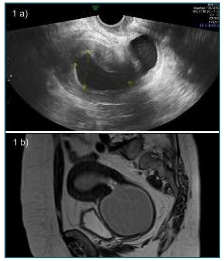
Figure 1 Transvaginal ultrasound (a) and parasagittal MRI (b) showing enlarged postmenopausal uterus with moderate cavity distension and severe distended cervical canal. Both structures are filled with hematic content. Maximum diameter of the uterine cavity - 32 mm; maximum diameter of the endocervical canal - 71 mm. No signs of focal lesions nor neoformation masses were recognized. Lesions probably secondary to upper vaginal stenosis. 

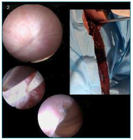
Figure 2 Office hysteroscopy details: a 5Fr scissors was used to perform a star-shaped incision in the exact location of the external cervical os, identified by the “blue behind the white” sign and allowed the passage into the cervical canal, creating an adequate external cervical os, which drained one liter of old blood. Cavity inspection and biopsy revealed atrophic endometrium. 

The patient underwent an office hysteroscopy with vaginoscopic approach. A complete obliteration of the external cervical os was observed (Figure 2). Stenosis was solved using endoscopic scissors and one liter of old blood was evacuated (Figure 2). The procedure was completed without complications and the patient remains under surveillance, with no recurrence of the clinical condition.

This case demonstrates a successful management of a voluminous hematometra and cervical stenosis with office hysteroscopy with vaginoscopic approach without anesthesia. This minimally invasive procedure represents the gold standard approach in these settings1),(2. With direct visualization, it is possible to overcome the stenosis while reducing the risk of injury3.

Author’s contribution

António de Pinho has contributed substantially to the collection and analysis of data, writing of the manuscript and final approval of the version to be published. Catarina Estevinho and Cristina Oliveira have contributed substantially to the critical review of the manuscript and final approval of the version to be published.

Conflicts of interest

The authors declare no conflicts of interest. No funding.

Statement of ethics

The authors declare that the procedures were followed according to the regulations established by the Clinical Research and Ethics Committee and to the Helsinki Declaration of the World Medical Association updated in 2013.

The authors declare having followed the protocols in use at their working center regarding patients’ data publication.

Informed consent

Obtained.

References

1. Di Spiezio Sardo A, Di Carlo C, Salerno MC, Sparice S, Bifulco G, Guida M, Nappi C. Use of office hysteroscopy to empty a very large hematometra in a young virgin patient with mosaic Turner's syndrome. Fertil Steril. 2007 Feb;87(2):417.e1-3. doi: 10.1016/j.fertnstert.2006.04.055. Epub 2006 Nov 21. PMID: 17118367. [ Links ]

2. Bettocchi S, Selvaggi L. A vaginoscopic approach to reduce the pain of office hysteroscopy. J Am Assoc Gynecol Laparosc. 1997 Feb;4(2):255-8. doi: 10.1016/s1074-3804(97)80019-9. PMID: 9050737. [ Links ]

3. Vitale SG, De Angelis MC, Della Corte L, Saponara S, Carugno J, Laganà AS, Török P, Tinelli R, Pérez-Medina T, Ertas S, Urman B, Angioni S. Uterine cervical stenosis: from classification to advances in management. Overcoming the obstacles to access the uterine cavity. Arch Gynecol Obstet. 2023 Jul 10. doi: 10.1007/s00404-023-07126-1. Epub ahead of print. PMID: 37428263. [ Links ]

Received: July 16, 2024; Accepted: August 26, 2024

Correspondence to António de Pinho E-mail: antonioapdepinho@hotmail.com

Creative Commons License This is an open-access article distributed under the terms of the Creative Commons Attribution License