Serviços Personalizados
Journal
Artigo
Indicadores
-
 Citado por SciELO
 -
 Acessos
Links relacionados
-
 Similares em
SciELO
Compartilhar
Angiologia e Cirurgia Vascular
versão impressa ISSN 1646-706X
Angiol Cir Vasc vol.15 no.2 Lisboa jun. 2019
ARTIGO ORIGINAL
Carotid endarterectomy: guidelines versus real-world practice
Endarterectomia carotídea: aplicação as guidelines na prática clínica
Gabriela Teixeira1, Pedro Sá Pinto1, Ivone Silva1, João Gonçalves1, Sérgio Teixeira1, Duarte Rego1, Vítor Ferreira1, Inês Antunes1, Carlos Veiga1, Daniel Mendes1, Paulo Teles2, Arlindo Matos1, Rui Almeida1
1 Serviço de Angiologia e Cirurgia Vascular, Centro Hospitalar Universitário do Porto
2 Faculdade de Economia do Porto e LIAAD, Universidade do Porto
Endereço para correspondência | Dirección para correspondencia | Correspondence
ABSTRACT
Introduction: A number of guidelines for the management of carotid disease are available to help clinicians in therapeutic decision and provide useful guidance for the best care of the patients. They stated that carotid endarterectomy (CEA) has some benefit in symptomatic patients with carotid stenosis of 50-69% and is highly beneficial in stenosis of 70-99%, with mortality/ stroke rate less than 6%. The maximum benefit is observed when surgery is performed within two weeks after the cerebrovascular event. CEA in asymptomatic patients should be offered to patients with life expectancy higher than 5 years, stenosis of >70%, and perioperative complications should be less than 3%.
The aim of this study is to analyse our clinical practice, review treated patients and referral times and compare the outcomes of our institutional practice with published guidelines.
Materials and Methods: Retrospective study of patients undergoing carotid endarterectomy at Centro Hospitalar Universitário do Porto between 2010 and 2015.
Results: Between 2010 and 2015, 404 patients underwent CEA, 76% men, and mean age 69 years for both sexes. The degree of stenosis was usually evaluated by ultrasound. The use of complementary study with angio-CT was required in 20% and angiography in 2.2% of the cases. The majority of patients (54.2%) were symptomatic (stroke/ TIA <6 months). In patients with a cerebrovascular event diagnosed at our institution, the time to surgery was 10 days (median). All CEA were performed under general anaesthesia and for arterial closure, a Dacronpatch was systematically used. Shunt was used in 4.6% of cases (n=18). The mean surgery time was 101 minutes and the mean clamping time was 40 minutes. Reversal of antioagulation with protamine was performed in 48.3% patients. Morbimortality: 9 post-operative sequelae events (major stroke), 8 minimal / transient deficit (minor stroke) and 4 deaths. In symptomatic patients (n = 219), the rate of postoperative major stroke was 3.6%. In asymptomatic patients (n = 185), a major and a minor stroke was observed (1.1%). Other complications: haematoma 5.9% (n=24); infection 0.2% (n=1); peripheral nerve dysfunction 8.7% (n=35); false aneurysm 0.5% (n=2).
Conclusions: Our results are within the reference values. This study allowed us to know our reality, our ability to give an effective answer for our patients, and to serve as a basis for improving ways of acting.
Keywords: Carotid disease guidelines; Carotid disease symptomatic and asymptomatic; Carotid endarterectomy; Complications
RESUMO
Introdução: Atualmente estão disponíveis diversas guidelines sobre o tratamento da doença carotídea de modo a ajudar os médicos na sua decisão terapêutica e garantir o melhor tratamento a cada paciente. Estas afirmam que a endarterectomia carotídea tem algum benefício nos doentes sintomáticos com estenose carotídea de 50 a 69% e é altamente benéfica em doentes sintomáticos com estenose carotídea de 70 a 99%, sendo que a taxa de mortalidade ou AVC deverá ser inferior a 6%. Para um benefício máximo, a endarterectomia deverá ser realizada nas primeiras duas semanas após o evento cerebrovascular. Em doentes assintomáticos, a endarterectomia carotídea deverá ser oferecida a pacientes com esperança de vida superior a 5 anos, estenose carotídea > 70% e as complicações perioperatórias deverão ser inferiores a 3%.
O objectivo deste estudo é analisar a nossa prática clínica, rever os doentes tratados, tempos de referência e comparar os resultados da nossa instituição com os publicados nas guidelines internacionais.
Material e Métodos: Estudo retrospectivo dos pacientes submetidos a endarterectomia carotídea no Centro Hospitalar Universitário do Porto entre 2010 e 2015.
Resultados: Durante o período de 2010 a 2015, 404 pacientes foram submetidos a endarterectomia carotídea, 76% homens e uma média de idades de 69 anos para ambos os sexos. O grau de estenose carotídea foi normalmente determinado por ecodoppler, havendo necessidade de estudo complementar com angioTC em 20% e angiografia em 2,2% dos casos. A maioria dos doentes (54,2%) eram sintomáticos (AVC ou AIT < 6 meses). Os pacientes cujo diagnóstico de evento cerebrovascular foi realizado na nossa instituição foram operados após 10 dias (mediana). Todas as endarterectomias foram realizadas sob anestesia geral e o encerramento arterial foi feito, sistematicamente, com recurso a patchde Dacron. Foi usado shunt em 4,6% dos casos (n=18). O período médio de cirurgia foi de 101 minutos e o tempo de clampagem de 40 minutos. A hipocoagulação foi revertida intraoperatoriamente com protamina em 48,3% dos casos. A morbimortalidade observada foi de 9 AVC major (com sequelas), 8 eventos cerebrovasculares mínimos/ transitórios e 4 mortes. Nos doentes sintomáticos (n=219), a taxa de AVC major foi de 3,6%. Nos doentes assintomáticos (n=185), um AVC majore um minorforam registados (1,1%). Outras complicações: hematoma 5,9% (n=24); infeção 0,2% (n=1); disfunção de nervo periférico 8,7% (n=35); falso aneurisma 0,5% (n=2).
Conclusão: Os nossos resultados enquadram-se dentro dos valores de referência. Este estudo permite-nos conhecer a nossa realidade e a nossa capacidade de resposta, assim como compreender formas de melhorar o nosso modo de actuação.
Palavras-chave: Diretrizes para doenças carotídeas; Doença carotídea sintomática e assintomática; Endarterectomia de carótida; Complicações
Introduction
A number of guidelines for the management of carotid disease are available to help clinicians in therapeutic decision and provide useful guidance for the best care of the patients.
American(1) and European(2) guidelines stated that carotid endarterectomy (CEA) has some benefit in symptomatic patients with carotid stenosis of 50-69% and is highly beneficial in stenosis of 70-99%. The mortality/ stroke rate should be less than 6%. The maximum benefit is observed when surgery is performed within two weeks after the cerebrovascular event. CEA in asymptomatic patients should be offered to patients with life expectancy higher than 5 years, stenosis of >70%, and perioperative complications should be less than 3%. The documented incidence of haemorrhagic complications requiring re-exploration was 3.4%. Peripheral nerve injury prevalence was 4.2% for the recurrent laryngeal nerve, 3.8% for the hypoglossal nerve, 1.6% for the mandibular branch of the facial nerve, 0.2% for the glossopharyngeal nerve, and 0.2% for the spinal accessory nerve. And infection complicates 0.4-1.8% of procedures.
The aim of this study is to analyse our clinical practice, review treated patients and referral times and compare the outcomes of our institutional practice with published guidelines.
Methods
A retrospective cohort study was conducted on a consecutive series of patients undergoing carotid endarterectomy between January 2010 and December 2015, at Centro Hospitalar Universitário do Porto - Hospital de Santo António. Study eligibility criteria included patients submitted to carotid endarterectomy, with a documented carotid stenosis by ultrasound, angio-CT or angiography.
Data Collection and Description: All data was obtained from electronic patient records. The following data were recorded: demographic data (age and sex); co-morbidities (smoking status, diabetes mellitus, hypertension, dyslipidaemia and chronic kidney disease); risk factors for surgical endarterectomy - "high-risk" patients (contralateral carotid occlusion, New York Heart Association (NYHA) Grade III/IV Congestive Heart Failure (CHF), severe left ventricular dysfunction, acute myocardial infarction <6 months, unstable angina Class III/IV of the Canadian Cardiovascular Society (CCV) and severe lung disease); chronic medication; symptomatology in the presentation; hospital of referral; complementary diagnostic tests; length of surgery, length of clamp; type of anaesthesia, use of shunt, reversal of anticoagulation; time from the event to surgery; length of hospital stay; medication on discharge date and morbidity and mortality within 30 days after surgery.
Definitions: Patients with a history of focal neurological deficits with definitive or transient ischemic aetiology within a period of 6 months prior to the surgical procedure were considered symptomatic. According to the definition of WHO, World Health Organization, we distinguished patients with stroke when the neurological condition lasted more than 24 hours, transient ischemic attack (TIA) when the duration of the deficits was less than 24 hours(6). Patients who presented without a history of cerebral ischemia or with episodes greater than 6 months were considered asymptomatic. Those whose carotid stenosis was detected during the investigation of a dizziness, syncope or tinnitus, were considered as atypical symptomatology.
The postoperative complications included haematoma defined as cervical haematoma collection requiring surgical drainage; infection defined as development of local or systemic inflammatory signs, with a starting point in the surgical wound, requiring antibiotic therapy or surgical revision; cranial nerve injury defined as transient or definitive dysfunction of peripheral nerves in the postoperative period, usually characterized by tongue deviation, hoarseness or dysphagia; acute myocardial infarction defined as ischemic cardiac event with alterations of new electrocardiogram or with sustained elevation of cardiac enzymes; stroke defined as new neurological deficit in the postoperative period, with permanent sequelae; minor stroke defined as postoperative minimal neurological symptoms or deficits with complete recovery during the follow up.
Surgical procedure: All endarterectomies were performed by anteromedial approach and the conventional endarterectomy procedure was used. Patients received systemic heparinisation with unfractionated heparin and, in some cases, reversal with protamine. We used patch closure for all patients undergoing CEA. All procedures were performed under general anaesthesia, and cerebral perfusion monitoring was performed through retrograde arterial measurement by the internal carotid artery and regional cerebral oximetry (INVOS). In patients with a stump pressure < 40mmHg or a significant drop (>20%) in INVOS, a Javid's shunt was used. When necessary, fibrin sealants were used for haemostasis of the patch suture. All patients were left with a suction drain for the prevention of haematoma formation. Patients were kept under surveillance during 24 hours with invasive arterial pressures, in the Intermediate Care Unit or, exceptionally, in the recovery room of theatre. After this period, and in the absence of complications, the patients were transferred to the Vascular Surgery Unit.
Statistical analysis: Statistical analysis was performed using IBM SPSS® statistical software (version 22). The binary logistic regression model was used with estimates of odds ratio (OR) and 95% confidence interval (95% CI). Two-sided tests statistical significance was assumed for p<0.05.
Results
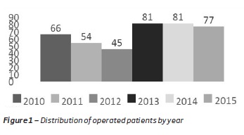
Over 6 years, 404 patients underwent CEA at Centro Hospitalar Universitário do Porto - Hospital de Santo António. The number of patients operated by year is shown in Figure 1.
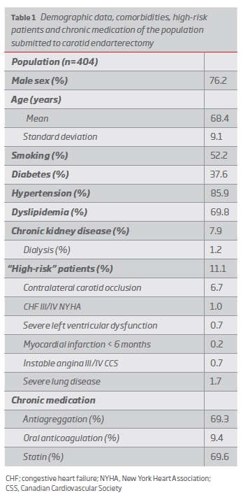
The patients submitted to CEA were mostly male (76.2%) and the mean age was approximately 69 years in each sex, (men, min: 47, max: 90; women, min: 48, max 89). The population characterization and comorbidities are summarized in Table 1.
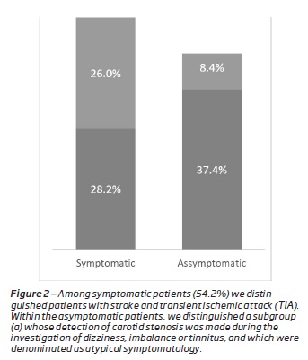
Most patients (54.2%) were symptomatic (stroke / TIA <6 months), with 28.2% having stroke and 26% with TIA. The remaining patients (45.8%) were asymptomatic. Within these, we distinguished those whose carotid stenosis was a finding in the investigation of dizziness, imbalances or dizziness, as represented in Figure 2.
The carotid arteriotomy was closed with a Dacron patch and all interventions underwent under general anaesthesia. Shunt was used in 4.6% of the cases (n = 18). The mean time to surgery was 101 minutes and the mean clamp time was 40 minutes.
Unfractionated heparin was given prior to arterial clamping in 100% of cases and reversion of anticoagulation with protamine was done in 48.3%.
The mean and median length of hospital stay were 8 and 5 days, respectively.
At discharge, 76.7% of the patients were antiaggregated with acetylsalicylic acid and 29% with clopidogrel; 93.6% of the patients were prescribed statin and 8.4% restarted previous oral anticoagulation.
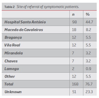
Regarding the site of referral of the 219 symptomatic patients, we only have the data of 168 (76.7%). Of these, 98 patients had a diagnosis of cerebrovascular event and surgical indication at Hospital de Santo António. In the others, the diagnosis of cerebral ischemic event was made in another hospital, and they were transferred to the Vascular Surgery Unit at Hospital Santo António, where they received surgical indication - Table 2.
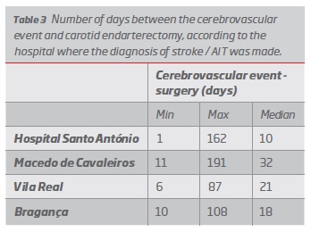
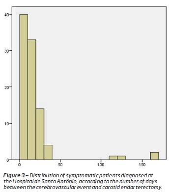
Regarding the time between the cerebrovascular event and the surgery, there is a large difference between the patients who were sent for evaluation by Vascular Surgery at the time of the acute event, and the patients referred by scheduled appointment. We present in Table 3 the reference times, in the form of minimum, maximum and median. It should be noted that patients whose diagnosis of the acute event was made in the Hospital de Santo António were the only ones whose time until surgery reached the gold period for surgery in symptomatic patients (<14 days) - Figure 3.
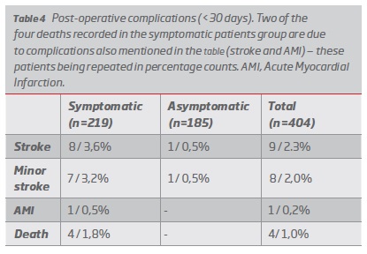
Postoperative morbidity and mortality are shown in Table 4. In the symptomatic patients, eight strokes, seven minor strokes (mild or transient neurological deficits), one acute myocardial infarction and four deaths were registered. In asymptomatic patients, a stroke with permanent sequelae and one transient neurological deficit was detected (1.1%). The deaths were secondary to one stroke with haemorrhagic transformation, one acute myocardial infarction, one cervical haematoma and one death of unidentified cause, in a patient found in cardiac arrest, two days after surgery, and whose aetiology was not possible to determine.
The other postoperative complications were: cervical haematoma requiring surgical revision in 5.9% of the patients (n = 24). Among patients with cervical haematoma, two required tracheostomy due to a compromised airway. The infection rate was 0.2% - one patient developed a wound abscess without patch involvement, and resolved with abscess drainage, seven days after CEA, and antibiotherapy - a methicillin-susceptible Staphylococcus aureus was isolated. Peripheral nerve injury was observed in 8.7% of the patients (n = 35), the vast majority with only transitional complaints. Reoperation (<30 days) was required in 6.4% of patients (n = 26), 24 per haematoma, one due to immediate postoperative neurological deficit and one per infection.
The surgery time was divided into two groups (d117 min vs > 117min), with the upper quartile time (percentile > 75%) being 117 minutes(7), and a logistic regression was performed in the search for the association between longer surgical time and the development of complications. None of the parameters was statistically significant, so it is not possible to state that the time of surgery > 117min predisposes to the occurrence of stroke, cranial nerve dysfunction or haematoma formation.
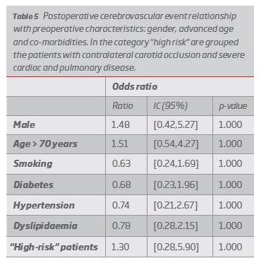
A statistical analysis was performed in an attempt to find risk factors for cerebrovascular event in the postoperative period. The following variables were compared: gender, age > 70 years, smoking, diabetes, hypertension, dyslipidaemia, chronic kidney disease and "high-risk" in the patients that had a postoperative cerebrovascular event. However, no statistically significant relationship was found - Table 5.
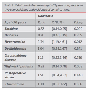
Patients younger and older than 70 years were compared in an attempt to understand whether advanced age would be a risk factor for carotid endarterectomy in our population. The only statistically significant differences were in the preoperative characteristics: the older population is less likely to be a smoker, are more hypertensive, and more likely to be in the "high-risk" group. No difference was found in the incidence of complications - Table 6.
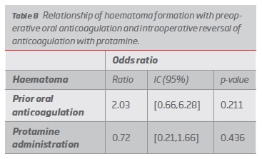
Finally, we analysed if the incidence of postoperative cervical haematoma requiring surgical revision was linked to prior oral anticoagulation or with reversal of anticoagulation (unfractionated heparin administered intraoperatively) with protamine. Despite the trend of increased numbers of haematomas in previously anticoagulated patients and an apparent protective role for protamine, odds ratio confidence intervals include the value 1 and the difference in proportions is not statistically significant - Table 8.
Discussion
Knowing that the main reason for performing carotid surgery - the prevention of an acute cerebrovascular event - is also one of its possible complications, it is necessary that the indications, limitations and contraindications are well defined, so that the benefits outweigh the risks. Thus, careful guidelines were created and successively updated so that the decision of surgery (conventional or endovascular) is made in the light of scientific evidence, with safety for the surgeon and for the patient(1-2). However the results presented in these guidelines, based on prospective, controlled studies, with selection criteria for surgeons in them, may be different from those of the "real world".
The latest guidelines(2) published by the European Society of Vascular Surgery (ESVS) describe, with level of evidence I, surgical indication for patients with symptoms of the carotid territory in the last 6 months and carotid stenosis of 50-99% in centres with mortality rate <less than 6%. For asymptomatic patients, surgical indication is indicated in patients with "medium surgical risk" and stenosis of 60 to 99%, with imaging characteristics associated with an increased risk of ipsilateral stroke in centres with a mortality / stroke rate of less than 3% and in patients with a life expectancy of more than 5 years. The results presented are within the proposed values. In the asymptomatic patients, the postoperative stroke rate was 0.5%, and no death was recorded. In symptomatic patients, the complication rate (major stroke, acute myocardial infarction and mortality rate) was 5.0%.
Holt et al, proposed that the results will be better in high volume specialized centres. In a meta-analysis that gathered 25 studies and more than 900,000 carotid endarterectomies, it documented significantly lower mortality and stroke rates in high-volume hospitals and defined 79 surgeries per year as the threshold value to achieve these results. However, in our study, we found that in the period 2010-2012 when the volume of surgeries in our hospital was lower, 6 cerebrovascular events were recorded (6/165, 3.6%), while in the period 2013 (11/241, 4.5%), which may lead to the presumption of other risk factors for complication beyond the volume of surgeries.
In an attempt to understand that other preoperative and intraoperative factors would be associated with incidence of complications, a comparative analysis was made. Patients with anatomic or clinical risk factors with a higher probability of complications following conventional surgery (death, stroke, MI or peripheral nerve injury) were classified as "high risk" patients, and in these cases, the surgical indication should be rethought, and carotid stenting considered besides endarterectomy. Based on the SAPPHIRE study(9), we considered "high-risk" patients: contralateral carotid occlusion, NYHA grade III / IV heart failure, severe left ventricular dysfunction, unstable angina III / IV CCS, and severe pulmonary disease. No significant value was found to relate this "high-risk" population to a greater number of postoperative complications, but this can be attributed to the small sample size (45 "high-risk" patients, 11.1%). No patient in our population had previous history of cervical radiotherapy, neck surgery or contralateral laryngeal nerve palsy, usually considered contraindications for endarterectomy.
Age does not appear to be a risk factor for carotid endarterectomy. Several meta-analyses have shown that advanced age is a risk factor for postoperative stroke after stenting, but not after carotid endarterectomy (CREST(10) and EVA-3S(11) for age over 70 years and SPACE(12) for over 80 years). Our study, although limited by the small sample of postoperative strokes (n = 17), did not find statistically significant differences in the incidence of postoperative stroke or other complications in the population older than 70 years, compared to the younger population.
Cervical haematoma is a common and dangerous complication as it can compromise the airway and endanger the lives of patients. Our haematoma rate (5.9%) is similar to previously described(3, 13); however, our study also confirms its severity and associated risks, with one death following a cervical haematoma.
Longer surgical times may be related to an increase in postoperative complications. Azis et al(7), in a review of the American College of Surgeons database with patients undergoing carotid endarterectomy between 2005 and 2007, associated a prolonged surgery time (> 140 min, corresponding to the> 75% percentile) (1.3% vs. 0.7%, P = 0.013), hospitalization time e 7 days (12.7% vs. 8.1%, P <0.001), postoperative pneumonia (1.6% vs. 0.9% P = 0.001), incapacity to wean the ventilator for more than 48 hours (1.8% vs. 0.6%, P <0.001) and need for reintervention (6.5% vs. 5.2%, P = 0.010). More recently, Perri et al(14), in an analysis of 26,327 carotid endarterectomies performed between 2012 and 2015 in 249 different centres, with an average surgical time of 114 minutes, demonstrated an association between cardiac complications and longer surgical time (OR 2.16; 95% CI, 1.59-2.95, P <0.001). In the retrospective study presented here, the mean surgical time was 101 minutes, and the statistical study, considering the longest surgical time corresponding to percentile> 75% (> 117 minutes) did not show a statistically significant association with the development of postoperative complications.
Carotid ultrasound for the study of syncope is not recommended(15). Despite the limited literature published on syncope and carotid stenosis suggesting their possible association with bilateral severe carotid stenosis(16-17), their relation was not recognized. The same is true for dizziness(18-19) and for tinnitus(20-21) with some work associating these symptoms with carotid stenosis. However, none of these symptoms appear in the most recent guidelines on carotid disease. Thus, given the frequency of these patients, and the absence of criteria to include them in the symptomatic patient group, we chose to create a category of atypical symptomatology within asymptomatic patients. In the work presented no vascular accident, with definitive or transient symptoms, was documented within the first 30 postoperative days of these patients.
Regarding the gold period for surgical intervention after cerebrovascular event, in our institution only patients whose stroke / TIA diagnosis was made at our hospital achieved a median of less than the indicated 14 days. In a more detailed analysis, we can see that it is not so much due to delays in the scheduling of patients, who usually receive the title of priority, but rather at referral level, especially at the level of the most remote districts. Thus, a more detailed analysis of referral and scheduling times will be worthwhile in order to understand the flaws and try to improve this point.
This study has several limitations, the main one related to its retrospective nature and its implications, namely the lack of some data (such as the origin of 23.3% of symptomatic patients). The reduced number of cerebrovascular complications in the postoperative period did not allow the achievement of statistical significance in the establishment of risk factors. Another important limitation is the follow-up time: complications are limited to the first 30 postoperative days.
Conclusions
This study allowed us to know our reality, our patients, our ability to give an effective answer, and serve as a basis for improving techniques and ways of acting. Although retrospective, and with no statistical power to establish cause-effect relationships between risk factors and complications, we were able to understood our results and fit them into the reference values, serving as a basis for further investigation and to improve detected failures - in particular referral times.
REFERENCES
1. Brott TG, Halperin JL, Abbara S, Bacharach JM, Barr JD, Bush RL, et al. 2011ASA/ACCF/AHA/AANN/AANS/ACR/ASNR/CNS/SAIP/SCAI/SIR/SNIS/SVM/SVS guideline on the management of patients with extracranial carotid and vertebral artery disease. A report of the American College of Cardiology Foundation/American Heart Association Task Force on Practice Guidelines, and the American Stroke Association, American Association of Neuroscience Nurses, American Association of Neurological Surgeons, American College of Radiology, American Society of Neuroradiology, Congress of Neurological Surgeons, Society of Atherosclerosis Imaging and Prevention, Society for Cardiovascular Angiography and Interventions, Society of Interventional Radiology, Society of NeuroInterventional Surgery, Society for Vascular Medicine, and Society for Vascular Surgery. Circulation. 2011;124(4):e54-130. [ Links ]
2. Naylor AR, Ricco JB, de Borst GJ, Debus S, de Haro J, Halliday A, et al. Editor's Choice - Management of Atherosclerotic Carotid and Vertebral Artery Disease: 2017 Clinical Practice Guidelines of the European Society for Vascular Surgery (ESVS). Eur J Vasc Endovasc Surg. 2018 Jan;55(1):3-81. [ Links ]
3. Kakisis JD, Antonopoulos CN, Mantas G, Moulakakis KG, Sfyroeras G, Geroulakos G. Cranial Nerve Injury After Carotid Endarterectomy: Incidence, Risk Factors, and Time Trends. Eur J Vasc Endovasc Surg. 2017 Mar;53(3):320-335. [ Links ]
4. Morales Gisbert SM, Sala Almonacil VA, Zaragozá García JM, Genovés Gascó B, Gómez Palonés FJ, Ortiz Monzón E. Predictors of cervical bleeding after carotid endarterectomy. Ann Vasc Surg. 2014 Feb;28(2):366-74. [ Links ]
5. Naylor AR, Payne D, London NJ, Thompson MM, Dennis MS, Sayers RD, Bell PR. Prosthetic patch infection after carotid endarterectomy. Eur J Vasc Endovasc Surg. 2002 Jan;23(1):11-6. [ Links ]
6. Aho K, Harmsen P, Hatano S, Marquardsen J, Smirnov V, Strasser T. Cerebrovascular disease in the community: results of a WHO Collaborative Study. Bull World Health Organ. 1980;58(1):113-30. [ Links ]
7. Aziz F, Lehman EB, Reed AB. Increased Duration of Operating Time for Carotid Endarterectomy Is Associated with Increased Mortality. Annals of vascular surgery. 2016;36:166-74. [ Links ]
8. Holt PJ, Poloniecki JD, Loftus IM, Thompson MM. Meta-analysis and systematic review of the relationship between hospital volume and outcome following carotid endarterectomy. European journal of vascular and endovascular surgery : the official journal of the European Society for Vascular Surgery. 2007;33(6):645-51. [ Links ]
9. JS JY, Wholey M, Kuntz R, Fayad P, Katzen B, Mishkel G, et al. Protected carotid-artery stenting versus endarterectomy in high-risk patients. N Engl J Med. 2004;351(15):1493-501. [ Links ]
10. Silver FL, Mackey A, Clark WM, Brooks W, Timaran CH, Chiu D, et al. Safety of stenting and endarterectomy by symptomatic status in the Carotid Revascularization Endarterectomy Versus Stenting Trial (CREST). Stroke. 2011;42(3):675-80. [ Links ]
11. Mas J-L, Trinquart L, Leys D, Albucher J-F, Rousseau H, Viguier A, et al. Endarterectomy Versus Angioplasty in Patients with Symptomatic Severe Carotid Stenosis (EVA-3S) trial: results up to 4 years from a randomised, multicentre trial. The Lancet Neurology. 2008;7(10):885-92. [ Links ]
12. Eckstein H-H, Ringleb P, Allenberg J-R, Berger J, Fraedrich G, Hacke W, et al. Results of the Stent-Protected Angioplasty versus Carotid Endarterectomy (SPACE) study to treat symptomatic stenoses at 2 years: a multinational, prospective, randomised trial. The Lancet Neurology. 2008;7(10):893-902. [ Links ]
13. Ferreira A, Vieira M, Sampaio S, Cerqueira A, Teixeira J. Predictors of neck bleeding after carotid endarterectomy: A 5 year revision. Angiologia e Cirurgia Vascular. 2016;12(1):12-9. [ Links ]
14. Perri JL, Nolan BW, Goodney PP, DeMartino RR, Brooke BS, Arya S, et al. Factors affecting operative time and outcome of carotid endarterectomy in the Vascular Quality Initiative. Journal of vascular surgery. 2017;66(4):1100-8. [ Links ]
15. Scott JW, Schwartz AL, Gates JD, Gerhard-Herman M, Havens JM. Choosing wisely for syncope: low-value carotid ultrasound use. Journal of the American Heart Association. 2014;3(4). [ Links ]
16. White RP, Markus HS. Impaired Dynamic Cerebral Autoregulation in Carotid Artery Stenosis. Stroke. 1997;28(7):1340-4. [ Links ]
17. Miran M, Suri M, Qureshi M, Ahmad A, Suri M, Basreen R, et al. Syncope in Patient with Bilateral Severe Internal Carotid Arteries Stenosis/Near Occlusion: A Case Report and Literature Review. J Vasc Interv Neurol. 2016;9(1):42-5. [ Links ]
18. Hsu LC, Chang FC, Teng MM, Chern CM, Wong WJ. Impact of carotid stenting in dizzy patients with carotid stenosis. Journal of the Chinese Medical Association : JCMA. 2014;77(8):403-8. [ Links ]
19. Cheng HL, Lin CJ, Soong BW, Wang PN, Chang FC, Wu YT, et al. Impairments in cognitive function and brain connectivity in severe asymptomatic carotid stenosis. Stroke. 2012;43(10):2567-73. [ Links ]
20. Carlin R, McGraw D, Anderson C. Objective tinnitus resulting from carotid artery stenosis. Journal of vascular surgery. 1997;25(3):581-3. [ Links ]
21. Terzi S, Arslanoglu S, Demiray U, Eren E, Cancuri O. Carotid Doppler ultrasound evaluation in patients with pulsatile tinnitus. Indian journal of otolaryngology and head and neck surgery : official publication of the Association of Otolaryngologists of India. 2015;67(1):43-7. [ Links ]
Endereço para correspondência | Dirección para correspondencia | Correspondence
Correio eletrónico: tgabrielateixeira@gmail.com (G. Teixeira).
Recebido a 30 de junho de 2019
Aceite a 23 de agosto de 2019


