




 Inglês (pdf)
 Artigo em XML
 Referências do artigo

 Tradução automática
 Enviar este artigo por email
 Citado por SciELO
 Similares em
SciELO

 Permalink

Introduction
Marfan Syndrome is a hereditary connective tissue disorder that affects multiple systems and presents with a characteristic phenotype. Cardiovascular involvement is common, manifesting as ascending aortic dilation, aortic insufficiency, aortic dissection, or extensive thoraco-abdominal aneurysms.1 Mortality associated with vascular complications is high and requires complex surgical treatment by highly experienced professionals. The gold standard for treating the thoracic or abdominal aorta, when necessary, is open surgery, with good long-term outcomes.2 However, given the thoraco-abdominal involvement in many cases of Marfan syndrome, patients often require multiple staged interventions. The use of endovascular approaches is often questioned because of the disease’s continuous evolution and long-term complications.3
Case report
We present the case of a 44-year-old male patient admitted to hospital in January 2011 with retrosternal pain and hypertension. The patient was diagnosed with Marfan syndrome, characterised by an elongated skeleton, pectus deformity, and an ophthalmological disorder. Computed tomography angiography (CTA) revealed a type A aortic dissection, prompting emergency surgery to replace the aortic valve (mechanical) and the ascending aorta. In subsequent years, the patient developed a type II thoraco-abdominal aortic aneurysm following residual dissection, and open repair was indicated. Nevertheless, in January 2014, the aneurysm ruptured at the level of the thoracic aorta, necessitating urgent open repair with an interposition graft (30 mm tube graft) in the descending thoracic aorta via a left thoracotomy, Figure 1.
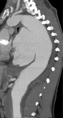
Figure 1 Computed tomography angiography revealing rupture of descending thoracic aorta in January 2014 (sagittal view)
One year later, in July 2015, a multidisciplinary team performed an open thoraco-abdominal repair with an interposition aorto-bi-iliac graft and individual bypasses to the visceral and renal arteries, Figure 2.
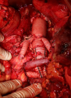
Figure 2 Intra-operative image of the thoraco-abdominal interposition graft, with individual bypass to the visceral and renal arteries
The patient recovered well from these procedures, but in September 2019, presented with thoracic pain and fever. Additionally, an abnormally elevated leucocyte count and increased C-reactive protein levels were identified, prompting further imaging that revealed a perigraft collection and air bubbles. Furthermore, a blood culture grew E. coli, and a positron emission tomography (PET) scan confirmed a diagnosis of prosthetic infection. Antibiotic therapy was initiated, stabilising the inflammatory/infectious process. During this period, progressive dilation of the last segment of the native aorta, the arch, was observed. The patient was discussed in a multidisciplinary aortic team, which decided that redo sternotomy and open repair of the arch carried prohibitive risk, and thus he was kept under conservative management.
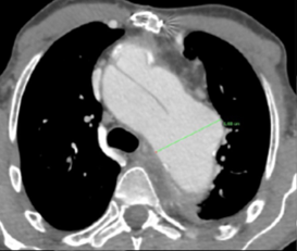
However, the aortic arch kept enlarging, reaching 7cm, Figure 3. Since open surgery was not an option, a compassionate endovascular strategy was planned, knowing the risks of endovascular repair in a connective tissue patient and given the high risk of endograft infection.
The case was discussed with the COOK® Planning Centre, and a custom-made arch endograft (COOK® Zenith) with two inner branches (one antegrade for the left carotid and one retrograde for the left subclavian) was planned, Figure 4. The device was built with a short tip to pass the mechanical valve and relied on the previous surgical grafts as proximal and distal landing zones (graft-to-graft repair). The left carotid and left subclavian were planned as target vessels to ensure sufficient proximal sealing length in the remaining native aorta and ascending graft without covering the coronary ostia, which was not possible with an inner branch for the innominate artery.
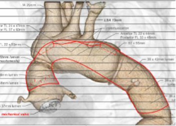
A staged repair was planned and performed, comprising cervical debranching followed by endovascular arch repair. Left carotid-to-right carotid and prosthetic-to-right subclavian bypasses were performed. Additionally, because the right vertebral artery originated directly from the arch, it was re-implanted onto the right common carotid artery.
The endovascular procedure was performed under general anaesthesia with fusion guidance. Access was obtained via surgical exposure of the right common femoral artery, the left common carotid artery, and the left brachial artery, together with a left percutaneous femoral artery access. The left ventricle was catheterised after passing the mechanical valve (bileaflet valve) through the side of one of the leaflets to avoid complete aortic insufficiency during endograft deployment (Figure 5). The main graft was then positioned through the mechanical valve and deployed under rapid pacing to reduce cardiac output. During this stage, after the device had been fully deployed under rapid pacing, the patient developed cardiorespiratory arrest (ventricular fibrillation), which was rapidly reversed with defibrillation without complications.
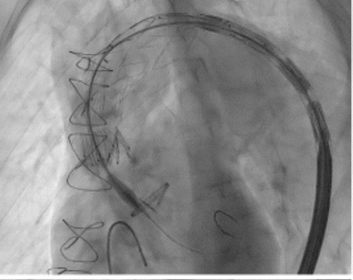
The tip of the delivery system is seen crossing the aortic mechanical valve
Catheterisation of the antegrade inner-branch was performed via the carotid access, and a covered balloon-expandable stent was delivered (2x Bentley® Begraft Aortic 12x59). The retrograde inner-branch was catheterised via the brachial access, and a through-and-through wire was established via the right femoral access. Due to the large size of the subclavian artery, covered balloon-expandable stents were used (2x Bentley® Begraft Aortic 14x59), which were advanced over the through-and-through wire from the femoral access. The final angiogram showed aneurysm exclusion and patency of all devices. The patient was discharged after 30 days, and a control CTA showed aneurysm exclusion and patency of all stented target vessels and the cervical debranching. He was kept on lifelong antibiotic treatment with amoxicillin + clavulanic acid and doxycycline.
The patient continues outpatient follow-up with regular appointments every 3 to 6 months. A new CTA was performed at 1 month, 3 months, 6 months, and 1 year after the last surgery. Currently, the patient undergoes regular imaging follow-up annually, with stable infection control and no aortic complications, maintaining a good quality of life.
Discussion
Open surgery remains the gold standard for patients with Marfan Syndrome, with good early and long-term outcomes,2 and the endovascular approach is considered by different authors as an option when open surgery is not feasible or as a bridging measure in urgent repairs.4 Complications associated with endovascular treatment typically involve the proximal and distal sealing zones, including a high risk of retrograde aortic dissection, type 1 endoleaks, and progressive aortic dilatation. Meanwhile, a recent multicentre study analysed the treatment of 171 patients with connective tissue disorders (83% Marfan patients) submitted to endovascular repair, including complex arch and thoraco-abdominal repairs. Results demonstrated high technical success (98.2%), very low peri-operative mortality (2.9%), and 5-year survival of 80.6% for Marfan patients.5 However, the study showed that 53.2% of patients required secondary procedures. Comparable results were reported by Gomez-Mayorga et al, with endovascular repair achieving similar outcomes when comparing patients with and without genetic aortopathies, with excellent short-term results but higher 2-year reintervention rates.6 These results call for close surveillance after endovascular repair in genetic aortopathies. However, endovascular treatment should probably be considered a viable option in the guidelines, especially in patients with previous repairs, where surgical grafts may be used as landing zones, avoiding highly complex redo surgeries.7
In our case, both proximal and distal landing zones were achieved using previous aortic grafts reducing the risks of sealing zone complications. The mechanical aortic valve, frequently considered a contraindication for endovascular aortic arch repair, was managed by careful planning, which included extensive right side cervical debranching to allow sufficient proximal aortic sealing length for graft delivery in addition to the short-tip delivery system and the previously described technique of catheterizing the side of on the leaflet (in bi-leaflet aortic valves) to avoid complete aortic insufficiency.8
Concerns obviously exist in our patient regarding long-term durability and risk of infection. However, with a 7 cm and growing aneurysm, we considered rupture to be imminent and after discussing the option with the patient, the endovascular solution with all of its caveats was considered the best option.
In conclusion, complex endovascular solutions can be an option in selected connective tissue patients, ideally with both proximal and distal landing zones in previous surgical grafts. These patients should be referred to centres with established aortic teams able to provide the full range of options including both modern open and endovascular solutions.

