SciELO - Scientific Electronic Library Online

 
vol.79 número4Duas Novas Mutações do Gene ATP2C1 em Doentes Portuguesas com Doença de Hailey-HaileyCriptococose Cutânea Primária em Paciente Imunocompetente: Um Relato de Caso índice de autoresíndice de assuntosPesquisa de artigos
Home Pagelista alfabética de periódicos  

Serviços Personalizados

Journal

Artigo

Indicadores

Links relacionados

  • Não possue artigos similaresSimilares em SciELO

Compartilhar


Revista da Sociedade Portuguesa de Dermatologia e Venereologia

versão impressa ISSN 2182-2395versão On-line ISSN 2182-2409

Rev Soc Port Dermatol Venereol vol.79 no.4 Lisboa dez. 2021  Epub 01-Jan-2022

https://doi.org/10.29021/spdv.79.4.1397 

Case Report

Topical Photodynamic Therapy as a Treatment Option for Periocular Bowen's Disease: A Case Report

Terapêutica Fotodinâmica Tópica como Opção de Tratamento de Doença de Bowen Periocular: Caso Clínico

Joel Reis1 

A. Machado1 

André Coelho2 

Virgilio Costa1 

Manuela Selores1  3  4  5 
http://orcid.org/0000-0001-8270-8317

1Department of Dermatology, Centro Hospitalar Universitário do Porto, Porto, Portugal

2Department of Anatomopathology, Centro Hospitalar Universitário do Porto, Porto, Portugal

3Department of Dermatology and Dermatology Research Unit, Centro Hospitalar Universitário do Porto, Portugal

4Instituto de Ciências Biomédicas Abel Salazar, University of Porto, Portugal

5Unit for Multidisciplinary research in Biomedicine, University of Porto, Portugal


ABSTRACT

Cutaneous squamous cell carcinoma in situ of the eyelid and periorbital skin malignancies is common and its management is demanding. Surgical excision is considered the first-line treatment, but these techniques have limitations. Topical photodynamic therapy is currently approved for the treatment of squamous cell carcinoma in situ in other areas, but the reports of its use in this sensitive anatomic location area are scarce. Herein we report the case of a 61-year-old-man with extensive periocular Bowen disease that was treated successfully with photodynamic therapy. Photodynamic therapy may be an option as neoadjuvant or curative therapy in selected cases, especially in sensitive anatomic locations where surgery might lead to distortion of the structures or where techniques as micrographically controlled surgery are not available.

KEYWORDS: Bowen's Disease/therapy; Photochemotherapy; Photosensitizing Agents/therapeutic use.

RESUMO

O carcinoma epidermóide in situ localizado na pálpebra e região periocular é comum e o seu tratamento é desafiante. A excisão cirúrgica é considerada opção terapêutica de primeira-linha, mas tem limitações. A terapia fotodinâmica tópica está atualmente aprovada para o tratamento do carcinoma epidermoide in situ em outras localizações, mas os relatos do seu uso em nesta zona anatómica são escassos. Apresentamos o caso de um homem de 61 anos, com um carcinoma epidermoide in situ de grandes dimensões localizado na região periocular tratado eficazmente com terapia fotodinâmica. A terapia fotodinâmica poderá ser uma opção terapêutica neoadjuvante ou curativa nos casos selecionados, sobretudo em áreas anatómicas complexas nas quais a cirurgia pode causar distorção das estruturas ou técnicas cirúrgicas com controlo micrográfico não estão disponíveis.

PALAVRAS-CHAVE: Doença de Bowen/tratamento; Fármacos Fotossensibilizantes/uso terapêutico; Fotoquimioterapia.

INTRODUCTION

Cutaneous squamous cell carcinoma (cSCC) comprises 5% - 10% of eyelid and periorbital skin malignancies. Surgical excision is considered the first-line treatment of primary cSCC, and micrographically controlled surgery (MCS) has been pointed as the preferred choice in this sensitive anatomic location.1,2Nevertheless, these are time-consuming procedures that are not readily available, require reconstructive procedure, and are associated with recurrence.1 Topical photodynamic therapy (PDT), using a photosensitizing agent - methyl aminolevulinate (Metvix®) - is currently approved for the treatment of squamous cell carcinoma in situ.3 Therefore, it is reasonable to consider topical PDT as an alternative or additional option when dealing with preinvasive disease at this sensitive anatomic location.

Herein, we report a case in which topical PDT was used in the pe-riocular region to reduce the size of Bowen disease, before surgery, but revealed to be the best option for curative intent. Our report high-points the benefits of an integrative therapeutic approach to fulfill each patient’s individual needs.

CASE REPORT

A 61-year-old man had a punch biopsy-proven Bowen disease. The present lesion was extensive, measuring 40*25 mm in diameter, involving the left medial canthus, part of the lower and upper lid, and nasal dorsum (Fig.1). The remaining physical exam was normal. Initially, PDT was performed to reduce the size of the lesion before surgery. The procedure included scrapping all the affected areas to remove surface scaling. Subsequently, methyl aminole-vulinate 160 mg/g (Metvix®) cream was applied for a period of 3 hours before the light activation. A 10 mm rim of clinically normal skin was included. A dressing held the cream in situ and an eye protector was placed to cover the eye. After 3 hours, the cream was removed. The Aktilite CL128 was used for 7 minutes and 24 seconds (37 J/cm2). Each course of PDT consisted of two treatments, 1 week apart.

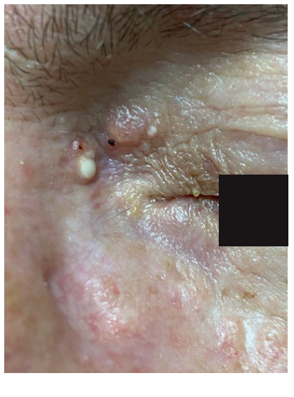
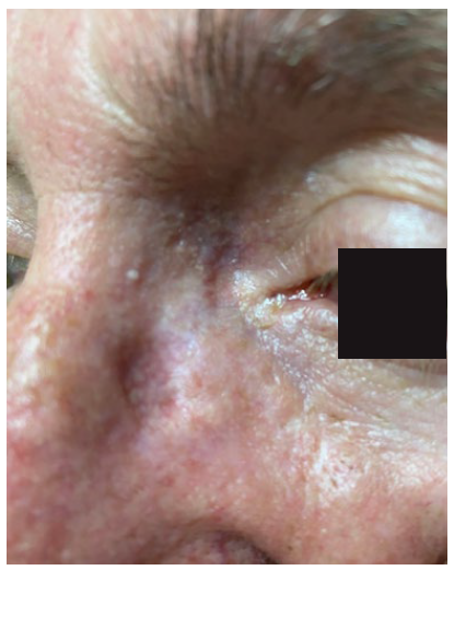
He underwent the first course of PDT in September of 2019. The reduction in the size of the lesion after the first treatment was significant. Despite the excellent results after the first course of PDT, the response was not complete, remaining a small papule in the upper eyelid (Fig. 2). He underwent another course of PDT in March of 2020. The only significant adverse effect reported was tolerable pain. The response was complete with an excellent aesthetic result. After 12 months of follow-up, no recurrence was observed (Fig. 3). A new skin biopsy ruled out residual disease (Fig. 4). He remains under follow-up.
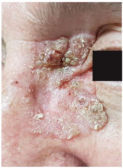
Figure 1 Skin-colored plaque, slightly desquamative, measuring 40*25 mm, involving the left medial canthus, lower and upper lid, and nasal dorsum. 
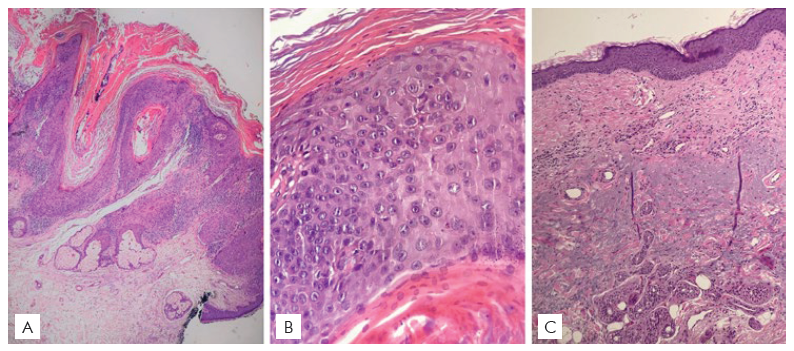
Figure 2 Result after the first course of PDT. A small papule remains in the upper eyelid. 

Figure 3 Result after the second course of PDT: a complete clinical response is visible.  

Figure 4 (A) Punch biopsy of a malignant proliferation of squamous cells in the epidermis and extending into follicular infundibula, with a verrucous pattern and associated with hyperkeratosis. (Hematoxylin-eosin stain; original magnification: x20). (B) The atypical squamous cells occupy the full thickness of the squamous layer, showing depolarization, irregular nuclear contour, increased nuclear/cytoplasmic ratio, and occasional binucleation. (Hematoxylin-eosin stain; original magnification: x40). (C) After treatment, the epidermis is flattened, with a regular squamous layer and normal cornification and without signs of malignancy. The papillary dermis is fibrotic, replacing and pushing down the elastotic fibers to the reticular dermis, consistent with post-treatment status. (Hematoxylin-eosin stain; original magnification: x40). 

DISCUSSION

Published studies concerning PDT for periocular tumors are scarce. Almost all obtain some response and up to 42% of patients maintain response in long-term follow-up.4 Yet, we should be aware that these results usually refer to recurrent/persistent tumors treated with PDT.4,5The recurrence usually occurs within the first two years of follow-up. These recurrences might respond to another course of PDT. Moreover, PDT as neoadjuvant treatment allows more conservative surgery.4,5Treatment seems to be as well tolerated as at other sites with no long-term sequelae.4,5Even with MCS for primary periocular Bowen disease, the reported 5-year local recurrence rate is 2.5%.2

Our case reinforces PDT role in Bowen disease at the periocular area used as treatment or as a way for pre-surgical tumor debulking, thus reducing surgical morbidity. The final aesthetic and functional result may be excellent. Further studies are needed to establish the role and long-term results of PDT in similar clinical scenarios.

Presentations/Apresentações

Part of this work has been presented at the “1º Congresso Vir-tual de Dermatologia e Venereologia” in November of 2020.

REFERENCES

1. Stratigos AJ, Garbe C, Dessinioti C, Lebbe C, Bataille V, Bastholt L, et al. European interdiscipli-nary guideline on invasive squamous cell carcinoma of the skin: Part 2. Treatment. Eur J Cancer. 2020;128:83-102. doi:10.1016/j.ejca.2020.01.008 [ Links ]

2. Nägeli MC, Ramelyte E, Dummer R. Cutaneous squamous cell carcinomas on special loca-tions: perioral, periocular and genital area. J Eur Acad Dermatology Venereol. 2019;33:21-4. doi:10.1111/jdv.15901 [ Links ]

3. Morton CA, Szeimies RM, Basset-Seguin N, Calzavara-Pinton P, Gilaberte Y, Haedersdal M, et al. European Dermatology Forum guidelines on topical photodynamic therapy 2019 Part 1: treatment delivery and established indications - actinic keratoses, Bowen's disease and basal cell carcinomas. J Eur Acad Dermatology Venereol. 2019;33:2225-38. doi:10.1111/jdv.16017 [ Links ]

4. Attili SK, Ibbotson SH, Fleming C. Role of non-surgical therapies in the management of periocular basal cell carcinoma and squamous intra-epidermal carcinoma: A case series and review of the literature. Photodermatol Photoimmunol Photomed. 2012;28:68-79. doi:10.1111/j.1600-0781.2011.00640.x [ Links ]

5. Casie Chetty N, Hemmant B, Skellett AM. Periocular photodynamic therapy for squamous intra-epidermal carcinoma. J Dermatolog Treat. 2014;25:516-8. doi:10.3109/09546634.2013.84 9793 [ Links ]

1© Author(s) (or their employer(s)) 2021 SPDV Journal. Re-use permitted under CC BY-NC. No commercial re-use. © Autor (es) (ou seu (s) empregador (es)) 2021 Revista SPDV. Reutilização permitida de acordo com CC BY-NC. Nenhuma reutilização comercial

2Conflicts of Interest: The authors have no conflicts of interest to declare. Financing Support: This work has not received any contribution, grant or scholarship. Confidentiality of Data: The authors declare that they have followed the protocols of their work center on the publication of data from patients. Patient Consent: Consent for publication was obtained. Provenance and Peer Review: Not commissioned; externally peer reviewed. Conflitos de Interesse: Os autores declaram a inexistência de conflitos de interes-se na realização do presente trabalho. Fontes de Financiamento: Não existiram fontes externas de financiamento para a realização deste artigo. Confidencialidade dos Dados: Os autores declaram ter seguido os protocolos da sua instituição acerca da publicação dos dados de doentes. Consentimento: Consentimento do doente para publicação obtido. Proveniência e Revisão por Pares: Não comissionado; revisão externa por pares

Received: April 11, 2021; Accepted: July 25, 2021

Corresponding Author: Joel Lopes dos Reis Address: Serviço de Dermatologia Centro Hospitalar do Porto Largo do Prof. Abel Salazar, 4099-001 Porto E-mail: joelreis@outlook.com

Creative Commons License This is an open-access article distributed under the terms of the Creative Commons Attribution License