Case Presentation
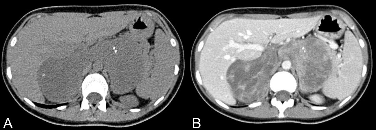
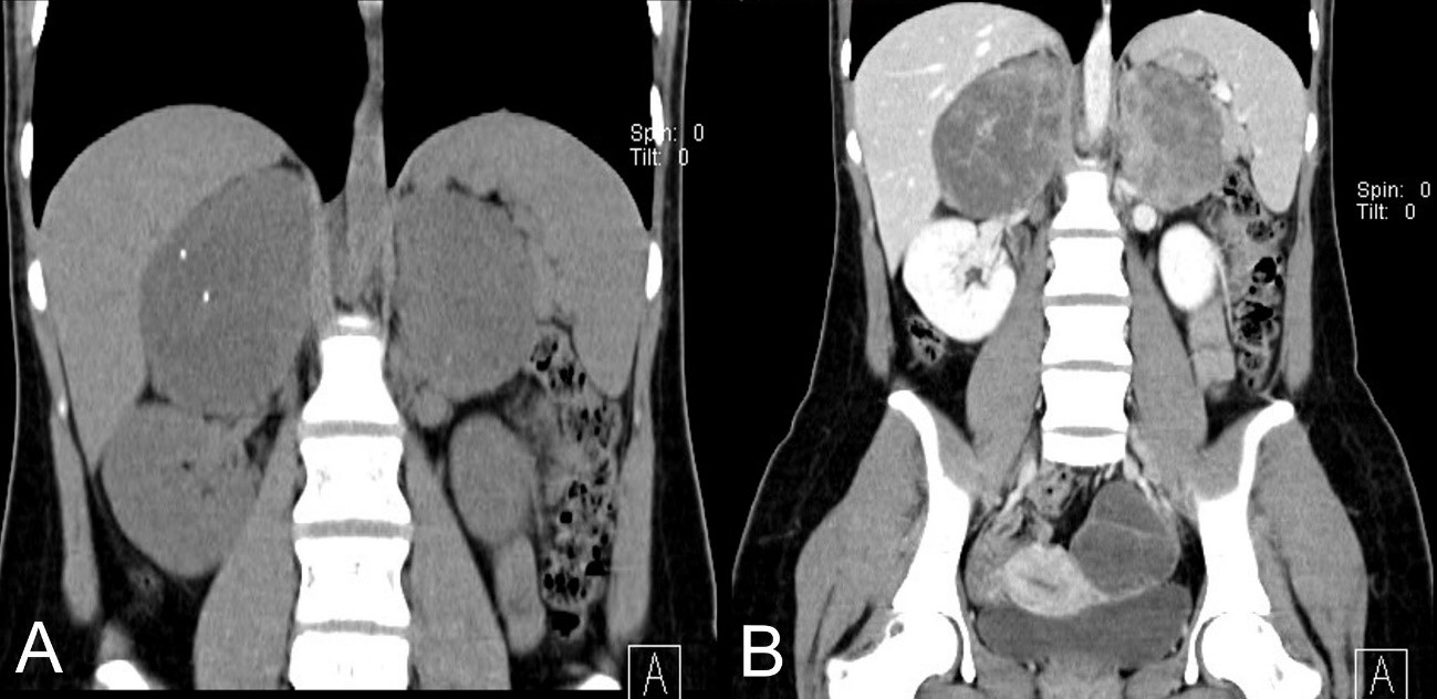
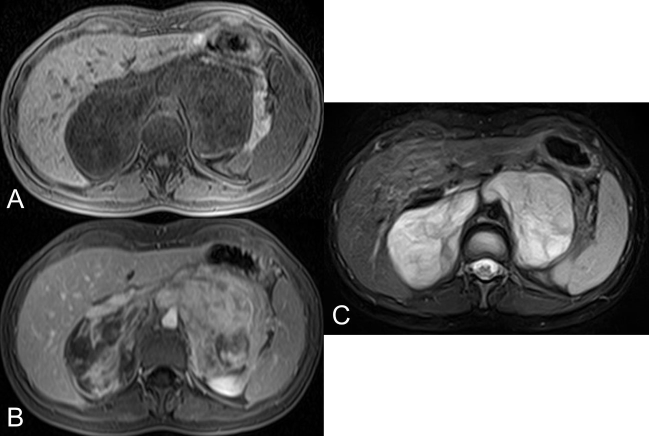
A 22-year-old female, previously healthy, without alcohol or smoking habits, was referred to our hospital after an emergency appendectomy in October 2019 at Centro Hospitalar da Cova da Beira (CHCB). During appendectomy, surgeons found a retroperitoneal mass. There was no previous knowledge of this retroperitoneal mass, and the patient had never had any abdominal symptoms before. Abdominal and pelvic computed tomography was initially performed to better characterize the mass, followed by magnetic resonance. Abdominal and pelvic CT revealed a heterogeneous soft tissue mass with some microcalcifications, measuring approximately 9.5 cm in the left adrenal area and 10 cm in the right adrenal, with heterogeneous enhancement after contrast administration, especially at the left (Fig. 1 and 2). Magnetic resonance imaging confirmed the existence of two heterogeneous masses involving the adrenal glands, which cross the midline and contact the liver, spleen-portal confluent, inferior vena cava, aorta, celiac trunk, superior mesenteric artery, renal vessels, and superior pole of both kidneys, with no other evident changes. Dependent on the left ovary, a complex cystic formation was observed, with several septa, measuring approximately 5 cm on the longest axis (Fig. 3). There are no adenopathies.
The patient underwent a biopsy of one of the masses, and the result was ganglioneuroma of the adrenal glands. There was a discussion of the case at a multidisciplinary meeting with doctors from different specialties, and the doctors decided to propose to the patient bilateral adrenalectomy, which she refused, opting for clinical follow-up that is still ongoing.

Figure 1: Axial soft tissue window, 1.2 mm slice thickness, non-enhanced CT (A), and contrast-enhanced CT (B).
Discussion
Ganglioneuromas are rare, well-differentiated benign neoplasms that originate from the neural crest and are mainly composed of mature Schwann cells, nerve fibers, and ganglion cells, and their malignant transformation is extremely rare 1,2. They were first described in 1870 by Lorentz and can arise anywhere along with the paravertebral sympathetic ganglia. Because ganglioneuromas originate from ganglion cells, they are part of a family of tumors that include neuroblastomas (which are highly malignant tumors) and ganglioneuroblastomas (which are tumors with intermediate characteristics) 1,2,3. They most often occur in the posterior mediastinum and retroperitoneum but can also be found in the cervical region 1,2,3.
The adrenal gland is one of the most affected sites, with about 20-30% of cases being ganglioneuromas affecting the gland unilaterally, with no description of bilateral cases 3,4. Ganglioneuromas only represent about 5% of tumors that affect the adrenal glands 3,4. These tumors arise mainly between the 4th and 5th decades of life, although those that affect the retroperitoneum and posterior mediastinum are more frequent in children and young adults, between 10 and 40 years of age, generally showing no gender preference, with a slight female prevalence 5. They are sporadic but may be associated with other pathologies such as type 1 neurofibromatosis, type 2 multiple endocrine neoplasias, and Turner syndrome 2,6. Most of these tumors are incidental findings on routine abdominal examinations for other reasons. The young woman never reported any abdominal symptoms apart from some menstrual pain and recently some acute pain and abdominal tenderness, related to acute appendicitis. In some cases, and due to the mass effect, that these tumors can cause, patients may report unspecific abdominal pain or discomfort 6.
Generally, this type of tumor does not cause hormone secretion, but when there is an association with pheochromocytoma or neuroblastoma, there may be some hormone secretion, namely catecholamines 7. Our patient didn’t have any analytical alteration. The diagnosis of this type of tumor is quite challenging, due to the absence of characteristic hormonal changes and because there are no specific imaging features that allow us to make the diagnosis, clearly distinguishing the ganglioneuroma from other solid adrenal masses such as adenoma, carcinoma, neuroblastoma, ganglioneuroblastoma or pheochromocytoma6,8. The role of histopathology is essential. A biopsy was performed and, combining the anatomopathological and immunohistochemical characteristics, ganglioneuroma was diagnosed.
Regarding the imaging features, CT does not have distinctive features, usually showing a homogeneous or slightly heterogeneous, well-defined, hypodense mass, with little or no enhancement after contrast administration, surrounding the great vessels without causing occlusion or compression 2,3,9,10. It is also possible to find punctiform calcifications, early linear septal enhancement, and late heterogeneous contrast uptake8,9. In this case, the CT findings were similar, with a heterogeneous mass, microcalcifications, and heterogeneous enhancement after contrast administration.
MRI generally shows a homogeneous mass with low signal intensity on T1 and a heterogeneous mass with a higher signal intensity on T2, which may be related to the combination of a myxoid matrix and several small ganglion cells 8,9. After contrast administration, a progressive and late enhancement is often observed, and an early or intense enhancement is not generally observed 2,8. The MRI features of our case are also similar, with a heterogeneous mass, lobulated contours, hyperintensity on T2, and hypointensity on T1, with multiple areas of heterogeneous enhancement after contrast.
Most ganglioneuromas (regardless of their size) are benign, with low recurrence or metastasis. There are sporadic reports of malignant cases with local lymph node involvement, which was not the case. Thus, the most common treatment is surgery (adrenalectomy with excision of the associated retroperitoneal mass). In the case of bilateral ganglioneuroma, the type of surgery should be bilateral adrenalectomy, with the resulting hormonal consequences 4,11,12,13.
Conclusion
The clinical and imaging diagnosis of ganglioneuromas is difficult, as we are dealing with a generally asymptomatic tumor that does not present pathognomonic imaging features. There are, however, some characteristics that can make us think about this possibility, namely: absence of hormonal changes; on CT, the presence of punctate calcifications, non-vascular involvement, and densities below 40 HU; on MRI the presence of a hypointense mass on T1 and hyperintense on T2, with weak late enhancement. The treatment of choice is surgery, which was recommended to our patient, but she did not accept it.


