


 Inglês (pdf)
 Artigo em XML
 Referências do artigo
 Enviar este artigo por email
 Citado por SciELO
  Similares em
SciELO
Similares em
SciELO 
 Permalink
Case Presentation
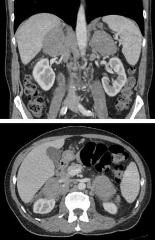
A 73-year-old man with no relevant past medical history and with complaints of asthenia, fever and weight loss over the past few months was referred for abdominal computer tomography after an incidental adrenal mass was detected on abdominal ultrasound. The CT scan (figure 1) revealed bilateral large solid adrenal masses with well-defined margins, homogeneous density and discrete contrast enhancement. Several enlarged paraaortic lymph nodes were also noted. Posterior laboratory studies revealed anemia and elevated LDH and β2-microglobulin. An ultrasound-guided biopsy of the left adrenal mass was performed revealing diffuse large B-cell lymphoma.
Discussion
Adrenal incidentalomas have been increasingly detected due to improvements in CT resolution, with some studies estimating a prevalence between 1.4% and 7.3%.1,2,3 Some features are suggestive of malignancy namely a diameter greater than 4 cm, irregular margins, inhomogeneous density, tumor calcification and high unenhanced CT attenuation values (>10 HU).4 Among the rarer causes of malignant adrenal lesions is lymphoma, which may be secondary or primary. Secondary adrenal lymphoma is more common (seen in approximately 5% of non-Hodgkin lymphomas5) and occurs when lymphoma spreads from another primary site to the adrenal glands, often presenting with asymmetrical adrenal involvement and associated disseminated disease.
Primary adrenal lymphoma (PAL), on the other hand, is a rare diagnosis with fewer than 200 cases reported in the literature.6 Its definition is not well established, but most authors have defined it as a histologically proven lymphoma in a patient with no prior history of lymphoma elsewhere and, if there is involvement of lymph nodes and/or other organs, then the adrenal lesions are unequivocally dominant.6 Its pathophysiology is poorly understood considering the lack of lymphatics in the adrenal parenchyma,7 with some authors suggesting ectopic hematopoietic tissue6 and others proposing an association with preexisting adrenalitis.8,9 Its most common histological subtype is the diffuse large B-cell lymphoma (78%).10 Demographically, it predominantly affects older men, with a male-to-female ratio of 1.8:1 and a median age of around 60 years old.6,7,8,9,10,11 The clinical presentation is variable, but B-symptoms, pain and fatigue seem to be present in the majority of patients as well as laboratory changes such as elevated LDH and decreased HDL.6,7,8,9,10,11 Treatment usually involves aggressive chemotherapy although the prognosis is poor.6
Its radiological appearance6,12,13 is non-specific and variable, appearing on CT as bilateral (about 70% of cases) solid, large and well-defined adrenal masses that may be hypovascular or have mild to moderate enhancement and that are not associated with calcifications. Similarly to our case, there may be involvement of other structures such as adjacent lymph nodes, emphasizing that for the diagnosis of PAL, the adrenal lesions must be the most prominent ones. On PET-CT these are metabolically active presenting avid glucose uptake.
On MRI, PAL tends to be iso/hypointense in T1 and hyperintense in T2 weighted images and have a variably heterogeneous enhancement pattern.6,12,13 Additionally, given their high cellularity, these also present restricted water diffusion with high signal on DWI. While the usual characteristics of PAL are sufficiently worrisome to warrant further testing, other possible diagnoses must be considered,10 especially in the context of bilaterally occurring adrenal masses such as pheochromocytoma, congenital adrenal hyperplasia, tuberculosis and metastatic disease.
In conclusion, PAL is a rare and aggressive entity affecting mostly older males and that should be considered in the differential diagnosis of bilateral, hypovascular and hypermetabolic bulky adrenal masses.

