


 Inglês (pdf)
 Artigo em XML
 Referências do artigo
 Enviar este artigo por email
 Citado por SciELO
  Similares em
SciELO
Similares em
SciELO 
 Permalink
Case Presentation
An 18-year-old male, with intellectual disability, presented with congenital blindness and a history of repeated fractures since early childhood, including a fracture of the right femur at 14 years of age. The patient is a refugee from a sub-developed country and no further medical history was available.
On clinical examination, there is marked scoliosis as well as bilateral microphthalmia and anophthalmia.
A typical skeletal survey was performed using conventional x-ray with bilateral projections of hands, forearms, humerus, feet, leg, femur, pelvis, spine, and skull.
Frontal and lateral skull radiographs revealed generalized cortical thinning as well as a reduction of the orbital spaces (Figure 1).
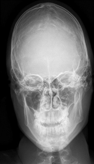
Figure 1: AP x-ray of the skull shows generalized cortical thinning as well as reduction of the orbital spaces.
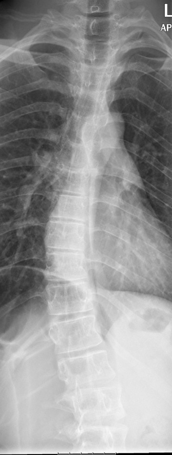
AP x-ray of the spine shows dorsal and lumbar dextroscoliosis (Figure 2).
AP x-ray of the forearms shows bowing of the radius and ulna, as well as thinning of the diaphysis and widening of the metaphysis (Figure 3).
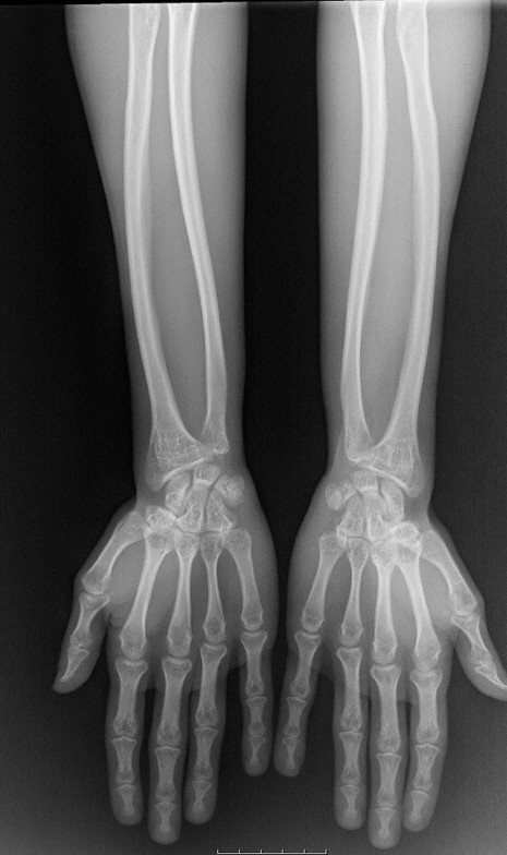
Figure 3: AP x-ray of the forearms shows thinning of the diaphysis and widening of the metaphysis of the ulna and radius with decreased density of these bones.
Also showing the bowing of the forearm bones.
Chilaiditi syndrome was observed.
Bone mineral density (BMD) was measured by dual energy X-ray absorptiometry (DXA), confirming osteoporosis.
Discussion
Osteoporosis-pseudoglioma syndrome (OPPG) is a rare autosomal recessive disorder of severe juvenile osteoporosis and congenital blindness.1 It is caused by inactivating mutations in the gene encoding low-density lipoprotein receptor-related protein 5 (LRP5), responsible for the Wnt/ β-catenin signaling pathway. To date, around 80 cases have been reported worldwide and the estimated prevalence is 1/2.000.000.1 A large proportion of cases emerges in populations with a high rate of consanguinity.
OPPG is characterized by intellectual disability, osteoporosis of bones and eye abnormalities. It is usually diagnosed in early childhood, with affected children displaying early onset blindness, severe osteoporosis, short stature and fractures.
Because most patients are blind before the age of 25, a bone examination should be performed on infants who present with eye abnormalities, such as, but not limited to, microphthalmia, microcornea, corneal clouding or vitreoretinal detachment to rule out this syndrome. Similarly, infants who present with severe bone abnormalities should undergo eye examination.2
On plain radiographs, patients usually manifest severe thinning of bones, bowing of extremities, and spinal deformities, such as fish mouth vertebra. One should be aware that radiological characteristics of the patients may present marked differences in the severity of the clinical phenotype and the degree of bone deformity.3
The final diagnosis is made via genetic testing.
Treatment options are regular bisphosphonates.
The main differential diagnosis for this disease was Osteogenesis imperfecta which was ruled out, as this patient had no hearing loss, blue sclera, joint laxity, contractures or teeth problems.
Radiologists should be aware of OPPG as a diagnostic possibility when facing osteoporosis in infants with eye abnormalities.

