SciELO - Scientific Electronic Library Online

 
vol.37 número1Tumor Fibroso Solitário Pélvico: Relato de um Caso Clínico com Revisão Imagiológica e Anatomopatológica da LiteraturaEdema dos tecidos moles em lactente: qual o diagnóstico? índice de autoresíndice de assuntosPesquisa de artigos
Home Pagelista alfabética de periódicos  

Serviços Personalizados

Journal

Artigo

Indicadores

Links relacionados

  • Não possue artigos similaresSimilares em SciELO

Compartilhar


Acta Radiológica Portuguesa

versão impressa ISSN 2183-1351versão On-line ISSN 2976-0763

Acta Radiol Port vol.37 no.1 Lisboa abr. 2025  Epub 30-Abr-2025

https://doi.org/10.25748/arp.33803 

Imagens de Interesse

Extraskeletal Mesenchymal Chondrosarcoma Heart Metastasis: A Rare Entity

Metástase Cardíaca de Condrossarcoma Mesenquimatoso Extra-Esquelético: Uma Entidade Rara

1Serviço de Radiologia, Centro Hospitalar e Universitário de Coimbra, Coimbra, Portugal


Abstract

Extraskeletal mesenchymal chondrosarcoma is an extremely rare neoplastic disease associated with high incidence of distant metastasis and poor prognosis, with heart metastasis being even more uncommon.

We present a case of a female patient, 62 years old, diagnosed with extraskeletal mesenchymal chondrosarcoma with disseminated metastatic disease for thirteen years, currently under second-line chemotherapy for three years. During her annual follow up with chest-abdominopelvic computed tomography (CT), an incidental heart metastasis was found, with no symptoms associated.

Key words: Chondrosarcoma; Mesenchymal; Heart.

Resumo

Condrossarcoma mesenquimatoso extra-esquelético é uma neoplasia extremamente rara, que se associa a uma incidência elevada de doença metastática à distância e prognóstico reservado, sendo a ocorrência de metastização cardíaca ainda mais incomum.

Apresentamos um caso de uma doente do sexo feminino, com 62 anos, diagnosticada com condrossarcoma mesenquimatoso extra-esquelético com metastização à distância desde há treze anos, presentemente sob tratamento de segunda-linha com quimioterapia desde há três anos. Durante o controlo evolutivo da doença, com tomografia computorizada anualmente, foi detectada metastização cardíaca incidental, sem sintomas associados.

Palavras-chave: Condrossarcoma; Mesenquimatoso; Coração.

Images in Case

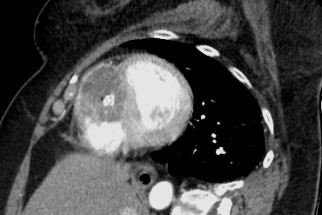
A female patient in her 60s has been followed up with annual chest-abdominopelvic computed tomography (CT) due to a clinical history of extraskeletal mesenchymal chondrosarcoma with disseminated metastatic disease. She was previously submitted to various surgical treatments, namely excision of the primary lesion located on the left thigh thirteen years before, as well as bilateral pulmonary and colic metastasectomy, left adrenalectomy and nephrectomy, splenectomy and caudal pancreatectomy in 2019. In a routine contrast-enhanced thoraco-abdominopelvic CT, a round, solid mass was found with heterogeneous attenuation, as well as internal calcifications, occupying most of the right ventricular chamber, due to heart metastasis from extraskeletal mesenchymal chondrosarcoma. It involves the right apex and almost two thirds of the ventricular septum, causing discrete bulging to the left.(Figs. 1, 2 and 3)

At the time of this diagnosis the patient was under a second-line treatment with trabectedin for three years. No symptoms were reported, so the patient remained under vigilance with chest-abdominopelvic CT once a year, as previously established.

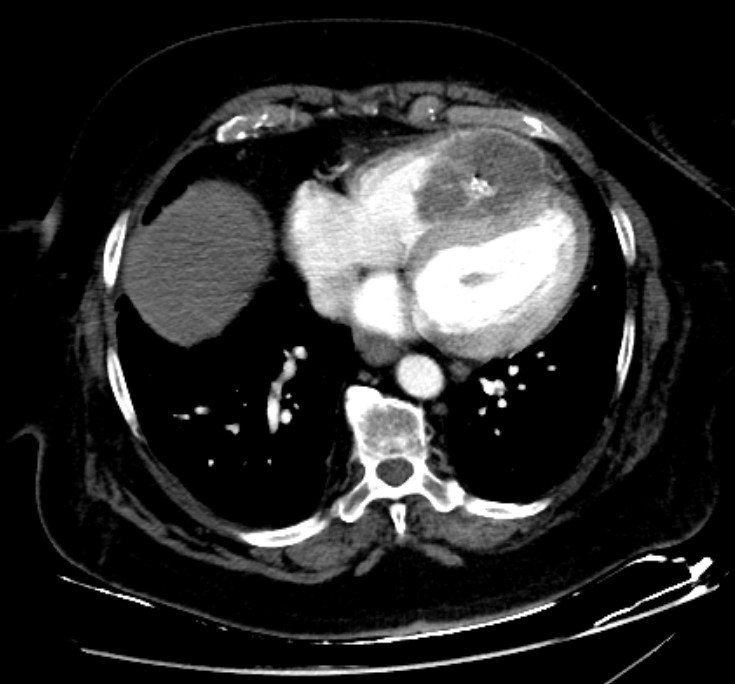
Figure 1: Axial contrast-enhanced CT image reveals a round, solid mass, with heterogeneous attenuation, as well as internal calcifications, occupying most of the right ventricular chamber, due to heart metastasis from extraskeletal mesenchymal chondrosarcoma. It involves the right apex and almost two thirds of the ventricular septum, causing discrete bulging to the left. 

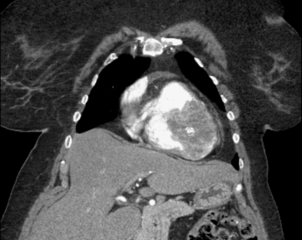
Figure 2: Coronal contrast-enhanced CT image shows the same round, solid mass, with heterogeneous attenuation, as well as internal calcifications, occupying most of the right ventricular chamber, in this image being more evident its apical location, as a result of heart metastasis from extraskeletal mesenchymal chondrosarcoma. 

Figure 3: Oblique reconstructed CT image following the heart short axis demonstrating ventricular septal involvement from the extraskeletal mesenchymal chondrosarcoma heart metastasis previously described. 

Discussion

Mesenchymal chondrosarcoma is an extremely rare neoplastic disease, comprising fewer than 2% of all chondrosarcomas, usually originating from bone or soft tissue, with an extraskeletal location in 30-50% of all cases .1,2Most commonly found in patients in their third decade, in contrast with conventional chondrosarcoma, which is more prevalent during the fifth to seventh decade of life,3 mesenchymal chondrosarcoma is associated with high incidence of distant metastasis, thus with poor prognosis.1,3

The standard therapy for heart metastasis should be surgery resection, as it aids with symptoms in addition to providing a histologic confirmation. Unfortunately, it is rarely possible, with chemotherapy and radiotherapy as the main options for these patients, often showing suboptimal efficacy.3

In this case we present a diagnosis of heart metastatic disease in a patient already under chemotherapy treatment, with no associated clinical manifestations, and for that reason remained with yearly follow up. Extraskeletal mesenchymal chondrosarcoma has a high risk of early dissemination, poor response to treatment and, consequently, it is associated with low rates of survival. Given the sparse therapeutic interventions, follow up with imaging studies is frequently recommended and local, as well as distant, recurrence should be actively searched by the Radiologist when evaluating patients with a clinical history of extraskeletal mesenchymal chondrosarcoma.3

References

1. Ghafoor S, Hameed MS,Tap WD. Mesenchymal chondrosarcoma: imaging features and clinical findings. Skeletal Radiology. 2021;50:333-41. doi: 10.1007/s00256-020-03558-x [ Links ]

2. White DW, Lya JQ, Beall DP, et al. Extraskeletal mesenchymal chondrosarcoma - case report. Journal of Clinical Imaging. 2003;27:187-90. doi: 10.1016/S0899-7071(02)00538-7 [ Links ]

3. Parmar C, Jojo A, Vachhani KC, et al. Primary chondrosarcoma of the heart. European Journal of Cardio-Thoracic Surgery. 2008;33:512-4. doi: 10.1016/j.ejcts.2007.11.029 [ Links ]

Ethical Disclosures

Financing Support: This work has not received any contribution, grant or scholarship.

Received: November 28, 2023; Accepted: March 07, 2024

Address Filipa Lisboa Bento, ULS Coimbra - Centro Hospitalar e Universitário de Coimbra, Praceta Mota Pinto, Celas, 3004-561 Coimbra, Portugal, e-mail: filipanlisboabento@gmail.com

Conflicts of interest: The authors have no conflicts of interest to declare.

Confidentiality of data: The authors declare that they have followed the protocols of their work center on the publication of data from patients.

Protection of human and animal subjects: The authors declare that the procedures followed were in accordance with the regulations of the relevant clinical research ethics committee and with those of the Code of Ethics of the World Medical Association (Declaration of Helsinki).

Creative Commons License This is an open-access article distributed under the terms of the Creative Commons Attribution License