




 Inglês (pdf)
 Artigo em XML
 Referências do artigo

 Tradução automática
 Enviar este artigo por email
 Citado por SciELO
 Similares em
SciELO

 Permalink


Introduction
Polyorchidism is an exceedingly rare genitourinary congenital anomaly resulting from an abnormal division of the genital ridge1 and defined by the presence of more than two testicles - supernumerary testicles - with the most common variant being triorchidism, which involves the presence of three testicles.2,3,4
It is diagnosed most often in adolescence and young adulthood.5,6
Most supernumerary testicles are located within the scrotal sac, mainly on the left side.3,6,7Still, they can be encountered in the inguinal canal or other locations such as the abdomen.3,6,7,8
Spermatogenesis is preserved in more than half of the supernumerary testicles3,9 and is impaired mainly in the undescended supernumerary testicles6 and those without epididymis or vas deferens.
Case report
A 19-year-old sexually active male presented to the emergency department complaining of left-sided scrotal pain for the past two days. The pain had a sudden onset and improved after taking acetaminophen, but persisted at a mild intensity. The patient reported increased urinary frequency and denied dysuria.
On physical examination, no scrotal inflammatory signs were observed, but a soft nodule, approximately 5 mm in size, was palpated in the left scrotal sac. A scrotal ultrasonography was therefore requested.
The ultrasonography revealed both the right and left primary testicles within their respective scrotal sacs, with normal morphology and dimensions, homogeneous echotexture, and normal vascularization on color Doppler imaging. However, in the left scrotal sac, an oval-shaped structure with a volume of 1.2 cc was identified, with an echotexture and vascularization identical to the primary testicles, suggesting the presence of a supernumerary testicle (Figure 1A and 1B). This supernumerary testicle had its own epididymis, which appeared enlarged (Figure 1C), a finding suggesting epididymitis. Both primary epididymides showed normal morphology and echogenicity.
Scrotal ultrasonography confirmed the diagnosis of polyorchidism, specifically triorchidism, with findings suggestive of epididymitis involving the epididymis of the left supernumerary testicle. This case may correspond to Type III or Type IV of Leung's classification, as the supernumerary testicle has its own epididymis. However, it was not possible to determine whether it has a proper vas deferens or if it is shared with the ipsilateral primary testicle.
As the supernumerary testicle was located within the scrotal sac, demonstrated normal gray-scale ultrasonographic characteristics, and had preserved vascular flow on Doppler evaluation, a conservative approach was adopted, and the patient was scheduled for follow-up scrotal ultrasonography within the following year. Given the diagnosis of epididymitis, empirical antibiotic treatment with ceftriaxone and azithromycin was initiated, alongside urine culture. The patient was also referred to his general physician for screening for sexually transmitted infections.
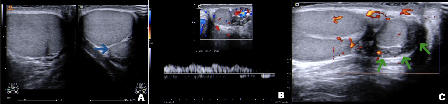
Figure 1: Triorchidism. Doppler ultrasonography images show one testicle in the right scrotal sac and two testicles in the left scrotal sac, the smaller one corresponding to a supernumerary testicle (blue arrow) (A). The supernumerary testicle has morphology, echotexture, and vascularization similar to the primary testicles (B). The epididymis of the supernumerary testicle (green arrows) is enlarged, suggesting epididymitis (C).
Discussion
Polyorchidism is often asymptomatic and typically identified incidentally during imaging evaluation or surgery for other scrotal conditions such as hydrocele, varicocele, epididymitis, testicular torsion, testicular tumors, cryptorchidism, and inguinal hernia.1,7,8,9
When symptomatic, it may present with scrotal pain, swelling, or a palpable mass.1,8
Supernumerary testicles can be mimicked by extra-testicular and para-testicular masses such as varicocele, lipoma, tumors, hydrocele, spermatocele, or inguinal hernia during physical examination.2,6,7,8
Due to the wide range of possible differential diagnoses for polyorchidism, physical examination alone is not sufficient to establish a definitive diagnosis. Imaging studies, such as ultrasonography and magnetic resonance imaging (MRI), are critical and have high accuracy in evaluating scrotal masses, often enabling diagnosis without surgical intervention or histological confirmation.12
Ultrasonography with color Doppler is the primary imaging modality for diagnosing polyorchidism, providing detailed images of scrotal contents and allowing for differentiation between intra- and extra-testicular masses.5 Supernumerary testicles present as oval-shaped and well-defined structures with a mediastinum, echotexture, and vascularity identical to those of the primary testicles.7,9 This imaging method is also valuable for excluding complications and allows for differential diagnosis of supernumerary testicles.
MRI is a supplementary tool to ultrasonography, providing superior soft tissue contrast and detailed anatomical information. It is beneficial in complex cases, when a definitive diagnosis is not possible using ultrasonography, when the supernumerary testicle is in unusual locations, or if malignancy is suspected.2,3,6,7 MRI findings generally correlate well with ultrasonography, depicting supernumerary testicle with signal intensities similar to the normal testicles (intermediate signal intensity on T1-weighted images and high signal intensity on T2-weighted images).2,5,6,7,8
According to Leung, polyorchidism can be classified into four types based on embryological and anatomical criteria5,6,10,13:
Type I: Supernumerary testicle without epididymis or vas deferens. Consequently, it has no reproductive potential.
Type II: Supernumerary testicle with a shared common epididymis and vas deferens with the ipsilateral testicle.
Type III: Supernumerary testicle with its own epididymis but a shared common vas deferens with the ipsilateral testicle.
Type IV: Supernumerary testicle with its own epididymis and vas deferens.
Types II and III are the most common forms of polyorchidism.1
Supernumerary testicles may be affected by pathologies otherwise observed in primary testicles; however, they are more predisposed to some complications, including infections, testicular torsion, malignancy, and impairment of spermatogenesis.
Supernumerary testicles are often associated with anatomical abnormalities, including absent or atypical communication with the epididymis and vas deferens. These abnormalities may impair normal drainage, resulting in spermatic stasis and potentially increasing the risk of secondary infections leading to epididymitis and orchiepididymitis.
Supernumerary testicles may have higher mobility because of a superior attachment of the tunica vaginalis and, consequently, an increased risk of testicular torsion, a condition usually presenting with acute scrotal pain requiring prompt surgical intervention.1,9,10
The prevalence of testicular malignancy in patients with polyorchidism is increased, reported in approximately 4-7% of supernumerary testicles, particularly when undescended, since cryptorchidism is considered a significant risk factor for testicular malignancy.1,3,7,10,11,12
Most supernumerary testicles preserve their reproductive potential.3,9,12 However, supernumerary testicles, particularly in cases of cryptorchidism, may present impaired spermatogenesis, which can be diminished or absent in approximately 11% and 26% of cases, respectively.6,11
Management of polyorchidism depends on the presence of symptoms and complications. Asymptomatic patients with normal imaging findings and patients with complications that do not threaten the viability of supernumerary testicles (e.g., epididymitis or orchiepididymitis) are managed conservatively and should undergo regular follow-up including annual physical examination, serum tumor marker measurement, and scrotal ultrasonography.6,8,10,12
Surgical intervention is indicated in cases where polyorchidism is complicated by torsion, malignancy, or results in significant discomfort.3,5,14 Given the increased mobility of supernumerary testicles, orchiopexy may be considered to prevent future testicular torsion.6 Orchiectomy of the supernumerary testicles must be performed in cases of malignancy and may be considered in cases of nonviable and atrophic testicles, testicles without reproductive potential (i.e. testicles presenting impaired spermatogenesis or testicles lacking a draining vas deferens), and undescended testicles, to reduce the risk of malignancy.3,6,7,10
Conclusion
Polyorchidism is a rare congenital anomaly with variable clinical presentations and potential complications. Diagnostic imaging, particularly ultrasonography and MRI, plays a key role in ensuring accurate diagnosis and effective monitoring. Management strategies should be tailored to the individual patient, weighing the risk of complications against the benefits of either surgical or conservative approaches.

