




 Inglês (pdf)
 Artigo em XML
 Referências do artigo
 Tradução automática
 Enviar este artigo por email
 Citado por SciELO
  Similares em
SciELO
Similares em
SciELO 
 Permalink

Case Presentation
A 44-year-old woman presented with pelvic and shoulder muscle weakness, along with malar erythema and periorbital edema, with onset two months before. The patient also complained of intermittent mild pruritus. She reported no cough or dyspnea at rest, but had mild exertional shortness of breath, which prompted complementary chest CT evaluation. Physical examination revealed scaly pink-to-violaceous papules on the interphalangeal and metacarpophalangeal joints, as well as violaceous erythema around the eyes. The blood test detected elevated creatine phosphokinase (CPK) levels of 5,238 U/L (reference range: 26-192 U/L). Magnetic resonance imaging (MRI) of the shoulders showed diffuse muscle edema involving the rotator cuff muscles (supraspinatus, infraspinatus, subscapularis, and teres minor), the deltoid (clavicular, acromial, and spinal portions), trapezius, teres major, and pectoralis major and minor, without architectural distortion (Figure 1). MRI of the hips demonstrated diffuse muscle edema involving the sartorius, tensor fasciae latae, iliacus, psoas major, iliopsoas, gluteus minimus and medius, piriformis, quadratus femoris, rectus femoris, obturator externus and internus, pectineus, vastus intermedius, and the adductors longus, brevis, and magnus (Figure 2). No significant fatty replacement was identified at the time of examination. Chest computed tomography (CT) was performed and was unremarkable. The diagnosis of dermatomyositis (DM) was established, and systemic corticosteroids were initiated, resulting in symptomatic improvement.
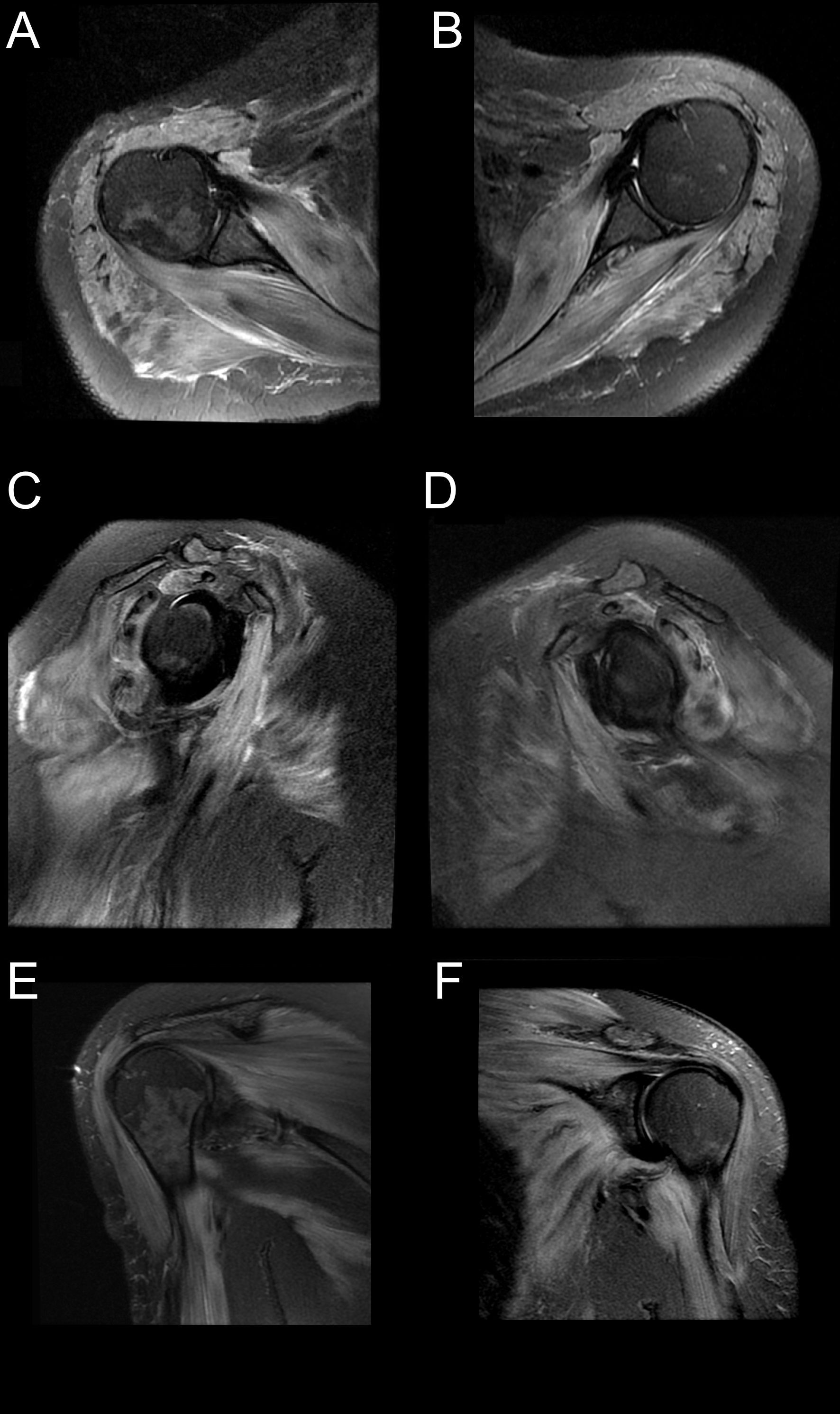
Figure 1: Fat-suppressed T2-weighted magnetic resonance imaging (MRI) of the shoulders. (A, C, E) Right shoulder - axial, sagittal, and coronal planes; (B, D, F) left shoulder - axial, sagittal, and coronal planes. Images show diffuse, bilateral muscle edema involving multiple muscles of the shoulder girdle. Findings demonstrate symmetric muscle involvement consistent with inflammatory myopathy.
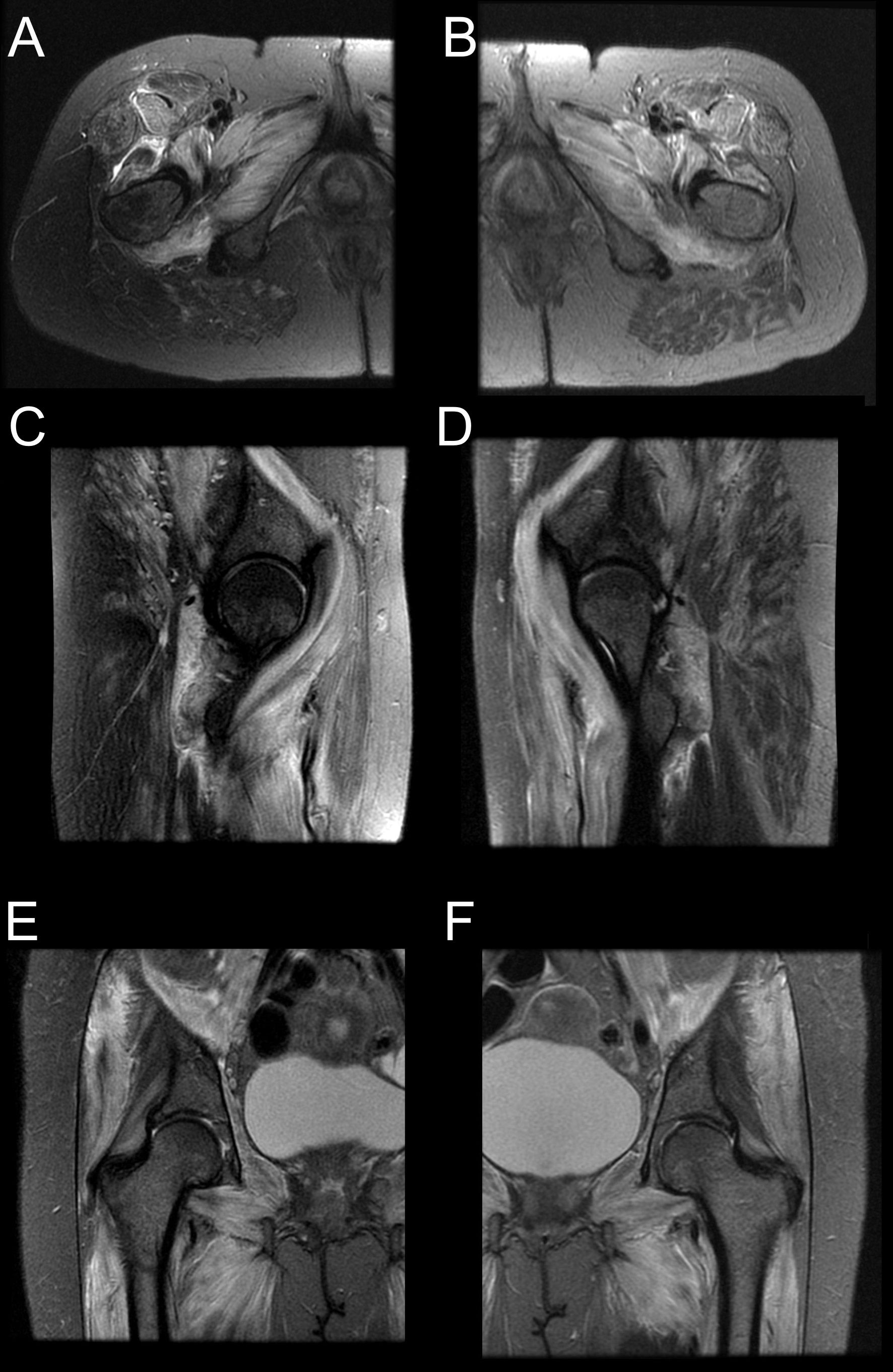
Figure 2: Fat-suppressed T2-weighted magnetic resonance imaging (MRI) of the hips. (A, C, E) Right hip - axial, sagittal, and coronal planes; (B, D, F) left hip - axial, sagittal, and coronal planes. Images show diffuse, bilateral muscle edema involving multiple muscles of the hip and pelvic girdles, demonstrating a symmetric pattern consistent with inflammatory myopathy.
Discussion
DM represents a complex interplay of cutaneous and systemic manifestations, significantly affecting patients' quality of life. In the case discussed, hallmark skin lesions such as heliotrope rash and Gottron's papules were evident, which are key diagnostic indicators of DM. These lesions are often associated with photosensitivity, a frequent trigger that exacerbates the inflammatory skin response. Although the mechanisms are not fully understood, ultraviolet (UV) radiation appears to amplify type I interferon activity via endogenous nucleic acid activation, worsening skin symptoms.1
The skin involvement in DM is not merely cosmetic; it can be debilitating due to severe pruritus and chronic lesions that persist despite control of the systemic disease. These symptoms often correlate poorly with muscle involvement, particularly in amyopathic dermatomyositis (ADM), complicating both diagnosis and treatment strategies. In the presented case, MRI demonstrated diffuse muscle oedema in both shoulder and hip girdles, and serum CK levels were markedly elevated. These findings are in keeping with classical dermatomyositis and differ from amyopathic forms, in which muscle enzymes and imaging are often normal or only minimally altered. This correlation between imaging and biochemical markers is supported by previous studies demonstrating that classical DM tends to show greater muscle involvement than ADM.2
Recent advances in imaging have expanded the understanding of inflammatory myopathies. Techniques like intravoxel incoherent motion MRI and fat quantification are proving valuable in evaluating disease activity and prognosis. Among advanced MRI techniques, intravoxel incoherent motion (IVIM) enables the separation of pure molecular diffusion from microcapillary perfusion without the need for contrast agents, which is particularly useful in inflammatory myopathies to assess disease activity and microvascular changes.3 Fat quantification techniques, such as Dixon-based sequences and proton density fat fraction (PDFF) mapping, allow accurate and reproducible measurement of intramuscular fat infiltration, supporting disease staging, monitoring of progression, and evaluation of therapeutic response. In the context of dermatomyositis, these methods can provide complementary information to conventional sequences, improving both diagnostic accuracy and prognostic assessment.4
Notably, the presence of anti-MDA5 antibodies and intramuscular lesions seen on MRI have been linked to more severe disease courses and may inform treatment decisions. Pulmonary involvement is a well-recognized systemic complication of dermatomyositis, especially in patients with anti-MDA5 antibodies, and may present with interstitial lung disease (ILD). On chest CT, ILD typically manifests as ground-glass opacities and/or consolidations, reflecting active inflammation or fibrosis. In our case, chest CT was normal, but even mild or nonspecific respiratory symptoms should prompt early imaging evaluation, as subclinical ILD can occur in up to 65% of patients with dermatomyositis.2,5
Environmental and drug-related triggers, including statins and hydroxyurea, have been implicated in disease onset or exacerbation, possibly through mechanisms involving DNA repair interference and type I interferon pathways. The treatment of skin manifestations in DM typically begins with topical corticosteroids or calcineurin inhibitors. In mild cases, hydroxychloroquine may be effective, whereas methotrexate is preferred in more extensive or disabling disease. Systemic corticosteroids, while effective for myositis, are not routinely recommended for cutaneous involvement due to inconsistent results and potential side effects.6 In refractory cases, biologic agents have emerged as potential therapeutic options for dermatomyositis. Rituximab, an anti-CD20 monoclonal antibody, has shown benefit in improving muscle strength and reducing cutaneous activity in patients unresponsive to conventional immunosuppression.7 Intravenous immunoglobulin (IVIG) is also recommended in certain scenarios, particularly for severe or rapidly progressive muscle weakness and dysphagia, with evidence of improved functional outcomes.8 Other agents, such as Janus kinase (JAK) inhibitors, are under investigation and have demonstrated promising results in reducing skin disease activity, especially in anti-MDA5-positive patients with concomitant interstitial lung disease.9
Overall, managing DM requires a multidisciplinary approach that integrates clinical, immunologic, and imaging data to tailor treatment, improve quality of life, and address both cutaneous and systemic complications.6
Dermatomyositis is also associated with an increased risk of malignancy, particularly within the first three years after diagnosis. The most commonly reported cancers include ovarian, lung, pancreatic, stomach, and colorectal carcinomas, as well as non-Hodgkin lymphoma.10 The pathophysiological link is thought to involve paraneoplastic immune mechanisms triggered by tumor antigens. Current guidelines recommend age- and sex-appropriate cancer screening at diagnosis and during follow-up, with a tailored approach based on clinical risk factors. Incorporating this screening into the multidisciplinary management of DM can lead to earlier detection and improved outcomes.11
Conclusion
This case illustrates the value of MRI in the diagnosis and assessment of dermatomyositis, providing detailed information on the extent of muscle involvement and enabling the detection of early changes that may not be clinically evident. The integration of imaging findings with laboratory and clinical data supports accurate diagnosis, guides management decisions, and helps monitor disease progression. Awareness of the systemic complications, including pulmonary involvement and increased malignancy risk, as well as the availability of emerging therapeutic options such as biologic agents, is essential for comprehensive patient care.

