




 Português (pdf)
 Artigo em XML
 Referências do artigo
 Tradução automática
 Enviar este artigo por email
 Citado por SciELO
  Similares em
SciELO
Similares em
SciELO 
 Permalink

Case Report
We present the case of a 50-year-old patient with recurrent painless haematuria and a history of unilateral renal agenesis with a single left pelvic kidney. Urine analysis confirmed haematuria, with no other significant findings. Computed tomography (CT) revealed an outpouching of the posterior bladder wall adjacent to the left ureterovesical junction containing a heterogeneous contrast-enhancing lesion, with a few internal calcifications (Figure 1). Magnetic resonance imaging (MRI) confirmed a posterior bladder diverticulum containing a solid lesion. The lesion was hypointense on T2-weighted images, showed contrast enhancement, and exhibited restricted diffusion (Figure 2). The diverticular wall appeared mildly thickened but intact, with no invasion of the diverticular neck or adjacent bladder wall. No signs of perivesical fat invasion or enlarged lymph nodes were observed. These findings were suggestive of a tumour arising within a Hutch diverticulum. The patient underwent diverticulectomy with ureteral reimplantation and histopathological examination confirmed the diagnosis of papillary urothelial carcinoma. Following surgery, adjuvant chemotherapy was initiated. Six months later, the patient developed intravesical tumour recurrence, which was managed with transurethral resection. The patient is currently receiving systemic therapy and remains under clinical and radiologic surveillance.
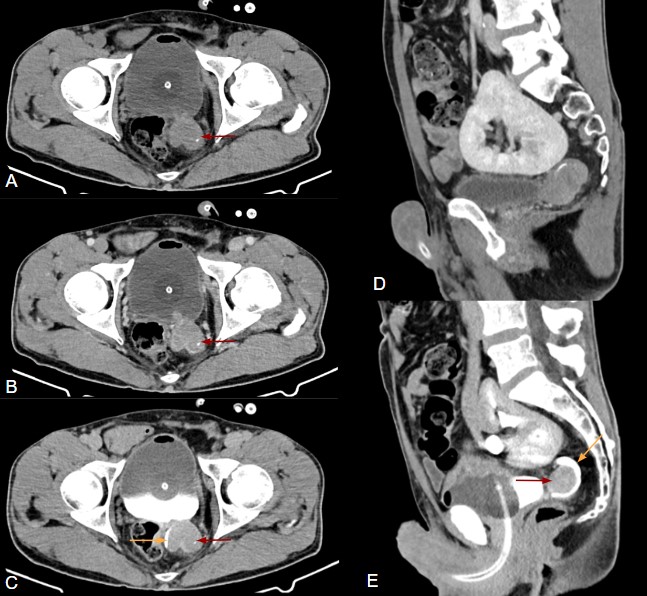
Figure 1: Pelvic CT axial (A - without contrast, B - venous phase, C - excretory phase) and sagittal (D - venous phase, E - excretory phase) images showing a solid contrast-enhancing lesion located within a bladder diverticulum adjacent to the left ureterovesical junction (red arrows), with a few internal calcifications. This lesion can be seen displacing the ureter posteriorly and medially (orange arrow). In the sagittal images (D and E), a pelvic kidney can also be seen.
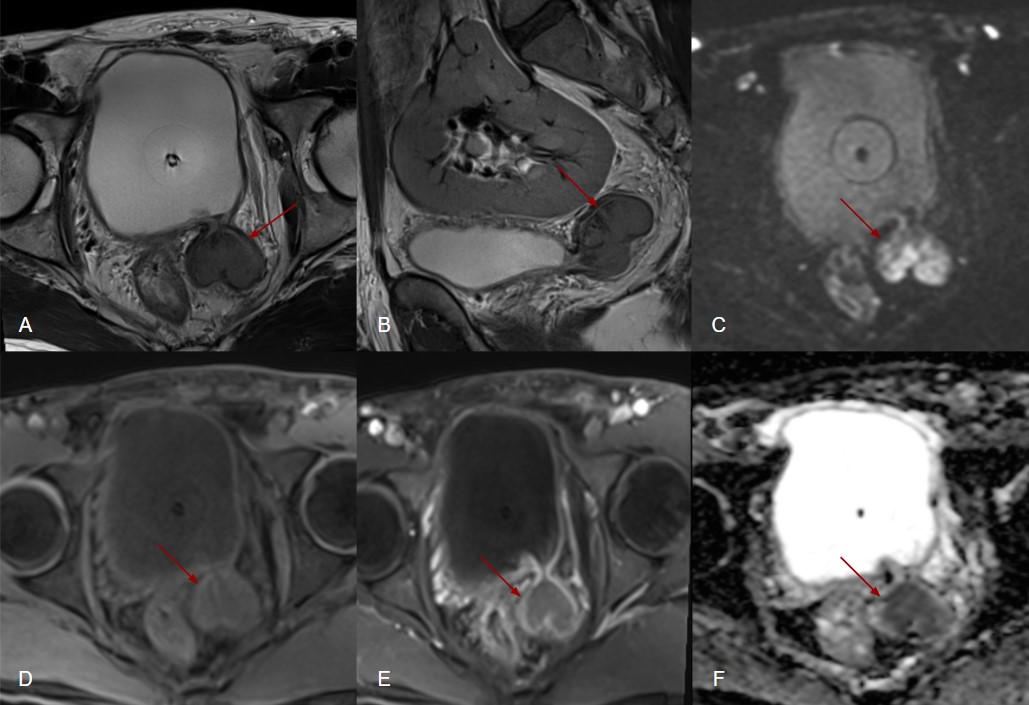
Figure 2: Pelvic MRI axial (A - T2-weighted, C - Diffusion-weighted, D - T1-weighted without contrast, E - T1-weighted venous phase, F - ADC map) and sagittal (B - T2-weighted) images showing a solid lesion located within a bladder diverticulum adjacent to the left ureterovesical junction (red arrows). This lesion is hypointense on T2-weighted images (A and B), exhibits restricted diffusion (C and F) and enhances after contrast administration (E). The wall of the diverticulum appears mildly thickened but intact, with no visible infiltration of the perivesical fat. No invasion of the neck of the diverticulum or adjacent bladder wall are seen.
Discussion
Hutch diverticula (HD) are rare congenital bladder diverticula thought to result from a weakness in the bladder wall near the ureterovesical junction, where incomplete muscle development and abnormal incorporation of the mesonephric duct allow gradual mucosal herniation under increased intravesical pressure. HD are more frequently seen in males.1,3
HD are often asymptomatic, but may present with symptoms such as recurrent urinary tract infections, vesicoureteral reflux, or, if large, compression or obstruction of adjacent structures.1,3
Intra-diverticular tumours are rare and can be challenging to diagnose. These tumours tend to be of a higher grade compared to other bladder tumours, with urothelial carcinoma (UC) being the most common histological type.1,3
The most common symptom in patients with intra-diverticular tumours is painless haematuria.2,3
On imaging, diverticula appear as outpouchings of the bladder wall. Their location and anatomical relationship to the vesicoureteral junction help distinguish different types, with HD typically being unilateral and arising from the posterior wall near the ureteral orifice.1,3
Intra-diverticular tumours can be identified on ultrasound as heterogeneous lesions with internal vascularity within a diverticulum. On CT and MRI, they present as solid lesions, occasionally containing calcifications, typically showing heterogeneous contrast enhancement and restricted diffusion on MRI. MRI is particularly valuable in cases of diagnostic uncertainty and for local staging.2,3
Cystoscopy is also fundamental, enabling direct evaluation of the bladder wall and targeted biopsy. HD can be difficult to access cystoscopically when the diverticular neck is narrow.1,2
Management of UC arising from HD generally involves a combination of surgery (such as diverticulectomy or cystectomy) and adjuvant therapy, depending on the stage of the disease. For small, low-grade tumours, transurethral resection may be an option.2,3
In conclusion, UC arising within HD is rare and can be a diagnostic challenge. Recognizing the potential for malignancy in these diverticula is essential to ensure early detection and better outcomes.

