




 Inglês (pdf)
 Artigo em XML
 Referências do artigo

 Tradução automática
 Enviar este artigo por email
 Citado por SciELO
 Similares em
SciELO

 Permalink


Case presentation
A 51-year-old female presented to the emergency department with acute-onset right-sided hemiparesis. Imaging revealed an occlusion of the proximal M1 segment of the left middle cerebral artery. The patient underwent urgent endovascular thrombectomy.
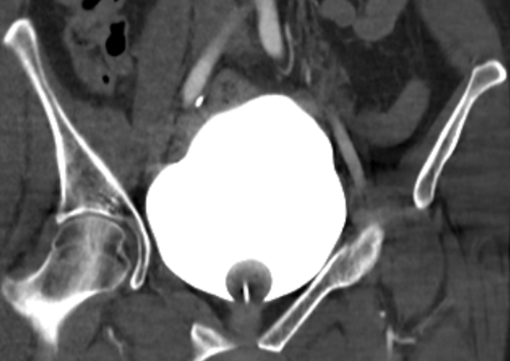
In the setting of the stroke, the patient sustained a ground-level fall and presented with right-sided flank pain and tenderness on palpation. After the emergent procedure, a contrast-enhanced computed tomography (CT) scan of the abdomen and pelvis was performed to assess for complications. Imaging obtained prior to administration of additional intravenous contrast demonstrated bilateral peripelvic contrast extravasation from prior angiography (figure 1). The urinary bladder was well-distended despite the presence of a correctly positioned indwelling catheter (figure 2). There was no evidence of renal or collecting system traumatic injury, calculi or structural abnormalities.
The patient remained hemodynamically stable and asymptomatic and conservative management was adopted. Follow-up imaging showed resolution of the previous findings.
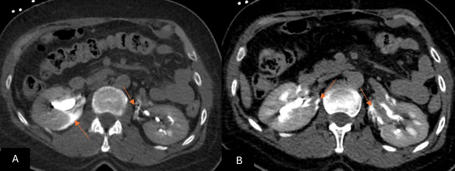
Figure 1: Abdominal CT before endovenous contrast administration showed bilateral contrast material extravasation at the level of renal pelves (arrows in A and B), probably from the recent angiographic procedure, consistent with forniceal rupture.
Discussion
Spontaneous renal pelvis rupture is a rare complication of acute urinary tract obstruction. It results from rapidly increasing intraluminal pressure within the collecting system, that outgrows the slow adaptative mechanisms seen in chronic hydronephrosis.1 The sudden elevation in pyeloureteral pressure exceeds the tensile limits of the forniceal wall, resulting in rupture with urine extravasation.1 The most common cause is obstructing ureteric calculi.1 Less common aetiologies include extrinsic compression, posterior urethral valves, ureteropelvic junction obstruction, malignancy, and iatrogenic causes.1
Iodinated contrast agents play a role in precipitating forniceal rupture, as they act as osmotic diuretics and may cause a transient increase in urine production and collecting system pressure.2
In the present case, bilateral contrast extravasation at the level of the renal pelves was identified on abdominal CT after an angiographic procedure. There was no evidence of ureteric obstruction and no structural renal or ureteral abnormality. Although the patient sustained a fall, it was a low-mechanism injury, with no CT evidence of traumatic lesions. While the precise pathophysiology remains speculative in this single case, despite the indwelling catheter being correctly positioned, the urinary bladder was markedly distended on CT, suggesting impaired drainage, which may occur transiently during procedural handling or high urine output states. A sudden, transient rise in intrapelvic pressure during the recent angiographic procedure may have contributed to forniceal rupture. It is worth noting that a few cases of spontaneous rupture have been reported in association with iodinated contrast administration, even in the absence of urinary tract obstruction.2
While the risk of complications such as perinephric urinoma, abscess formation, sepsis, or renal function deterioration is well documented,3 recent literature supports a conservative management approach in uncomplicated cases. Morgan et al.3 suggest that in the absence of infection, obstruction, or clinical deterioration, spontaneous resolution of forniceal rupture is common, and unnecessary antibiotic or surgical overtreatment should be avoided.

