


SciELO - Scientific Electronic Library Online

 
vol.39 número3 índice de autoresíndice de assuntospesquisa de artigos
Home Pagelista alfabética de periódicos  

Serviços Personalizados

Journal

Artigo

Indicadores

Links relacionados

  • Não possue artigos similaresSimilares em SciELO

Compartilhar


Acta Radiológica Portuguesa

versão impressa ISSN 2183-1351versão On-line ISSN 2976-0763

Acta Radiol Port vol.39 no.3 Lisboa set. 2027  Epub 31-Dez-2027

https://doi.org/10.25748/arp.44910 

Imagens de Interesse

Imaging Features of Waldenström Macroglobulinemia: A Case Report

Manifestações Imagiológicas da Macroglobulinemia de Waldenström: Relato de Caso

1 Serviço de Imagiologia, ULS de Viseu Dão-Lafões, Viseu, Portugal


Abstract

Waldenström macroglobulinemia is a rare subtype of non-Hodgkin lymphoma with bone marrow involvement and IgM overproduction. Patients may present with nonspecific symptoms or organ-related manifestations. Imaging, although not required for the diagnosis, can assess disease extent and monitor treatment.

We report a 54-year-old male with weight loss, dyspnea, cervical lymphadenopathy and elevated IgM. CT revealed widespread lymphadenopathy, hepatosplenomegaly, renal infiltration and pulmonary consolidations. Bone marrow biopsy confirmed the diagnosis. Treatment with bendamustine and rituximab led to near-complete resolution on follow-up CT.

This case highlights the diverse imaging features of Waldenström macroglobulinemia, including rare renal and pulmonary involvement and demonstrates the role of CT in disease evaluation and response monitoring.

Keywords: Waldenstrom macroglobulinemia; Lymphadenopathy; Computed tomography; Hematologic diseases; Rare diseases; Case reports.

Resumo

A macroglobulinemia de Waldenström é um subtipo raro de linfoma não-Hodgkin caracterizado pelo envolvimento da medula óssea e pela produção excessiva de IgM. Os doentes podem apresentar sintomas inespecíficos ou manifestações relacionadas com os órgãos afetados. Os exames de imagem, embora não sejam necessários para o diagnóstico, permitem avaliar a extensão da doença e monitorizar o tratamento.

Relatamos o caso de um homem de 54 anos com perda de peso, dispneia, adenomegalias cervicais e níveis elevados de IgM. A TC revelou adenomegalias generalizadas, hepatoesplenomegalia, infiltração renal e consolidações pulmonares. A biópsia da medula óssea confirmou o diagnóstico. O tratamento com bendamustina e rituximab conduziu à resolução quase completa das alterações na TC de seguimento.

Este caso evidencia a diversidade das manifestações imagiológicas da macroglobulinemia de Waldenström, incluindo o raro envolvimento renal e pulmonar e realça o papel da TC na avaliação da doença e no seguimento da resposta terapêutica.

Palavras-chave: Macroglobulinemia de Waldenstrom; Linfadenopatia; Tomografia computorizada; Doenças hematológicas; Doenças raras; Relatos de caso.

Case Presentation

We present the case of a 54-year-old male, a former smoker with a history of chronic cough, dyspnea, weight loss and asthenia. The physical exam revealed cervical lymphadenopathy. Laboratory tests revealed anemia, leukopenia and elevated IgM levels.

Contrast-enhanced CT of the neck, thorax, abdomen and pelvis demonstrated widespread lymphadenopathy (Figure 1), including a large retroperitoneal lymphomatous mass (Figure 2). In the pulmonary parenchyma, there were peribronchovascular ground-glass opacities and consolidation in the lower lobes (Figure 1). Additionally, a soft-tissue mass infiltrating the renal hila and perinephric fat was observed (Figure 3). The liver and spleen were enlarged.

The results of the bone marrow biopsy, integrated with the clinical and laboratory findings, confirmed the diagnosis of Waldenström macroglobulinemia (WM), a subtype of lymphoplasmacytic lymphoma (LPL).

Immunochemotherapy with bendamustine and rituximab was initiated. Follow-up CT showed near-complete resolution of the imaging findings.
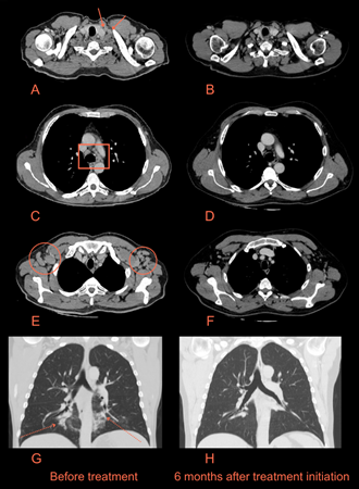
Figure 1: CT images of the neck and thorax before treatment (A, C, E, G) and six months after immunochemotherapy initiation (B, D, F, H). Note the cervical (arrows on A), mediastinal (rectangle on C) and axillary (circles on E) lymphadenopathy and foci of consolidation (dashed arrows on G) with surrounding ground-glass opacities. The CT performed six months after treatment showed no cervical or axillary lymphadenopathy and resolution of the pulmonary findings. 
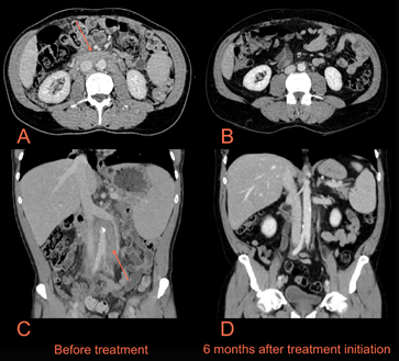
Figure 2: CT images of the abdomen before treatment (A, C) and six months after immunochemotherapy initiation (B, D). A lymphomatous mass surrounding the inferior vena cava and aorta could be seen (arrows on A and C). Follow-up CT showed resolution of the mass, with residual mild fat stranding surrounding the great vessels. 
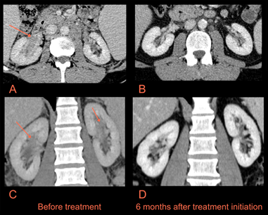
Figure 3: Close-up CT images of the kidneys before treatment (A, C) and six months after immunochemotherapy initiation (B, D). There was soft tissue extending into the renal sinuses (arrows on A and C), representing infiltration by neoplastic cells. Follow-up CT showed resolution of these findings. 

Discussion

LPL is a B-cell neoplasm comprised of small lymphocytes, plasmacytoid lymphocytes and plasma cells and is usually associated with a serum monoclonal protein. WM is the most common subtype of LPL and occurs when the serum monoclonal protein is IgM and when there is bone marrow involvement. Elevated IgM levels can also be associated with other conditions, so a bone marrow biopsy is necessary for the diagnosis. Although not specific, most WM patients have a MYD88 gene mutation.

The incidence of WM is approximately 3.8 per million, making it a rare disease.1 The most common symptoms are constitutional and can be related to the infiltration of different organ systems by abnormal blood cells and blood hyperviscosity due to IgM hyperproduction.

Even though imaging is not required for the diagnosis of WM, it can have an important role in the diagnosis and patient management. Due to its multisystemic nature, WM can virtually affect any organ and lead to imaging findings in any system. Bone marrow involvement is the most frequent imaging manifestation, showing abnormal signal intensity on MRI of the spine.2 Extra-medullary manifestations are less common and may include lymphadenopathy (frequently in the abdominal region), splenomegaly and hepatomegaly.3 Rare extramedullary involvement may include renal or perinephric infiltration, nonspecific pulmonary manifestations such as consolidation or ground-glass opacities and, less frequently, involvement of other organ systems such as the gastrointestinal, musculoskeletal and central nervous systems.

All the previously described CT findings of our patient, some of them very uncommon, correspond to known manifestations of WM. Given the clinical context and concurrent disease findings, pulmonary lesions were attributed to WM without invasive biopsy. Although the role of imaging in WM is still uncertain, in our patient it contributed to the diagnosis, helped determine the extent of disease and was important in monitoring treatment response.

References

1. Wang H, Chen Y, Li F, Delasalle K, Wang J, Alexanian R, et al. Temporal and geographic variations of Waldenstrom macroglobulinemia incidence: a large population-based study. Cancer. 2012;118:3793-800. [ Links ]

2. Moulopoulos LA, Dimopoulos MA, Varma DG, Manning JT, Johnston DA, Leeds NE, et al. Waldenström macroglobulinemia: MR imaging of the spine and CT of the abdomen and pelvis. Radiology. 1993;188:669-73. [ Links ]

3. Thomas R, Braschi-Amirfarzan M, Laferriere SL, Jagannathan JP. Imaging of Waldenström macroglobulinemia: a comprehensive review for the radiologist in the era of personalized medicine. AJR Am J Roentgenol. 2019;213:W248-W256. [ Links ]

Ethical Disclosures

Financing Support: This work has not received any contribution, grant or scholarship.

Received: January 10, 2026; Accepted: February 08, 2026

Address João R. Monteiro, Serviço de Imagiologia, ULS de Viseu Dão-Lafões, Av. Rei Dom Duarte, 3504-509 Viseu, Portugal, e-mail: joaoprmonteiro@hotmail.com

Conflicts of interest: The authors have no conflicts of interest to declare.

Confidentiality of data: The authors declare that they have followed the protocols of their work center on the publication of data from patients.

Protection of human and animal subjects: The authors declare that the procedures followed were in accordance with the regulations of the relevant clinical research ethics committee and with those of the Code of Ethics of the World Medical Association (Declaration of Helsinki).

Creative Commons License This is an open-access article distributed under the terms of the Creative Commons Attribution License