SciELO - Scientific Electronic Library Online

 
vol.9 número3Colecistite Aguda Alitiásica: A Propósito de uma Causa Inusitada“Pseudo-Geyser Sign” como Primeira Apresentação de Artrite Sética do Ombro índice de autoresíndice de assuntosPesquisa de artigos
Home Pagelista alfabética de periódicos  

Serviços Personalizados

Journal

Artigo

Indicadores

Links relacionados

  • Não possue artigos similaresSimilares em SciELO

Compartilhar


Gazeta Médica

versão impressa ISSN 2183-8135versão On-line ISSN 2184-0628

Gaz Med vol.9 no.3 Queluz set. 2022  Epub 30-Set-2022

https://doi.org/10.29315/gm.v1i1.546 

Casos Clínicos

Neoformação Renal: Um Diagnóstico a Considerar

Renal Neoformation: A Diagnosis to Consider

João Miranda1 
http://orcid.org/0000-0003-4896-2201

Paula Coelho2 

Céu Rosinha3 

Eduarda Marques4 

1. Serviço de Pediatria, Centro Hospitalar de Vila Nova de Gaia/Espinho, Vila Nova de Gaia, Portugal.

2. Serviço de Imagiologia, Centro Hospitalar de Vila Nova de Gaia/Espinho, Vila Nova de Gaia, Portugal.

3. Serviço de Cirurgia Pediátrica, Centro Hospitalar de Vila Nova de Gaia/Espinho, Vila Nova de Gaia, Portugal.

4. Unidade de Nefrologia Pediátrica, Serviço de Pediatria, Centro Hospitalar de Vila Nova de Gaia/Espinho, Vila Nova de Gaia, Portugal.


Resumo

Os granulomas renais relacionados com material de sutura são raros e habitualmente achados acidentais em exames imagiológicos.

Um adolescente de 12 anos, em seguimento após pieloplastia de Anderson-Hynes 7 anos antes, realizou ecografia renovesical que descreve formação nodular sólida, possivelmente na dependência do rim esquerdo. Posteriormente realizada uro-ressonância, que evidenciou lesão de características inespecíficas, possivelmente neoformação com componente fibroso predominante à periferia e mixomatoso central. A biópsia percutânea foi morfologicamente compatível com processo inflamatório crónico xantogranulomatoso. O exame anatomopatológico da lesão excisada cirurgicamente descreveu lesão de esboço nodular constituída por células histiocíticas com participação frequente de células gigantes de tipo corpo estranho e reação granulomatosa em relação com material exógeno de tipo fio de sutura. Mantém-se assintomático desde então.

Os pseudo-tumores granulomatosos de corpo estranho são difíceis de diagnosticar devido à ausência de características imagiológicas específicas distintivas de neoplasias, requerendo exame histológico para confirmação do diagnóstico.

Palavras-chave: Criança; Granuloma; Reação a Corpo Estranho; Rim

Abstract

Renal granulomas related to suture material are rare and usually accidental findings in imagiological studies.

A 12-year-old boy, followed-up after Anderson-Hynes pyeloplasty 7 years before, was submitted to an ultrasonography that described a solid nodular lesion, possibly in the dependence of the left kidney. Subsequently, magnetic resonance imaging was performed, which reported a lesion with non-specific features, suggesting neoformation with predominant fibrous peripheric component and myxomatous center. Percutaneous biopsy was morphologically compatible with chronic xanthogranulomatous inflammatory process. Anatomopathological examination of the surgical excised lesion described histiocytic cells with frequent participation of foreign body type giant cells and granulomatous reaction in relation to exogenous suture-like material. He remains asymptomatic since then.

Foreign body granulomatous pseudotumors are difficult to diagnose regarding the absence of specific imagiological features distinctive from neoplasms, requiring histological examination to confirm the diagnosis.

Keywords: Child; Foreign-Body Reaction; Granuloma; Kidney

Introduction

Renal foreign body granuloma are infrequent findings, particularly those ones related to suture material. Despite being sometimes symptomatic, they are much more frequently an incidental finding in a radiological examination. Its differential diagnosis is hampered by the lack of specificity of its radiological features.

Case report

A 12-year-old boy is being followed after having been submitted to an Anderson-Hynes pyeloplasty 7 years before. During his follow-up, it is detected a 4 mm calculus in the left kidney as well as 10 mm cortical cyst in the right kidney. At this point, it is also described in a follow-up ultrasonography a solid nodular lesion, possibly in the dependence of the left kidney, with a hypoecogenic periphery and hyperecogenic center (Fig. 1).
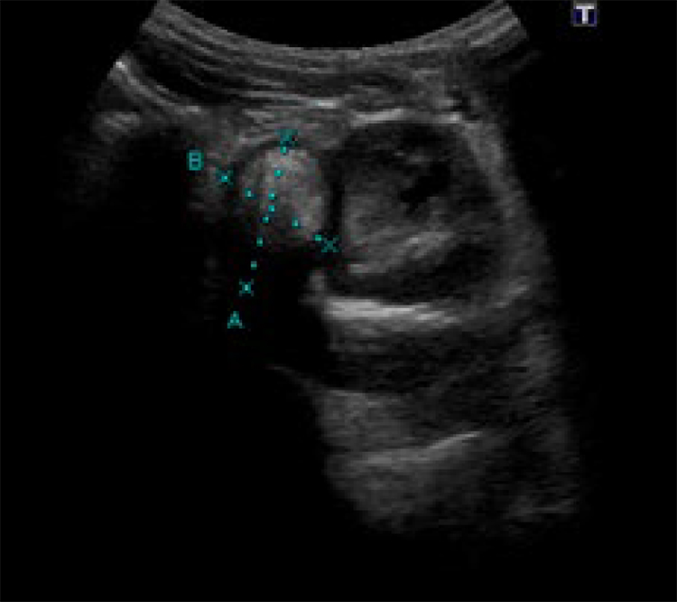
Figure 1:  . Ultrassonography: A solid nodule in close contact with the left kidney, 26 mm in diameter, hypoechogenic in the periphery, with a central hyperechogenic component, which suggested the presence of fine calcifications or fat.  

Subsequently, magneticresonance imaging (MRI) is performed, which reports a lesion with non-specific features, measuring 3.6x2.2x3.1 cm, showing peripheral hyposignal in all sequences suggesting fibrous component and central hypersignal in T2, with discrete contrast enhancement (Fig.s 2A and 2B).
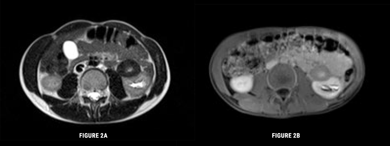
Figure 2: A) Magnetic ressonance imaging. Axial T2. Lesion of nonspecific characteristics, with a diameter of 3.6x2.2x3.1 cm, admitting to be a neoformation with fibrous component in the periphery. B) Magnetic ressonance imaging. T1FS Gd: After intravenous gadolinium. The lesion enhances less than the renal parenchyma and reveals slight central late enhancement. 

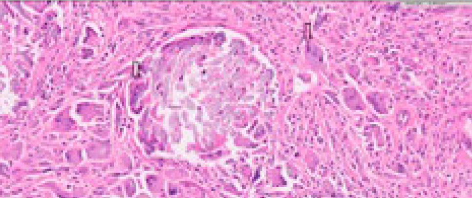
A percutaneous biopsy reveals a morphological picture compatible with chronic xanthogranulomatous inflammatory process. The lesion was submitted to surgical removal. After surgical removal of the lesion, anatomopathological examination describes histiocytic cells with frequent participation of foreign body type giant cells and granulomatous reaction in relation to exogenous suture-like material (Fig. 3).

Figure 3: Granulomatous lesion with foreign body giant cells (HE20x). Giant cells - arrows. 

The final diagnosis was renal granuloma due do foreign body material. The suture material used in the pyeloplasty was polydioxanone suture (PDS →), which a sterile synthetic absorbable monofilament suture is made from the polyester p-dioxanone.

The patient was discharged briefly after surgery and remains asymptomatic since then.

Discussion

Renal granulomas are very rare situations, most of which are due to surgical gauze left inadvertently inside the abdominal cavity1 or due to the ingestion of foreign material, causing erosion from the gastrointestinal tract through the urinary tract.1,2 The cases related to suture are even more exceptional and are due to the persistence or delay of absorption of the suture material used for closure of the pyelocalicial system and nephrostomies, being usually accidental findings.

This entity may present in a variety of forms, namely: purulent percutaneous fistula, pseudotumors and lithiasis.1 Clinical manifestations may appear early or late in the postoperative period, depending on the type of inflammatory reaction involved.1 There are cases described of surgical complication due to late persistence of suture, from 3 to 20 years after surgery.2-4 Its formation is due to a nodular, chronic reaction with cell-mediated inflammation to a persistent foreign agent such as suture material.3

The main differential diagnoses are tumors, abscesses, pseudo-cysts and hematomas.1

Although, computerized tomography and MRI are the elective imaging tests of choice for diagnostic approach, they may not be able to distinguish a foreign body granuloma unambiguously from other entities,1 especially from tumors,3 requiring a more invasive approach through percutaneous biopsy and surgical excision. Therefore, the diagnosis is usually established by the examination of the excised tissue.

Conclusion

Renal foreign body granulomas are a difficult entity to diagnose, given the lack of clinical specificity and imaging findings. The most relevant factor for diagnosis is a high index of suspicion, especially in patients with a history of nephro-urological surgery.3

References

1. Coelho RF, Mitre AI, Srougi M. Intrarenal foreign body presenting as a renal calculus. Clinics. 2007;62:527-8. doi: 10.1590/s1807-59322007000400021. [ Links ]

2. Singh DV, Swami YK, Pratap Y, Rana S, Wani SM. Foreign body in the kidney: an unusual case and its management. Cent European J Urol. 2014;66:497-500. doi: 10.5173/ceju.2013.04.art30. [ Links ]

3. Dogra PN, Tandon S, Ansari MS, Anupama, Chopra P . Suture foreign body granuloma masquerading as renal neoplasm. Int Urol Nephrol. 2005;37:27-9. doi: 10.1007/s11255-004-6707-8. [ Links ]

4. Kurz DA, Mucksavage P. The Endoscopic Removal of Eroded Foreign Bodies in the Ureter. J Endourol Case Rep. 2016;2:30-2. doi: 10.1089/cren.2016.0009. [ Links ]

Authors contribution/Contribuição autoral

JM and EM: Design and preparation of the article, review literature, drafting and revising the manuscript

PC: Review literature, drafting and revising the manuscript

CR: Scientific review of the manuscript

JM e EM: Projeção e preparação do artigo, revisão da literatura, redação e revisão do manuscrito

PC: Revisão da literatura, redação e revisão do manuscrito

CR: Revisão científica do manuscrito

Responsabilidades éticas

Conflitos de interesse: autores declaram a inexistência de conflitos de interesse na realização do presente trabalho.

Fontes de financiamento: Não existiram fontes externas de financiamento para a realização deste artigo.

Confidencialidade dos dados: Os autores declaram ter seguido os protocolos da sua instituição acerca da publicação dos dados de doentes.

Consentimento: Consentimento do doente para publicação obtido.

Proveniência e revisão por pares: Não comissionado; revisão externa por pares.

Ethical disclosures

Conflicts of interest: The authors have no conflicts of interest to declare.

Financing support: This work has not received any contribution, grant or scholarship.

Confidentiality of data: The authors declare that they have followed the protocols of their work center on the publication of data from patients.

Patient consent: Consent for publication was obtained.

Provenance and peer review: Not commissioned; externally peer reviewed.

Recebido: 02 de Dezembro de 2021; Aceito: 06 de Janeiro de 2022; : 17 de Janeiro de 2022; Publicado: 30 de Setembro de 2022

*Corresponding Author/Autor Correspondente: João Miranda ENT#091;joaocsmiranda@gmail.comENT#093; Rua Francisco Sá Carneiro, 4400-129 Vila Nova de Gaia, Portugal

Creative Commons License Este é um artigo publicado em acesso aberto sob uma licença Creative Commons