Introduction
Congenital hemangiomas (CH) are rare, benign vascular tumors typically located in the head, neck or limbs. Unlike infantile hemangiomas, they are present at birth and have their proliferative phase in utero.1,2 The three types of CH include the rapidly involuting (RICH), that involutes completely/nearly completely at 14 months; the noninvoluting (NICH), that does not resolve spontaneously and the partially involuting (PICH).1-3 Diagnosis is based on history and physical examination. Ultrasonography, MRI, arteriography and biopsy may be helpful when the diagnosis is uncertain.1,4 Complications include ulceration, bleeding, scarring, atrophy, thrombocytopenia and high-output cardiac failure.1,3 Treatment depends on tumor size, complications and tendency to spontaneous involution.1,4
Case Report
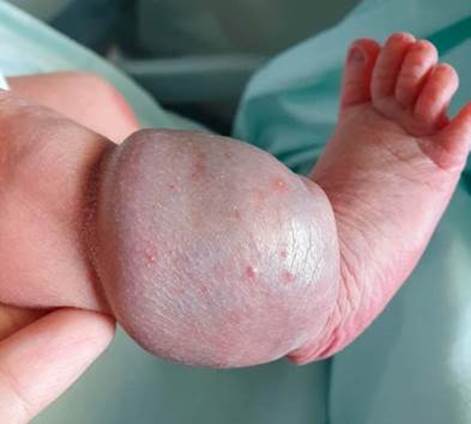
We report the case of a full-term female neonate, born by eutocic delivery, with a birth weight of 3175 g. Pregnancy was uneventful, with normal ultrasounds. Physical examination at birth revealed a raised, violaceous, soft-tissue mass at the right lateral malleolus, warm to touch, with prominent red small vesicles, measuring 3x4 cm (Fig. 1) and inguinal adenopathies in the ipsilateral limb. Doppler ultrasound revealed a well-defined hyperechogenic nodular image, with homogeneous texture and diffuse vascularity. There were no internal calcifications, and it did not extend to the profound plans. Magnetic resonance imaging (MRI) was performed for better characterization, revealing a benign tumoral vascular lesion (hemangioma or venous malformation), making the possibility of angiosarcoma or kaposiform hemangioendothelioma unlikely. Electrocardiogram, echocardiogram and transfontanelar ultrasound were normal. At four months, she has normal growth and development, and the lesion is undergoing rapid involution (discoloration and reduction in volume), with areas of skin redundancy, atrophy and changes in texture with persistent scattered veins and telangiectasias (Fig. 2).

Figure 1: At birth: Soft-tissue mass, raised, violaceous, warm to touch, with prominent red small vesicles, measuring 3x4 cm and inguinal adenopathies in the ipsilateral member.


