


 Inglês (pdf)
 Artigo em XML
 Referências do artigo
 Enviar este artigo por email
 Citado por SciELO
  Similares em
SciELO
Similares em
SciELO 
 Permalink
Introduction
Phytophotodermatitis is a nonimmunologic, phototoxic reaction caused by topical or oral exposure to photosensitizing plant-derived agents, followed by exposure to long-wavelength ultraviolet radiation (UVA).1
This dermatitis is most often seen after exposure to furocoumarins, which are potentially photosensitizing substances, also known as psoralens (mostly 5-methoxypsoralen).
They consist of botanical compounds constitutively present in certain plants, including fruits, leaves and roots.2 The most involved families are Compositae, Umbelliferae, and Rutaceae families, with the most frequent fruits causing this dermatitis including Persian limes and lemons (Rutaceae family); celery, parsley, carrots (Umbilliferae family) a nd fi gs (Moraceae family).3,4 It was described that the main psoralens present in limes are bergapten and psoralen and that the rind includes a greater amount, when compared to the pulp.5,6 When alone, they are innocent but might induce dermatitis if combined with sunlight exposure.
After experiencing UVA radiation, the photosensitizing agents are capable of inducing cell damage in the epidermis, resulting in photosensitive dermatitis. These photochemical reactions may appear in all skin types and are typical in gardeners, as well as in bartenders who manipulate limes while working.3 Although photosensitivity induced by topical agents is relatively rare in the pediatric population, its frequency has increased in the last few years due to the widespread use of those agents in the ecosystem. Plant-induced photosensitivity is the most important type involving children.7
We describe a lime-induced phytophotodermatitis case, appearing in two siblings.
Case report
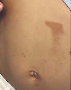
A 6-year-old girl presented with a 5-day history of brownish linear plaques on the left side of her face, abdomen and knees (Figs. 1 and 2). The lesions were not pruritic and the patient experienced a mild burning sensation. The patient had no history of medication intake or similar episodes. After careful history taking, it was found that the child had been squeezing limes while cooking a lime pie and got posterior UVA radiation exposure, as they had been on a beach vacation.
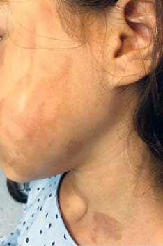
Figure 1: The affected area exhibits an erythematous patch with linear streaks, which later leads to hyperpigmentation, as observed on the face and neck.
Her brother had similar lesions, after the same type of exposure. Since there were only mild symptoms, the diagnostic hypothesis and its expected course were explained to the child and her parents. Moisturizing cream application and reinforced sun protection were recommended, together with covering the affected skin for the next 2-3 days and washing any clothes in contact with the limes. Both family members lesions healed spontaneously after nearly a month, with no scars or hyperpigmentation.
Discussion
Phytophotodermatitis should be hypothesized in the case of a child with a burn reaction, eruption and swelling of the sun-exposed skin, after short-term sun exposure. Clinical manifestations normally include erythema, edema, and blisters 12 to 36 hours after exposure to trigger compounds. Lesions are more frequently nonpruritic and may be associated with pain or burning sensation. The eruptions then place post-inflammatory hyperpigmentation that can take a few days to years to resolve spontaneously. Systemic symptoms are rare and may normally be due to sunburn.8,9 The bizarre configurations of the injuries, reflecting how the juice has dripped on the skin, can mimic a burn-like wound and also be categorized as trauma or child abuse in younger patients.10,11 On behalf of this, proper identification of the lesions nature becomes essential. To access accurate diagnosis, exhaustive history, and physical examination are crucial.
The skin examination should contain details concerning the lesions’ distribution, morphology, timing of onset and duration, as well as a history of interaction with potential photosensitizers and a family history of photosensitivity. It is also important to do the differential diagnosis between this disease, being nonimmunologic and arbitrary, and allergic contact or photoallergic contact dermatitis, which are immunologic responses, only present when there is previous sensibilization. In the latter, unexposed areas tend to be saved.12 A skin biopsy or phototesting is generally not necessary.
In this case, there was no severe reaction and slightly symptomatic hyperpigmentation happened instead, a clinical presentation that was consistent with previous reports. It looks like this happens especially in lime-induced phytophotodermatitis, according to the extent of both fruit and sun exposure, as well as in darker skin, which was also the case (the patient had a skin type IV, according to the Fitzpatrick Scale).13
The treatment depends on the severity of the skin damage, but it mostly relies on discontinuing the exogenous agent exposure, avoiding the sun for 8 to 72 hours and the use of a broad-spectrum sunscreen. After contact with the plants, immediate washing should be performed and emollients should be applied. In moderate to severe cases, topical or systemic steroids may be required.14
Conclusion
Lime-induced phytophotodermatitis is a self-limited phototoxic inflammatory eruption that is still weakly recognized. The diagnostic approach in children is frequently determined by meticulous history and objective examination. The onset of a rash in sun-exposed areas, following direct contact with both potentially photosensitizing substances and UV radiation, should create the hypothesis of phytophotodermatitis.
Should the disease go unrecognized, and given the agents' ubiquity, it can lead to a delayed diagnosis. It is required to raise awareness of this disease, so we ensure the safety of gardeners, chefs, and children. The typical clinical manifestations can allow for a diagnosis based solely on the physical exam. Early acknowledgment is crucial to diminish longstanding complications, often related to insufficient sun protection. Additionally, the tranquilization of the child’s family is extremely important, regarding the potential misdiagnosis of any kind of physical abuse. Physicians should be prepared to identify this illness.

