SciELO - Scientific Electronic Library Online

 
vol.11 número4Fenómeno Harlequin: um Recém-Nascido com Duas coresDilemas Éticos no Idoso em Cuidados Paliativos índice de autoresíndice de assuntosPesquisa de artigos
Home Pagelista alfabética de periódicos  

Serviços Personalizados

Journal

Artigo

Indicadores

Links relacionados

  • Não possue artigos similaresSimilares em SciELO

Compartilhar


Gazeta Médica

versão impressa ISSN 2183-8135versão On-line ISSN 2184-0628

Gaz Med vol.11 no.4 Queluz dez. 2024  Epub 27-Dez-2024

https://doi.org/10.29315/gm.v1i1.829 

Medicine or surgery imaging

Metastatic Squamous Cell Carcinoma: A Rare Presentation

Carcinoma Espinocelular Metastizado: Uma Apresentação Rara

1. Unidade de Cuidados de Saúde Personalizados de Cantanhede, ACES Baixo-Mondego, Cantanhede, Portugal.

2. Serviço de Dermatologia do Hospital Distrital da Figueira da Foz, Figueira da Foz, Portugal.

3. Serviço de Radiologia do Hospital Distrital da Figueira da Foz, Figueira da Foz, Portugal


Palavras-chave: Carcinoma de Células Escamosas/secundário; Metástases Linfáticas; Neoplasias da Pele

Keywords: Carcinoma, Squamous Cell/secondary; Lymphatic Metastasis; Skin Neoplasms

An 86-year-old woman, a nursing home resident presented to a dermatology consultation with an ulcerated/necrotic lesion on the inferior lip and right labial commissure, extending into the cervical region, with purulent discharge (Fig. 1) and hemilateral facial edema (right side), alongside multiple lymphadenopathies.

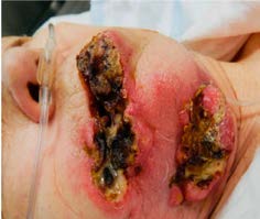
Figure 1: Tumoral lesion of the inferior lip, extending into the cervical region, with purulent discharge 

The cutaneous biopsy confirmed that it was a poor differentiated squamous cell carcinoma (SCC) and the staging by computed tomography (CT) scan (Fig. 2) revealed a carcinomatous lesion with a necrotic center and skin fistulation, on the right side of the inferior lip, that cover all the right hemiface, submandibular and sublingual areas, from jaw angle to the carotid space.

Figure 2: Skull CT scan - sagittal plane: tumor masses 

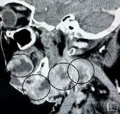
Also showed laryngeal invasion and jaw fracture. Necrotic-cystic lymph nodes were also observed in the carotid, cervical and supraclavicular chains, bilaterally.

The patient was referred to palliative care and passed away two weeks later.

The cutaneous SCC is a malignant tumor,1,2 usually noninvasive and with high cure rates, what highlights the exceptional nature of this case.3,4

Approximately 3% to 5% of cutaneous SCC cases metastasize to locoregional or distant lymph nodes.

Larger primary lesions and smaller cell divisions, as the location in a sun-exposed area and the immunocompromised state of the patient are associated with increased rates of tumor metastasis and recurrence.

In addition to this, there are several risk factors associated with metastases and recurrence: the size of the lesion (greater than 2 cm in greatest dimension), the thickness of the lesion (greater than 2 mm), the high-risk anatomic location (lip), and the poor differentiation confirmed by the biopsy.

With this case report, we intend to reinforce the importance of early referral of patients, regardless of their general physical or cognitive status.

Although the patient was not responsive, timely recognition, diagnosis and treatment could have influenced the outcome, giving her a dignified and serene end of life.

References

1. Waldman A, Schmults C. Cutaneous squamous cell carcinoma. Hematol Oncol Clin North Am. 2019;33:1-12. doi: 10.1016/j.hoc.2018.08.001. [ Links ]

2. Lam JKS, Sundaresan P, Gebski V, Veness MJ. Immunocompromised patients with met- astatic cutaneous nodal squamous cell carcinoma of the head and neck: poor outcome unrelated to the index lesion. Head Neck 2018;40:985-92. doi: 10.1002/hed.25069. [ Links ]

3. Ishitsuka Y, Hanaoka Y, Tanemura A, Fujimoto M. Cutaneous Squamous Cell Carcinoma in the Age of Immunotherapy. Cancers. 2021;13:1148. doi: 10.3390/cancers13051148. [ Links ]

4. Harris BN, Bayoumi A, Rao S, Moore MG, Farwell DG, Bewley AF. Factors Associated with Recurrence and Regional Adenopathy for Head and Neck Cutaneous Squamous Cell Carcinoma. Otolaryngol Head Neck Surg. 2017;156:863-9. doi: 10.1177/0194599817697053. [ Links ]

Declaração de contribuição/contributorship statement

BP: Write the article.

RM: Plan and design the analysis

SM: Data analysing

3All authors approved the final version to be published

BP: Escrever o artigo

RM: Planear e desenhar a análise

SM: Analisar os dados

7Todos autores aprovaram a versão final a ser publicada

Responsabilidades éticas

Conflitos de interesse: Os autores declaram a inexistência de conflitos de interesse na realização do presente trabalho.

Fontes de financiamento: Não existiram fontes externas de financiamento para a realização deste artigo.

Confidencialidade dos dados: Os autores declaram ter seguido os protocolos da sua instituição acerca da publicação dos dados de doentes.

Consentimento: Consentimento do doente para publicação obtido.

Proveniência e revisão por pares: Não comissionado; revisão externa por pares.

Ethical disclosures

Conflicts of interest: The authors have no conflicts of interest to declare.

Financing support: This work has not received any contribution, grant or scholarship.

Confidentiality of data: The authors declare that they have followed the protocols of their work center on the publication of data from patients.

Patient consent: Consent for publication was obtained.

Provenance and peer review: Not commissioned; externally peer reviewed.

Received: September 25, 2023; Accepted: September 15, 2024

Autor Correspondente/Corresponding Author: Beatriz Paupério [bpauperio@hotmail.com] ORCID ID: http://orcid.org/0009-0005-2755-2897

Creative Commons License This is an open-access article distributed under the terms of the Creative Commons Attribution License