Serviços Personalizados
Journal
Artigo
Indicadores
-
 Citado por SciELO
 -
 Acessos
Links relacionados
-
 Similares em
SciELO
Compartilhar
GE-Portuguese Journal of Gastroenterology
versão impressa ISSN 2341-4545
GE Port J Gastroenterol vol.25 no.1 Lisboa fev. 2018
https://doi.org/10.1159/000479590
CLINCIAL CASE STUDY
Signet Ring Cell Carcinoma, Ileal Crohn Disease or Both? – A Case of Diagnostic Challenge
Carcinoma de Células em Anel de Sinete do Íleon, Doença de Crohn ou Ambos? – Desafio Diagnóstico
Joana R. Carvalhoa, Joana Tavaresb, Inês Goularta, Paula Moura dos Santosa, Emília Vitorinob, Cristina Ferreirab, Fátima Serejoa, José Velosaa
Departments of aGastroenterology and bPathology, CHLN – Hospital de Santa Maria, Lisbon, Portugal
* Corresponding author.
ABSTRACT
Signet ring cell carcinoma is a rare form of adenocarcinoma that predominantly affects the stomach. Signet ring cell carcinoma originated from the ileum is extremely rare and the prognosis is poor. We present a case of small bowel obstruction with features suggesting Crohn disease of the ileum. The symptoms were chronic diarrhea and abdominal pain with a family history of inflammatory bowel disease. The patient underwent surgery and histopathology revealed both aspects of signet ring cell carcinoma and Crohn disease of the ileum. An association between long-standing inflammation and the development of this subtype of tumor has been proposed but there are no established surveillance guidelines for small bowel neoplasm in inflammatory bowel disease.
Keywords: Signet ring cell carcinoma, Crohn disease, Ileum
RESUMO
O carcinoma de células em anel de sinete é uma forma rara de adenocarcinoma que afeta predominantemente o estômago. A forma primária do íleon é muito rara e apresenta mau prognóstico. Apresentamos um caso de doença de Crohn do íleon cuja forma de apresentação cursou com quadro oclusivo associado a adenocarcinoma de células em anel de sinete do íleon. A doente apresentava história prévia de diarreia crónica e tinha história familiar de doença inflamatória. Foi submetida a ressecção cirúrgica e os achados histopatológicos revelaram a presença de aspetos compatíveis com doença de Crohn do íleon e adenocarcinoma de células em anel de sinete. Foi proposta uma associação com esta forma rara de tumor e inflamação de longa duração, contudo, não existem recomendações estabelecidas de vigilância de neoplasia na doença inflamatória intestinal com atingimento ileal.
Palavras-Chave: Carcinoma de células em anel de sinete, Doença de Crohn, Íleon
Introduction
Small intestinal malignancies are extremely rare accounting for 0.1–0.3% of all malignancies [1]. Signet ring cell carcinoma (SRCC) is a rare subtype of adenocarcinoma that most often arises in the stomach but may affect other organs, including the pancreas, breast, urinary bladder, ovarian, lung, esophagus, and large intestine [2,3]. It is an epithelial malignancy with cells resembling signet rings, as they contain large amounts of mucin, which pushes the nucleus to the cell periphery [1]. It represents one fourth of gastric cancers but in other locations has a very low incidence (<1%) [2]. The survival rate is poor (20–30% at 5 years) [1]. Only a very small number of cases of SRCC of the ileum in Crohn disease (CD) have been reported in the literature [1, 3–5].
It is consensual that the risk of developing small bowel adenocarcinoma is greater in patients with CD than in the general population; however, the exact magnitude and the etiologic mechanism of the increased risk are difficult to determine [5–8]. Sometimes the patients who develop an adenocarcinoma are those with small bowel CD but most often there is a combination of those with both small and large bowel CD [5]. We present a case of intestinal obstruction caused by SRCC as a first manifestation of small bowel CD.
Clinical Case
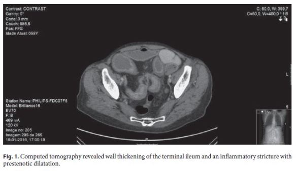
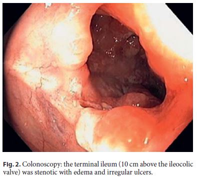
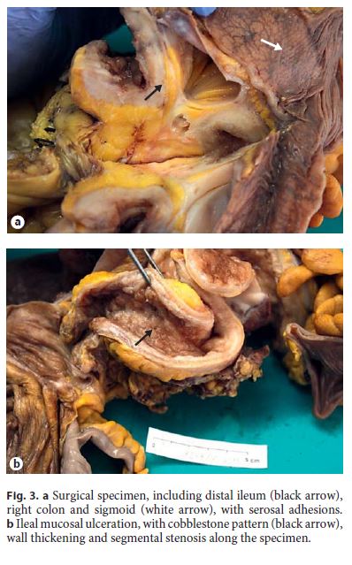
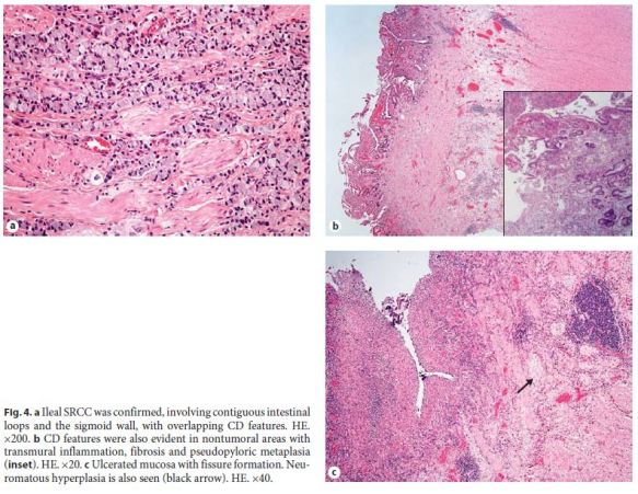
A 58-year-old female was admitted because of watery diarrhea without blood and diffuse abdominal pain of 3 months duration. She had lost 10 kg during this period. She was an active smoker and had a sister diagnosed with CD. Physical examination revealed a diffusely painful abdomen with augmented peristaltic sounds and no rebound tenderness. Laboratory tests showed normochromic normocytic anemia (hemoglobin 11.2 g/dL) and elevation of inflammatory markers (leukocytes 12,110 cells/mL, Creactive protein 0.6 mg/dL). Additional investigation with the Interferon Gamma Release Assay, cytomegalovirus DNA, blood and stool cultures, Epstein-Barr virus, human immunodeficiency virus, and hepatitis C and hepatitis B virus serologies was negative. We performed abdominal computed tomography that revealed wall thickening of the terminal ileum and an inflammatory stricture with prestenotic dilatation (Fig. 1). Colonoscopy revealed no abnormal finding in the rectum and all colonic segments; the terminal ileum (10 cm above the ileocolic valve) was stenotic with edema and irregular ulcers (Fig. 2). Multiple biopsies were performed on both segments. Given the strong suspicion of intestinal inflammatory disease of the ileum (possibly CD), the patient started intravenous prednisolone; the abdominal pain and diarrhea improved and she was discharged to outpatient consultation. Later, the histopathologic study of the biopsies of the terminal ileum revealed infiltration by SRCC, with lymphatic invasion; the colonic and rectal biopsies revealed mild nonspecific inflammation. The patient was readmitted. She had lost more than 3 kg in 3 weeks. Upper endoscopy with multiple biopsies of the stomach was performed and the presence of gastric tumor was excluded. CA 19–9 was 113 U/mL (normal range: 37 U/mL) and CA 125 was normal. Mammography, breast ultrasound, and body computed tomography excluded disease in other locations. The patient was submitted to surgical segmental ileal resection; a sigmoidectomy was also performed because of technical difficulties due to fibrotic adherences between the terminal ileum and sigmoid colon. The specimen included distal ileum, right colon, and sigmoid with extensive serosal adhesions throughout; mucosa presented with ulcerations in a cobblestone pattern; there was wall thickening and segmental stenosis (Fig. 3a, b). Histological examination confirmed SRCC, infiltrating both the ileum and sigmoid wall, admixed with CD features; transmural inflammation, neuromatous hyperplasia, and fibrosis were seen in areas with tumor but also in areas without malignant cells; in this one, there was also fissure ulceration and pseudopyloric metaplasia of the mucosa (Fig. 4a–c). The final TNM staging was T4N2M0. Five months after the diagnosis, the patient is on chemotherapy with oxaliplatin and 5-FU.


Discussion
Many reports have documented that adenocarcinoma of the small bowel is a complication of CD [6]. As there are only a small number of cases of SRCC of the ileum in CD, this association has not been clearly established. Patients with long-standing small bowel CD are thought to have an increased risk of small bowel carcinoma [6–8]. Many postulated risk factors surfaced as a result of observed trends across case reports such as previous strictureplasty and excluded/bypassed bowel segments [6, 8]. The risk of developing small bowel carcinoma was also found to be much higher in patients whose disease was confined to the small bowel than in the patients with ileocolic CD [9]. Protective factors against the development of small bowel carcinoma in CD have been less frequently studied, but it has been proposed that small bowel resection and prolonged use of salicylates may be protective [10]. In our case, the only identified risk factor was the location of CD, confined to the small bowel. Uncommonly, in this case, the intestinal bowel obstruction due to malignancy was the first manifestation of CD. The presence of pseudopyloric metaplasia, transmural lymphoid aggregates, neuromatous hyperplasia, and wall fibrosis unrelated with maliga nancy are indicators of a long-standing disease that had not been diagnosed and treated, although she did not report any previous gastrointestinal symptoms.
Obstruction is the most common presenting manifestation in small bowel carcinoma [6], as seen in this case. Less frequently, it presents with hemorrhage, fistula, or perforation [6, 8]. Unfortunately, these symptoms are hard to differentiate from those of CD exacerbation, which partly explains the majority of diagnoses being made at the time of operation or postoperatively [1, 5, 6].
The treatment of choice is wide resection of the small bowel segment harboring the carcinoma as well as resection of the corresponding mesentery and lymph nodes. Evidence regarding the value of adjuvant chemotherapy for small bowel carcinoma is sparse and mostly consists of small retrospective reviews [6]. In about 50% of the cases of small bowel carcinomas, the tumor is poorly differentiated and produces mucin, which is associated with worst prognosis [5]. Despite the medical advancements, over the decades, there has been a lack of significant improvement in prognosis [6]. In this context, there is a need to elucidate screening modalities to facilitate earlier diagnosis and treatment of small bowel neoplasm in patients with CD of the ileum. However, no screening method has been found to be particularly useful [6].
This case is particularly interesting because the first manifestation of CD of the ileum was this rare type of neoplasm. The diagnosis was challenging and the treatment options were limited. More studies and surveillance guidelines for small bowel neoplasm in CD are awaited.
References
1 Deuskar J, Joshi P, Adbe U, Railkar A: Signet ring cell carcinoma of the ileum: a case report and review of literature. Internet J Surg 2009;25:1. [ Links ]
2 Dogan S, Celikbilek M, Eroglu E, Yagbasan A, Can Sezgin G, Gursoy S, et al: Signet ring cell carcinoma mimicking ileal Crohns disease. Gastroenterol Insights 2013;5:7–8. [ Links ]
3 Paparo F, Piccardo A, Clavarezza M, Piccazzo R, Bacigalupo L, Cevasco L, et al: Computed tomography enterography and 18F-FDG PET/CT features of primary signet ring cell carcinoma of the small bowel in a patient with Crohns disease. Clin Imaging 2013;37:794–797. [ Links ]
4 Kim JS, Cheung DY, Park SH, Kim HK, Maeng IH, Kim SY, et al: A case of small intestinal signet ring cell carcinoma in Crohns disease. Korean J Gastroenterol 2007;50:51–55. [ Links ]
5 Valério F, Cutait R, Sipahi A, Damião A, Leite K: Cancer in Crohns disease: case report. Rev Bras Coloproct 2006;26:443–446. [ Links ]
6 Cahill C, Gordon PH, Petrucci A, Boutros M: Small bowel adenocarcinoma and Crohns disease: any further ahead than 50 years ago? World J Gastroenterol 2014;20:11486–11495. [ Links ]
7 Jess T, Winther KV, Munkholm P, Langholz E, Binder V: Intestinal and extra-intestinal cancer in Crohns disease: follow-up of a population-based cohort in Copenhagen County, Denmark. Aliment Pharmacol Ther 2004;19:287–293. [ Links ]
8 Ramos CR, Guillén P, Palomo MJ: Adenocarcinoma de intestino delgado y enfermedad de Crohn. Rev Esp Enf Digest 1997; 89:321–324. [ Links ]
9 Von Roon AC, Reese G, Teare J, Constantinides V, Darzi AW, Tekkis PP: The risk of cancer in patients with Crohns disease. Dis Colon Rectum 2007;50:839–855. [ Links ]
10 Piton G, Cosnes J, Monnet E, Beaugerie L, Seksik P, Savoye G, et al: Risk factors associated with small bowel adenocarcinoma in Crohns disease: a case-control study. Am J Gastroenterol 2008;103:1730–1736. [ Links ]
Statement of Ethics
The authors have no conflicts of interest to declare.
Disclosure Statement
This study did not require informed consent nor review/approval by the appropriate ethics committee.
* Corresponding author.
Dr. Joana R. Carvalho
Department of Gastroenterorology, Hospital de Santa Maria
Avenida Professor Egas Moniz
PT–1649-035 Lisbon (Portugal)
E-Mail joana.rita.carvalho@gmail.com
Received: April 6, 2017; Accepted after revision: May 26, 2017


