Serviços Personalizados
Journal
Artigo
Indicadores
-
 Citado por SciELO
 -
 Acessos
Links relacionados
-
 Similares em
SciELO
Compartilhar
GE-Portuguese Journal of Gastroenterology
versão impressa ISSN 2341-4545
GE Port J Gastroenterol vol.25 no.3 Lisboa jun. 2018
https://doi.org/10.1159/000485104
CLINICAL CASE STUDY
The Challenging Acute Buried Bumper Syndrome: A Case Report
Síndrome da campânula interna com apresentação precoce
Juliana Pinhoa, Diogo Libâniob, Pedro Pimentel-Nunesb, Mário Dinis-Ribeirob
aGastroenterology Department, Centro Hospitalar Tondela-Viseu, Viseu, and bGastroenterology Department, Instituto Português de Oncologia do Porto, Porto, Portugal
* Corresponding author.
ABSTRACT
Percutaneous endoscopic gastrostomy (PEG) is the preferred route of feeding and nutritional support in patients requiring long-term enteral nutrition. Major complications related to the procedure are rare. Buried bumper syndrome is a late major complication, occurring in 0.3–2.4% of patients. Although considered a late complication, it can rarely occur in an acute setting early after the procedure. We present the case of an early buried bumper syndrome, presenting 1 week after PEG tube placement, with local stoma infection associated with an infected cavity within the abdominal wall with feeding content, successfully managed with antibiotic therapy and PEG tube repositioning through the original track.
Keywords: Buried bumper syndrome, Percutaneous endoscopic gastrostomy, Complication
RESUMO
A gastrostomia endoscópica percutânea (PEG) é a via preferencial de alimentação e suporte nutricional nos doentes com necessidade de nutrição entérica a longo prazo. As complicações major associadas ao procedimento são raras. A síndroma da campânula interna é uma complicação major tardia, que ocorre em 0.3 a 2.4% dos casos. Apesar de ser considerada uma complicação tardia, pode ocorrer precocemente com uma apresentação aguda dias após o procedimento. Os autores descrevem o caso de uma síndroma da campânula interna precoce após 1 semana da colocação de PEG, com infeção associada do estoma e formação de loca na parede abdominal com conteúdo alimentar, abordado com sucesso com antibioterapia e reposicionamento da sonda através do trajeto original, sem necessidade de remoção.
Palavras-Chave: Síndrome campânula interna, gastrostomia endoscópica percutânea, complicação
Introduction
Percutaneous endoscopic gastrostomy (PEG) is a widely used method of nutrition delivery for patients with long-term insufficiency of oral food intake, with a high technical success rate [1, 2]. Although generally considered to be a safe procedure, complication rates vary from 0.4 to 22.5% of cases, with minor complications being three times more frequent [2]. Complications that follow this procedure include dislodgement, dysfunction, peristomal infection, and aspiration [3]. The buried bumper syndrome (BBS) is an uncommon and late complication, which can result in gastrointestinal bleeding, perforation, peritonitis, and intra-abdominal or abdominal wall abscesses that can lead to poor outcomes [1, 3]. Although considered a late complication developing months to years after placement, it can also occur in an acute setting (within 30 days after insertion) [1]. These cases are usually considered not suitable for a conservative and endoscopic approach due to the immature stoma tract [1]. We report a case of an early-onset BBS, successfully managed with PEG tube repositioning through the original track, without the need for removal.
Case Report
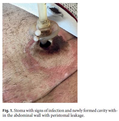
A 57-year-old male patient with a past medical history of locally advanced squamous cell oropharyngeal carcinoma under adjuvant chemoradiotherapy was submitted to PEG placement (24 Fr, Cook Medical, transnasal endoscopic approach), due to dysphagia and poor nutritional status. The placement was made by the Ponsky method, without immediate complications, and the external bumper was at 10 mm from the skin surface. One week after the procedure, the patient presented to the emergency department with peristomal leakage. On examination, stoma showed no signs of infection, but the external bumper was more than 1 cm from the skin surface. Adjustment of the external bumper was performed and the patient was discharged. One week later, the patient was readmitted because of abdominal pain and persistent peristomal leakage, exacerbated by food infusion through the tube. Laboratory examination showed 2,000 white blood cells/μL (72% neutrophils) and an elevated C-reactive protein level (142.5 mg/L). On examination, the stoma showed signs of infection and peristomal leakage with a newly formed cavity within the abdominal wall filled with feeding content (Fig. 1) and the internal bumper was juxtaposed to the skin surface under the cavity. Flushing the tube with water exacerbated the patient complaints. Given the previous difficult endoscopic approach at PEG placement and the patient being reluctant to endoscopy performance, a less invasive procedure was attempted. A guide wire was first passed through the PEG tube into the gastric cavity without resistance; using the guide wire to make traction, the tube was pulled back into the stomach through the original track, without complications. Peristomal swab analysis was performed. Fluoroscopy image with contrast injection through the tube after the procedure showed contrast in the stomach.

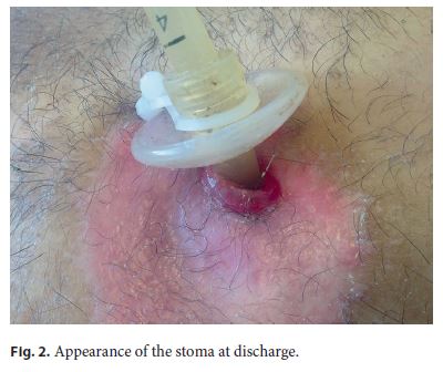
The patient was admitted to the gastroenterology department and started on parenteral nutrition and antibiotic therapy with piperacillin-tazobactam on admission, with progressive clinical improvement, reduction of peristomal leakage and local inflammatory signs, and reduction of the cavity within the abdominal wall. The PEG tube remained freely mobile into the stoma, and the peristomal swab analysis revealed Proteus mirabilis and Serratia marcescens sensible to piperacillin-tazobactam. Seven days after admission, given the clinical improvement and decrease in laboratory inflammatory parameters (C-reactive protein and white blood cells count), PEG tube feeding was restarted without local pain or signs of PEG tube malfunctioning. The patient completed 15 days of antibiotic therapy and was discharged 18 days after admission. At discharge, the stoma showed no signs of infection or peristomal leakage, without any difficulties in PEG tube feeding (Fig. 2).

Discussion
PEG is a well-established method of nutrition delivery, with a high technical success rate [1, 2]. PEG was introduced in 1980 by Gauderer et al. [4] as an alternative to open surgery. Although considered a safe and effective procedure, PEG complication rates vary from 0.4 to 22.5% of cases, being classified as major or minor, early or late [2]. Minor complications are three times more frequent, with peristomal infection being the most common [1, 5]. Major complications like BBS, bleeding, perforation, aspiration, and tumor seeding are rare [3].
BBS represents a severe complication, occurring in 0.3–2.4% of patients as a consequence of excessive tension of the tissue between the external and internal bumpers, leading to ischemic necrosis of the gastric wall and subsequent migration of the tube toward the abdominal wall [1, 3, 5]. Patients with malignancy, poor initial nutritional status (body mass index <20) and under systemic corticosteroids therapy or chemoradiation have a higher risk of BBS [1]. BBS can be complicated by gastrointestinal bleeding, perforation, peritonitis, intra-abdominal and abdominal wall abscesses or phlegmon, and these complications can lead to fatal outcomes [1, 3]. Inability to insert food or liquids, loss of patency and peristomal leakage are considered to be a typical symptomatic triad [1].
Although it is usually described as a late complication, presenting months to years after placement, it can also occur in an acute setting (less than 30 days after placement) [1, 6]. Migration to the skin level has been described in case reports as early as 6–9 days after insertion [7, 8]. In our patient, it occurred 1 week after the procedure; even though initially not recognized as a BBS, attending to the rapid evolution with stoma infection and a newly formed cavity within the abdominal wall with feeding content 1 week later, it is highly probable that migration of the internal bumper, even if partial, occurred 1 week after PEG placement. Early diagnosis is therefore very important and prompt management is required to avoid further complications [6]. Removal of the PEG tube is the most widely described treatment for BBS, either by endoscopy or surgery [1, 3]. A conservative approach, the cut and leave strategy, has been described for patients with high surgical risk and worse prognosis [1]. In the acute setting of BBS, conservative and endoscopic approach cases are usually not suitable because of the immature stoma tract, but the choice of treatment depends on the patient and the depth of disc migration [1]. In our case, the patient was successfully managed with PEG tube repositioning through guide wire and traction and antibiotic therapy without complications, allowing the maintenance of PEG feeding and an adequate nutritional support.
References
1 Cyrany J, Rejchrt S, Kopacova M, Bures J: Buried bumper syndrome: a complication of percutaneous endoscopic gastrostomy. World J Gastroenterol 2016;22:618–627. [ Links ]
2 Itkin M, DeLegge MH, Fang JC, McClave SA, Kundu S, dOthee BJ, et al: Multidisciplinary practical guidelines for gastrointestinal access for enteral nutrition and decompression from the Society of Interventional Radiology and American Gastroenterological Association (AGA) Institute, with endorsement by Canadian Interventional Radiological Association (CIRA) and Cardiovascular and Interventional Radiological Society of Europe (CIRSE). Gastroenterology 2011;141:742–765. [ Links ]
3 Rahnemai-Azar AA, Rahnemaiazar AA, Naghshizadian R, Kurtz A, Farkas DT: Percutaneous endoscopic gastrostomy: indications, technique, complications and management. World J Gastroenterol 2014;20:7739–7751. [ Links ]
4 Gauderer MW, Ponsky JL, Izant RJ Jr: Gastrostomy without laparotomy: a percutaneous endoscopic technique. J Pediatr Surg 1980;15:872–875. [ Links ]
5 Schrag SP, Sharma R, Jaik NP, Seamon MJ, Lukaszczyk JJ, Martin ND, et al: Complications related to percutaneous endoscopic gastrostomy (PEG) tubes. A comprehensive clinical review. J Gastrointestin Liver Dis 2007;16:407–418. [ Links ]
6 Lucendo AJ, Friginal-Ruiz AB: Percutaneous endoscopic gastrostomy: an update on its indications, management, complications, and care. Rev Esp Enferm Dig 2014;106:529–539. [ Links ]
7 Afifi I, Zarour A, Al-Hassani A, Peralta R, El-Menyar A, Al-Thani H: The challenging buried bumper syndrome after percutaneous endoscopic gastrostomy. Case Rep Gastroenterol 2016;10:224–232. [ Links ]
8 Geer W, Jeanmonod R: Early presentation of buried bumper syndrome. West J Emerg Med 2013;14:421–423. [ Links ]
Statement of Ethics
This study did not require informed consent or review/approval by the appropriate ethics committee.
Disclosure Statement
The authors have no conflicts of interest to disclose.
* Corresponding author.
Dr. Juliana Pinho
Gastroenterology Department, Centro Hospitalar Tondela/Viseu
Avenida Rei Dom Duarte
PT–3504-509 Viseu (Portugal)
E-Mail julianapinho18@gmail.com
Received: September 5, 2017; Accepted after revision: November 8, 2017


