SciELO - Scientific Electronic Library Online

 
vol.29 número5Abordagem endoscópica de quistos de duplicação duodenal sintomáticos: a propósito de dois casos clínicosRecuperação endoscópica bem sucedida de um fio-guia fraturado retido no ducto pancreático índice de autoresíndice de assuntosPesquisa de artigos
Home Pagelista alfabética de periódicos  

Serviços Personalizados

Journal

Artigo

Indicadores

Links relacionados

  • Não possue artigos similaresSimilares em SciELO

Compartilhar


GE-Portuguese Journal of Gastroenterology

versão impressa ISSN 2341-4545

GE Port J Gastroenterol vol.29 no.5 Lisboa out. 2022  Epub 02-Jan-2023

https://doi.org/10.1159/000516674 

Endoscopic Snapshot

Pancreatogastric Fistula with Fish-Mouth Sign

Fístula pancreatogástrica com sinal de boca de peixe

Sara Santos1 

Rita Saraiva1 

Mário Oliveira1 

Diana Carvalho1 

Gonçalo Ramos1 

1Centro Hospitalar de Lisboa Central, Lisbon, Portugal


We report a case of a 72-year-old female referred to a gastroenterology consultation for a finding of main pancreatic duct (MPD) dilation. The patient was a nonsmoker and had a mean daily alcohol intake of 5 g/day. Her medical history was positive for several episodes of pancreatitis 15 years previously, with biliary lithiasis, for which she underwent laparoscopic cholecystectomy. Subsequently, she had 2 other episodes of pancreatitis, including an infected pancreatic fluid collection (PFC), that underwent conventional endoscopy-guided transgastric drainage at another institution. The patient remained asymptomatic for 14 years.

A computed tomography (CT) scan and magnetic resonance cholangiopancreatography (MRCP) revealed irregularity and dilation of the MPD (8 mm in the tail and 26 mm in the body) and a communication with the gastric antrum, with no communication with the cephalic segment or ampulla of Vater. Blood tests were normal, including CEA and CA19.9 levels.

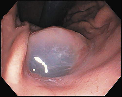
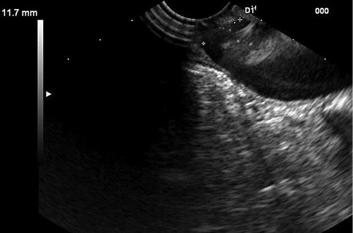
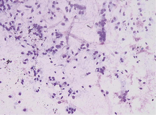
A presumptive diagnosis of disconnected MPD with a gastric fistula due to previous cystic drainage was made, but endoscopic ultrasound (EUS) was performed to exclude a main-duct intraductal papillary mucinous neoplasm (MD-IPMN). Endoscopically, a fistulous orifice 10 mm wide in the gastric antrum was apparent (Fig. 1), with a gelatinous, high-viscosity content which could not be removed despite vigorous lavage. A gastroscope was used to intubate the fistulous tract, but progression was not possible due to the viscous content. The EUS showed a globally dilated MPD (40 mm in the body) with a loss of interface with the gastric wall in the isthmus, 10 mm in diameter (Fig. 2). Fine-needle aspiration (FNA) of the MPD with a 19-gauge needle was performed, revealing a very thick mucinous content. Cytologic analysis confirmed high-grade IPMN, and the patient was referred to surgery (Fig. 3).

Fig. 1 Endoscopic findings of fistulous orifice with mucinous content. 

Fig. 2 Endoscopic ultrasound showing fistulous tract to the stomach. 

Fig. 3 Cytology. 

Although IPMN can spontaneously fistulize to adjacent organs [1, 2], in this patient, it most likely resulted from a prior drainage procedure. This case underlines the importance of a correct workup prior to endoscopic transmural drainage of PFCs to safely exclude cyst neoplasm, especially in the setting of repeated episodes of pancreatitis. Although the best approach to IPMN fistulization is yet to be defined, a MD-IPMN is a high-risk condition and should be referred to surgery [3].

References

1. Ftériche FS, Chebbi F, Bedioui H, Kchir N, Ammous A, Ksantini R, et al. Tumeur intracanalaire papillaire mucineuse du pancréas dégénérée et fistulisée dans l'estomac, le duodénum, et le côlon. Ann Chir. 2006;131(2):118-20. [ Links ]

2. Patel A, Allen A, Kuwahara J, Wadsworth T, Loeffler DM, Xie KL. Intraductal papillary mucinous neoplasm complicated by a gastropancreatic fistula. Radiol Case Rep. 2018;14(3):320-3. [ Links ]

3. Tanaka M, Fernández-Del Castillo C, Kamisawa T, Jang JY, Levy P, Ohtsuka T, et al. Revisions of international consensus Fukuoka guidelines for the management of IPMN of the pancreas. Pancreatology. 2017;17(5):738-53. [ Links ]

1Statement of Ethics The patient was provided with a copy of the manuscript and signed a consent form to publish the case.

Funding Sources There was no funding.

Received: January 08, 2021; Accepted: March 29, 2021

Corresponding author Sara Santos saramendezleiria@hotmail.com

Conflict of Interest Statement There were no conflicts of interest.

Author Contributions S.S.: writing of the manuscript and management of the patient. R.S., D.C., and M.O.: patient management. G.R.: patient management and editing of the paper

Creative Commons License This is an open-access article distributed under the terms of the Creative Commons Attribution License