SciELO - Scientific Electronic Library Online

 
vol.30 número5Estratégias de indução de infliximab na agudização de colite ulcerosa grave: uma série de casos e revisão da literatura índice de autoresíndice de assuntosPesquisa de artigos
Home Pagelista alfabética de periódicos  

Serviços Personalizados

Journal

Artigo

Indicadores

Links relacionados

  • Não possue artigos similaresSimilares em SciELO

Compartilhar


GE-Portuguese Journal of Gastroenterology

versão impressa ISSN 2341-4545

GE Port J Gastroenterol vol.30 no.5 Lisboa out. 2023  Epub 01-Dez-2023

https://doi.org/10.1159/000526854 

Clinical Case Study

Severe Hypercholesterolemia Mediated by Lipoprotein X in an Immunosuppressed Patient: A Case Report

Hipercolesterolemia grave mediada por lipoproteína X em doente imunossuprimido: caso clínico

Cristiana Sequeira1 
http://orcid.org/0000-0001-5568-3732

Mariana Coelho1 

Inês Costa Santos1 
http://orcid.org/0000-0003-3332-8590

Sara Ramos Lopes1 

João Mangualde1 

Ana Paula Oliveira1 

1Gastroenterology Department, Centro Hospitalar de Setúbal, Setúbal, Portugal


Abstract

Cholestatic liver diseases may be associated with increased plasmatic cholesterol due to an abnormal lipoprotein - lipoprotein X (LpX). Correcting the underlying cause of cholestasis is the critical treatment of LpX-associated hypercholesterolemia without any proven benefit from conventional lipidlowering agents. In some situations, plasma exchange may apply to prevent associated complications, such as hyperviscosity syndrome. The authors present the case of a 44-year-old man with orbital inflammatory pseudotumor on prednisolone, admitted due to hepatocellular and cholestatic lesion and severe hypercholesterolemia. Laboratory investigation established that hepatitis E virus was responsible for liver injury and showed that LpX mediated the severe hypercholesterolemia. Reduction of the immunosuppressive load contributed to virus clearance. The consequent resolution of cholestasis and cholesterol removal by plasmapheresis allowed lipid profile normalization. The authors report the first case of LpX-associated hypercholesterolemia in a patient with hepatitis E-induced cholestasis and revisit the role of the liver in lipid metabolism.

Keywords: Lipoprotein X; Hypercholesterolemia; Hepatitis E

Resumo

As doenças hepáticas colestáticas podem associar-se a um aumento do colesterol à custa de uma lipoproteína anómala, a lipoproteína X (LpX). Os agentes hipolipemiantes convencionais não apresentam benefício nesta entidade, pelo que o tratamento da hipercolesterolemia associada de LpX baseia-se na correção da causa subjacente da colestase. A plasmaferese pode ser necessária para evitar complicações, como a síndrome de hiperviscosidade. Os autores apresentam o caso de um homem de 44 anos com antecedentes de pseudotumor inflamatório da órbita sob prednisolona, admitido por lesão hepatocelular e colestática e hipercolesterolemia grave. A investigação laboratorial permitiu estabelecer a hepatite E aguda como responsável da lesão hepática e mostrou que a hipercolesterolemia grave foi mediada pela LpX. A redução da carga imunossupressora facilitou a eliminação do vírus da hepatite E. A consequente resolução da colestase coadjuvada pela remoção de colesterol por plasmaferese, permitiu a normalização mantida do perfil lipídico. Os autores relatam o primeiro caso de hipercolesterolemia associada a LpX em contexto de colestase induzida pelo vírus da hepatite E, e revisitam a importância do fígado no metabolismo dos lípidos.

Palavras Chave: Lipoproteína-X; Hipercolesterolemia; Hepatite E

Introduction

Hypercholesterolemia is a common finding in clinical practice. However, few conditions are associated with total cholesterol (TC) values greater than 1,000 mg/dL, primarily in patients with homozygous familial hypercholesterolemia and family lecithin acyltransferase deficiency [1, 2]. As the liver plays a pivotal role in lipid and lipoprotein metabolism, cholestatic liver diseases are associated with high levels of an abnormal lipoprotein with a density similar to very-low-density lipoprotein and low-density lipoprotein but different composition, the lipo-protein X (LpX) [3]. Recognition of this entity is of ut-most importance as lipid-lowering agents are ineffective, and definitive treatment requires correcting the underlying cause of cholestasis [4].

Case Presentation

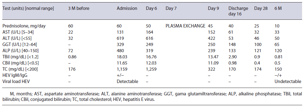
A 44-year-old Caucasian male was admitted to our gastroenterology department due to a 1-week clinical picture of jaundice, choluria, asthenia, and anorexia. He denied fever, abdominal pain, pruritus, weight loss, nausea, and vomiting. His medical history was remarkable for orbital inflammatory pseudotumor treated with prednisolone (60 mg/day) in the last 3 years. He has no family history of early-onset coronary artery disease or hyperlipidemia. Three months prior, his routine workup showed regular lipid panel and liver enzymology (shown in Table 1). There was no relevant epidemiological background, such as recent travel and eating uncooked meat. The patient denied alcohol or drug consumption. The physical exam only revealed jaundice; the remaining examination excluded encephalopathy, ascites, xanthelasma, or xanthomas.

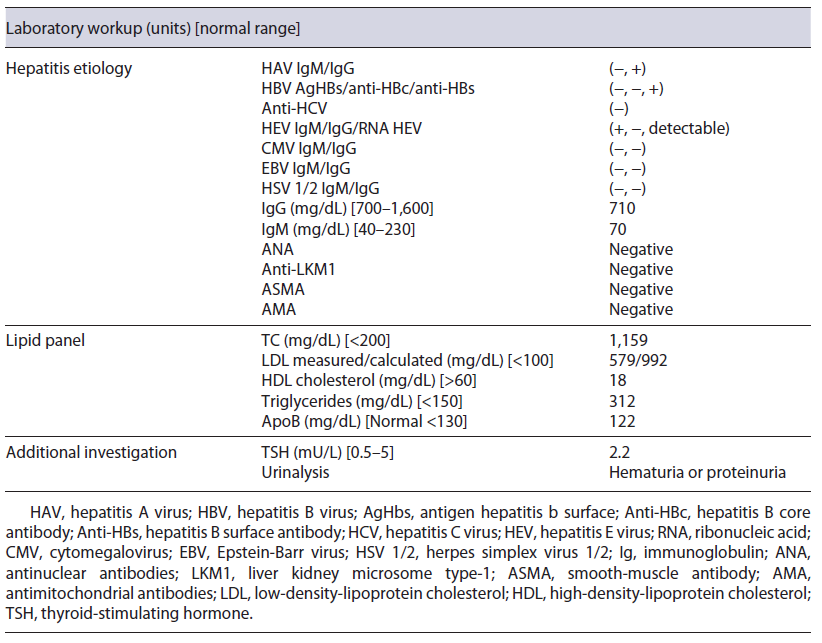
Initial blood investigations revealed elevated hepatic enzymes displaying a mixed pattern (ALT >15 times and ALP > four times the upper limit of the reference range; with R factor 4.5) and severe hypercholesterolemia (TC >1,000 mg/day) (shown in Table 1). Blood counts, albumin, glycemia, and coagulation tests were unremarkable. Abdominal computed tomography excluded biliary obstruction. Moreover, the diagnostic workup showed acute hepatitis E virus (HEV) features, excluding the remaining viral and autoimmune etiologies (shown in Table 2). A percutaneous liver biopsy denoted cholestasis, mixed lobular inflammatory infiltrate, and focal hepatocellular necrosis. We decided to taper prednisolone with close ophthalmologic surveillance; we did not start anti-lipemic medications.
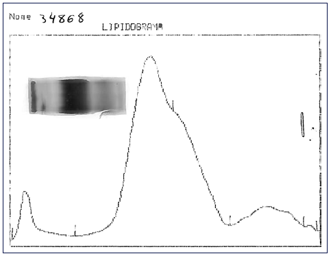
Table 1 Evolution of laboratory results over the time 

Fig. 1 Lipid electrophoresis: application point corresponds to the fraction with low protein content. 

Further investigation of the hyperlipidemia ruled out hypothyroidism and nephrotic syndrome (shown in Table 2), and we excluded primary causes due to the new onset and the absence of family history. We hypothesized hypercholesterolemia secondary to LpX due to cholestatic viral hepatitis. This diagnosis was supported by TC elevation without proportional elevation of apolipoprotein B (ApoB) and a calculated LDL cholesterol higher than measured (shown in Table 2). A nonquantitative fasting lipoprotein electrophoresis showed a band at the application site with slow migration, suggesting the presence of low density and protein fraction, such as Lp-X (Fig. 1).

Table 2 Additional laboratory workup 

Given the persistently high lipid levels, which could trigger complications related to hyperviscosity syndrome, the patient underwent plasma exchange, with a marked decrease in plasmatic cholesterol (shown in Table 1), after one single session. Along with the tapering of corticosteroids, we observed a favorable clinical course, with a complete resolution of liver tests (shown in Table 1).

No new ophthalmological alterations were detected, the lipid profile remained normal (without anti-lipemic drugs), and we exclud-ed chronic HEV infection.

Discussion

The liver is a critical organ in lipid and lipoprotein metabolism. In 1969, Seidel et al. [5] described for the first time a lipoprotein rich in phospholipid and free cholesterol in patients with cholestasis; due to its unknown origin, it was denominated as Lp-X. LpX is an abnormal lipoprotein with a density similar to very-low-density lipo-protein and LDL but different lipid and apolipoprotein compositions [3, 5, 6]. It is rich in phospholipids and free cholesterol (80-90%) and poor in cholesterol esters (<5%), triglycerides, and proteins. Unlike LDL, it contains albumin dissolved in its aqueous core. The apolipo-proteins encountered on its surface comprise Apo AI, E, and C, although never ApoB [3, 5, 6].

The pathogenesis is poorly understood. Biologically, cholesterol is eliminated by conversion to bile acids [3]; however, when biliary stasis develops, there is progressive retention of bile salts in hepatocytes, which stops the production of new bile acids accumulating cholesterol within the liver cells [3, 7, 8]. Subsequently, there is a reflux of cholesterol into the circulation [4]. LpX is associated with cholestatic liver diseases, including primary biliary cholangitis [4], bile duct obstruction, graft versus host disease in liver transplant recipients [9], deficiency of lecithin acyltransferase enzyme [1], granulomatous hepatitis [10], and other causes of hepatitis [11].

HEV infection is considered one of the common causes of acute fecal-oral-transmitted hepatitis in developing countries [12]. Still, in developed countries, HEV is estimated to account for less than 1% of cases of acute viral hepatitis despite being an often-overlooked cause. Evidence suggests that autochthonous transmission is zoonotic due to undercooked pigs and deer [13, 14]. HEV has an incubation period of 3-8 weeks, followed by a prodromal phase and acute icteric hepatitis lasting for days to several weeks. Acute HEV is usually clinically silent [13]. Marked cholestatic hepatitis with persistent jaundice and elevation of serum alkaline phosphatase is uncommon. In immunocompromized patients, anti-HEV IgM may be falsely negative; therefore, the gold standard for diagnosing acute HEV is the detection of HEV RNA in biological samples [13]. Acute HEV infection is usually self-limiting in immunocompetent patients and requires only supportive treatment. However, in immunocompromized patients, primarily reported in solid organ transplant recipients [15], HEV may not be cleared spontaneously and causes chronic hepatitis. Reducing the dosage of immunosuppressive therapy helps eliminate HEV [16, 17], so we reduced the immunosuppressive load with clinical improvement and no evolution to chronic HEV. Ribavirin is the treatment of choice in chronic HEV (14).

Cholesterol values greater than 1,000 mg/dL may be encountered with the presence of the LpX fraction [7, 18]. Methods for its direct determination are poorly available in conventional laboratories [18], so knowledge about the composition of this lipoprotein may be helpful in diagnosis. LpX does not contain ApoB, so it is frequent to find an excessive elevation of TC that is not accompanied by a proportional elevation of ApoB (increased TC/ApoB ratio) [4], as in our patient. A discrepancy between LDL-C calculated by the Friedewald formula and measured supports the presence of LpX [3, 4, 18]. In our patient, the calculated LDL cholesterol was 992.8 mg/dL, whereas the measured one was 579 mg/dL; this is because LpX interferes with the precipitation of chylomicrons, preventing their correct measurement. Demonstrating the presence of LpX can be done by lipoprotein electrophoresis using a commercially available kit [4, 18]; a low protein fraction may exist near the application point [18], as we observed in our case patient. Other alternative techniques for LpX determination have also been described, for example, agarose electrophoresis, non-denaturing polyacrylamide gradient gel electrophoresis, and nuclear magnetic resonance spectroscopy [18].

Apo-B’s absence results in LpX uptake by hepatocytes instead of macrophages, so liver dysfunction drives lower clear-up LpX from the plasma. Therefore, correction of the underlying cause is pivotal. Lipid-lowering agents such as statin and ezetimibe will not affect cholesterol levels in this setting and carry potential toxicity [4]. Lipoprotein apheresis is a temporary measure usually reserved for complications such as hyperviscosity syndrome, xanthomata, and cholesteroloma [4, 18, 19] when underlying cause resolution is not feasible or inadequate. The benefit appears to be greater with plasmapheresis than with lipoprotein apheresis [20]. Given that our patient had persistently high cholesterol levels and the high immunosuppressive burden delayed the resolution of the cause of cholestasis (lower viral clearance of HEV), he successfully and safely underwent plasmapheresis.

Thus, considering our case report, in routine practice, abnormalities in the lipid panel should raise suspicion about the presence of LpX, especially in patients with hypercholesterolemia and cholestasis. Recognizing this condition allows the correct therapeutic management and the prevention of complications. We describe the first case reported in the literature of LpX-associated hypercholesterolemia due to acute hepatitis E, responding to spontaneously eliminating HEV after weaning immu-nosuppressive therapy, aided by concomitant plasmapheresis (one session).

Acknowledgments

We thank Maria Elisa Micaelo Alves from the Faculdade de Farmácia da Universidade de Lisboa for performing the lipoprotein electrophoresis and supporting the diagnosis of this entity.

References

1. Norum KR, Remaley AT, Miettinen HE, Strøm EH, Balbo BEP, Sampaio CATL, et al. Lecithin: cholesterol acyltransferase - symposiumon 50 years of biomedical research from its discovery to latest findings. J Lipid Res. 2020 Aug; 61(8): 1142-9. [ Links ]

2. Wang A, Richhariya A, Gandra SR, Calimlim B, Kim L, Quek RGW, et al. Systematic review of low-density lipoprotein cholesterol apheresis for the treatment of familial hypercholesterolemia. J Am Heart Assoc. 2016 Jul 6; 5(7): e003294. [ Links ]

3. Fellin R, Manzato E. Lipoprotein-X fifty years after its original discovery. Nutr Metab Cardiovasc Dis. 2019 Jan; 29(1): 4-8. [ Links ]

4. Kattah L, Gómez A, Gutiérrez S, Puerto K, Moreno-Pallares ED, Jaramillo A, et al. Hypercholesterolemia due to lipoprotein X: case report and thematic review. Clin Med Insights Endocrinol Diabetes. 2019 Jan; 12: 1179551419878687 [ Links ]

5. Seidel D, Alaupovic P, Furman RH. A lipoprotein characterizing obstructive jaundice. I. Method for quantitative separation and identification of lipoproteins in jaundiced subjects. J Clin Invest. 1969 Jul 1; 48(7): 1211-23. [ Links ]

6. Seidel D, Alaupovic P, Furman RH, McConathy WJ. A lipoprotein characterizing obstructive jaundice. II. Isolation and partial characterization of the protein moieties of low density lipoproteins. J Clin Invest. 1970 Dec 1; 49(12): 2396-407. [ Links ]

7. Jankowski K, Wyzgal A, Wierzbicka A, Tronina O, Durlik M, Pruszczyk P. Rapid normalization of severe hypercholesterolemia mediated by lipoprotein X after liver transplantation in a patient with cholestasis: a case report. Acta Biochim Pol. 2015; 62(3): 621-3. [ Links ]

8. Sörös P, Böttcher J, Maschek H, Selberg O, Müller MJ. Lipoprotein-X in patients with cirrhosis: its relationship to cholestasis and hypercholesterolemia. Hepatology. 1998 Nov; 28(5): 1199-205. [ Links ]

9. Zidan H, Lo S, Wiebe D, Talano J, Alemzadeh R. Severe hypercholesterolemia mediated by lipoprotein X in a pediatric patient with chronic graft-versus-host disease of the liver. Pediatr Blood Cancer. 2008 Jun; 50(6): 1280-1. [ Links ]

10. Yon JL, Anuras S, Wu K, Forker EL. Granulomatous hepatitis, increased platelet aggregation, and hypercholesterolemia. Ann Intern Med. 1976 Feb 1; 84(2): 148-50. [ Links ]

11. Phatlhane DV, Zemlin AE. Severe hypercholesterolemia mediated by lipoprotein X in a patient with cholestasis. Ann Hepatol. 2015; 14(6): 924-8. [ Links ]

12. Xin S, Xiao L. Clinical manifestations of hepatitis E. In: Wang Y, editors. Hepatitis E Virus. Advances in Experimental Medicine and Biology. Dordrecht: Springer Netherlands; 2016. Vol. 948. p. 175-89. [ Links ]

13. Dalton HR, Kamar N, Baylis SA, Moradpour D, Wedemeyer H, Negro F. EASL clinical practice guidelines on hepatitis E virus infection. J Hepatol. 2018 Jun; 68(6): 1256-71. [ Links ]

14. Adlhoch C, Avellon A, Baylis SA, Ciccaglione AR, Couturier E, de Sousa R, et al. Hepatitis E virus: assessment of the epidemiological situation in humans in Europe, 2014/15. J Clin Virol. 2016 Sep; 82: 9-16. [ Links ]

15. Kamar N, Selves J, Mansuy J-M, Ouezzani L, Péron J-M, Guitard J, et al. Hepatitis E virus and chronic hepatitis in organ-transplant recipients. N Engl J Med. 2008 Feb 21; 358(8): 811-7. [ Links ]

16. Kamar N, Abravanel F, Selves J, Garrouste C, Esposito L, Lavayssière L, et al. Influence of immunosuppressive therapy on the natural history of genotype 3 hepatitis-E virus infection after organ transplantation. Transplantation. 2010 Feb 15; 89(3): 353-60. [ Links ]

17. Kamar N, Garrouste C, Haagsma EB, Garrigue V, Pischke S, Chauvet C, et al. Factors associated with chronic hepatitis in patients with hepatitis E virus infection who have received solid organ transplants. Gastroenterology. 2011 May; 140(5): 1481-9. [ Links ]

18. Cwiklinska A, Mickiewicz A, Kowalski R, Kortas-Stempak B, Kuchta A, Mucha K, et al. Detection of lipoprotein X (LPX): a challenge in patients with severe hypercholesterolaemia. J Med Biochem. 2020; 39(3): 283-9. [ Links ]

19. Nemes K, Åberg F, Gylling H, Isoniemi H. Cholesterol metabolism in cholestatic liver disease and liver transplantation: from molecular mechanisms to clinical implications. World J Hepatol. 2016; 8(22): 924-32. [ Links ]

20. Heinl RE, Tennant HM, Ricketts JC, Rice CR, Robinson CB, Sandesara PB, et al. Lipoprotein-X disease in the setting of severe cholestatic hepatobiliary autoimmune disease. J Clin Lipidol. 2017 Jan; 11(1): 282-6. [ Links ]

1Statement of Ethics The patient gave written informed consent for the publication of this manuscript. Aside from age and sex, identifying information was removed, and the images provided were anonymized to protect patient confidentiality.

3Funding Sources The authors declare that there was no source of funding.

5Data Availability Statement All data generated or analyzed during this study are included in this article and its online supplementary material. Further inquiries can be directed to the corresponding author.

Received: April 10, 2022; Accepted: August 10, 2022

Correspondence to: Cristiana Sequeira, c.afonso.sequeira@gmail.com

Conflict of Interest Statement The authors have no conflicts of interest to disclose.

Author Contributions Cristiana Sequeira performed the material preparation, data collection, and first draft of the manuscript. All the authors read and approved the final manuscript.

Creative Commons License This is an open-access article distributed under the terms of the Creative Commons Attribution License