Gastric cancer constitutes an important health problem. Despite a gradual decline of incidence and mortality in recent decades, and despite improvements in prevention, diagnosis, and therapeutic option, the burden remains high. In the particular case of Portugal, an intermediate to high incidence of gastric cancer is reported [1]. Earlier detection is therefore crucial since most patients with early gastric cancer (EGC) can be cured by endoscopic resection.
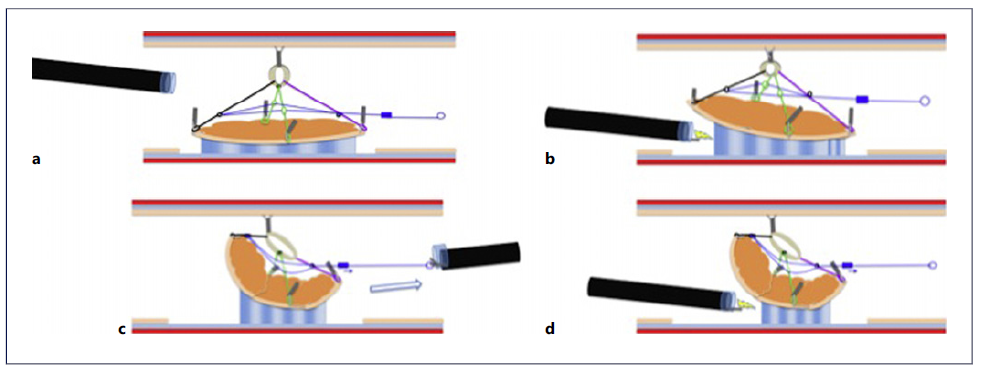
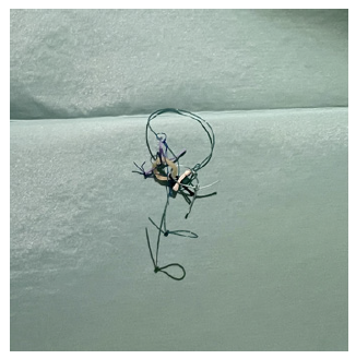
Endoscopic submucosal dissection (ESD) is currently recommended as the first-line therapy for EGC [2]. Nevertheless, a major barrier to the expansion of ESD in the West is the technical difficulty and risk of complications associated with the procedure. Several traction devices and techniques have been described to increase the ease, speed, and safety of this technique [3] but with many limitations that can preclude its use, especially the fixed amount of traction [4]. We describe the use of a new adjustable traction device (A-TRACT 4) in the endoscopic management of a gastric lesion (Fig. 1, 2).

Fig. 1 Schematic representation of the A-TRACT 4 device. a After complete circumferential incision and trimming, fixation of the loops of the device in 4 cardinal points using clips is performed. b Dissection is started. c After around one-third of the lesion is dissected, traction begins to decline, and we tighten the device by pulling out the loop into the operating channel with a rat tooth forceps. d Increased traction allows faster and safer finishing of the dissection.
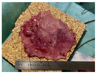
An 87-year-old male was referred due to a large gastric lesion (Paris IIa+Is with 10 cm), located in the anterior, superior, and posterior face of the antrum (previous biopsies with high-grade dysplasia) (Fig. 3). After multi-disciplinary discussion, endoscopic resection was attempted (online suppl. Video; for all online suppl. material, see https://doi.org/10.1159/000530828). Circumferential incision was performed using Dual-Knife® (Olympus, Tokyo) after submucosal injection with glycerol. ESD was started, but due to difficulties in accessing the submucosa, A-TRACT was used. Initially, fixation of the loops of the device in 4 cardinal points using clips was performed. Subsequently, another clip was used to attach the rubber band to the opposite wall, and ESD was continued. In the central part of the lesion, the device was tightened using grasping forceps in order to bring all the anchoring points of the device closer to the traction point between themselves and to the rubber band, which allowed a reestablishment of optimal traction. The procedure was completed in 120 min with complete en bloc resection of the lesion, without complications (Fig. 4, 5). At the end of the procedure, the device was removed together with the lesion using a snare. Histopathology revealed an adenocarcinoma pT1a R0 (very low-risk resection). The patient remains under endoscopic surveillance, without evidence of recurrence in the follow-up.
ESD remains a challenging technique, in spite of the positive and growing impact of training and experience dissemination [5]. In this regard, we report the use of a new device that can be helpful to increase the speed and safety of the procedure, allowing an easier access and visualization of the submucosa. Previously, other traction techniques such as clip and line, clip and snare, and S-O clip have been described to overcome the difficulties of gastric ESD [3]. Compared to these techniques, the use of adjustable traction overcomes a main limitation of previous traction devices that was loss or incorrect positioning of traction during the procedure. Moreover, the traction can be dynamically and more than once adjusted throughout the procedure, without need to remove the scope or reposition the traction device. Nevertheless, it is important to state that this device needs a precise sequence of instructions to install it properly, as was previously reported [6].
Although the A-TRACT was previously described in colonic lesions [7,8],to our knowledge,this is the first case report of the use of an adjustable traction device for ESD of a gastric lesion. This technique is a promising and innovative addition to the therapeutic armamentarium for the treatment of EGC.


