SciELO - Scientific Electronic Library Online

 
vol.31 número4Tumor desmóide após gastrectomia vertical: caso clínico e revisão da literaturaMucosectomia em banda de tumor neuroendócrino duodenal índice de autoresíndice de assuntosPesquisa de artigos
Home Pagelista alfabética de periódicos  

Serviços Personalizados

Journal

Artigo

Indicadores

Links relacionados

  • Não possue artigos similaresSimilares em SciELO

Compartilhar


GE-Portuguese Journal of Gastroenterology

versão impressa ISSN 2341-4545

GE Port J Gastroenterol vol.31 no.4 Lisboa ago. 2024  Epub 30-Set-2024

https://doi.org/10.1159/000534300 

Clinical Case Study

Schwannoma of Common Bile Duct: A Clinico-Radiologic Diagnostic Quagmire - A Case Report

Schwanoma da via biliar biliar comum: Um diagnóstico clinico-patologico volúvel

Shilpi Thakur1 

Nihar Ranjan Dash2 

Adarsh Barwad1 

Prasenjit Das1 

Kumble S. Madhusudhan3 

Rajni Yadav1 

1Department of Pathology, All India Institute of Medical Sciences, New Delhi, India;

2Gastrointestinal Surgery, All India Institute of Medical Sciences, New Delhi, India;

3Radiodiagnosis, All India Institute of Medical Sciences, New Delhi, India


Abstract

Background:

Schwannomas are benign nerve sheath tumors that are extremely rare in the biliary tract. A comprehensive review of literature enumerated ap-proximately 30 case reports of schwannoma in the biliary tract tree and porta hepatis region.

Case Presentation:

We report a case of a 40-year-old female who presented with abdominal pain. Imaging revealed a mass at the porta hepatis extending from the portal bifurcation till the hilum encasing the main portal vein and abutting the right portal vein. Differentials of carcinoma, lymphoma, and mesenchymal tumor were kept. Ultrasound-guided biopsy of the mass showed a benign nerve sheath tumor, immunopositive for S100. The histopathological evaluation of the excised mass confirmed the origin of mass in the common bile duct.

Conclusions:

Our case highlights that schwannomas, though benign, can mimic a carcinoma or lymphoma if present at a rare site such as bile ducts. An exhaustive clinical and radiological workup with diligent histopathological evaluation is mandatory in dealing with such rare cases as radical surgery and chemotherapy can be avoided in such patients.

Keywords: Schwannoma; Benign nerve sheath tumor; Common bile duct; Porta hepatis

Resumo

Introdução:

Os schwannomas são tumores benignos da bainhas nervosas, que são extremamente raros ao nível das vias biliares. Uma revisão abrangente da literatura enumerou cerca de 30 casos de schwannomas com envolvimento da árvore biliar e da região da Porta Hepatis.

Apresentação do caso:

Relatamos um caso de uma doente de 40 anos que apresentava dor abdominal. A imagem revelou uma massa que se prolonga desde a bifurcação da veia porta até ao hilo hepático, com “encasement” da veia porta principal e “abutement” da veia porta direita. Foram considerados os diagnósticos diferenciais de carcinoma, linfoma e tumor mesenquimatoso. A biópsia guiada por ecografia da massa mostrou um tumor benigno da bainha nervosa, imunopositivo para o S100. A avaliação histopatológica da massa excisada confirmou a sua origem na via biliar comum.

Conclusões:

O nosso caso realça que os schwannomas, embora benignos, podem imitar um carcinoma ou linfoma se estiverem presentes num local raro, como os canais biliares. Um trabalho clínico e radiológico exaustivo com uma avaliação histopatológica diligente é obrigatória para orientar com casos tão raros, em que a cirurgia radical e a quimioterapia podem ser evitadas.

Palavras Chave: Schwannoman; Tumor benigno da bainha nervosa; CBD; Porta hepatis

Background

Schwannomas are benign encapsulated nerve sheath tumors, usually attached to peripheral nerves and arise from differentiated Schwann cells. Mostly, they are sporadic, while some are associated with syndromes such as neurofibromatosis type 2, schwannomatosis, or Carney’s complex [1]. These are spindle cell tumors that generally occur in the upper limbs, head, and neck, followed by the trunk and flexor surfaces of the lower extremities. Approximately 0.2% of all gastrointestinal (GI) tumors are constituted by schwannomas [2]. The commonest site is the stomach followed by the colon, cecum, and rectum and rarely the jejunum [3]. GI schwannomas are uncommon and usually occur in sixth to seventh decade [4]. Herein, we report a rare case of schwannoma of the common bile duct (CBD) presenting as a porta hepatis mass. Few case reports of porta hepatis schwannomas arising from CBD, hepatoduodenal liga-ment, hepatic vein, or artery have been published in the literature.

Case Presentation

A 40-year-old female, presented with the chief complaint of pain in the abdomen for the past 10 months. Pain was located in the right upper quadrant, dull in nature with no aggravating factors. There was no history of fever, jaundice, vomiting, and upper or lower GI bleeding. The patient did not report any altered bowel habits, loss of appetite, or weight. Patient had no medical comorbidities. The general physical and abdominal examination was unremarkable. Complete hemogram and renal function tests were within normal limits. SGOT and SGPT were 26 and 25 IU/L, respectively. Serum bilirubin was 1.2 mg% while alkaline phosphatase was 186 IU/L.

Ultrasonography (USG) of the abdomen showed a hypoechoic mass at the porta abutting the right portal vein and the main portal vein (shown in Fig. 1). The liver was normal with no intrahepatic biliary radicle dilation and showed a normal echotexture. No ascites was reported. Contrast-enhanced computed tomography abdomen revealed a mass at the porta extending from the portal bifurcation till the hilum encasing the main portal vein and abutting the right portal vein (shown in Fig. 1). Common hepatic artery was free. Contrast-enhanced magnetic resonance imaging (MRI) abdomen showed a mass at the porta abutting the right portal vein, main portal vein, right hepatic artery, and the common hepatic artery (shown in Fig. 1). The mass was extending between the head of the pancreas and the inferior vena cava. Vertically, the mass was extending between the hilum and retropancreatic region. Endoscopic ultrasound was also done which revealed a mass from the superior mesenteric vein/portal vein confluence till the hilum (shown in Fig. 2). Fat planes with the main portal vein were maintained. Based on overall clinical and imaging findings, possibilities of carcinoma, lymphoma, and mesenchymal tumor were considered.

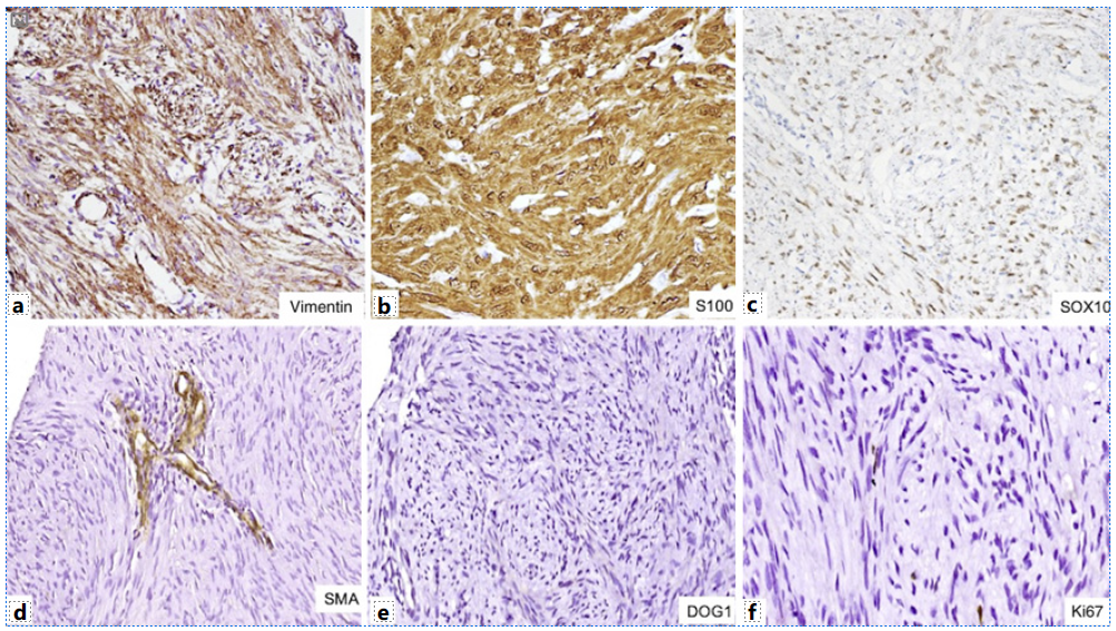
USG-guided biopsy was performed on the mass which showed a benign nerve sheath tumor, immunopositive for S100 (shown in Fig. 3). Following the above investigations, the patient was undertaken for surgery. En bloc excision of the mass and CBD with Roux-en-Y hepatico-jejunostomy was performed. The resection specimen was submitted to the department of pathology. Gross examination showed an encapsulated, circumscribed, yellow-white firm tumor measuring 7.5 cm in maximum dimension (shown in Fig. 3). On microscopy, it was a biphasic tumor composed of hypercellular areas with fascicular arrangement of spindle cells and palisades (Verocay bodies) along with myxoid hypocellular areas and focal hyalinization. The spindle-shaped tumor cells contained moderately ill-defined cytoplasm, wavy tapering nuclei with fine granular nuclear chromatin, and inconspicuous nucleoli. In addition, there were many interspersed blood vessels with hyalinized walls along with lymphoid infiltrate at the tumor periphery (shown in Fig. 3). No significant nuclear pleomorphism, mitosis, or necrosis was noted. On immunohistochemistry, the tumor cells were immunopositive for vimentin, S100, and SOX10 while they were negative for CK, CD34, smooth muscle actin, CD117, DOG1, myogenin, ALK, STAT6, CD21, and HMB45. Ki67 proliferation index was less than 1% (shown in Fig. 4). Based on the immunohistomorphological profile, the tumor was diagnosed as schwannoma. Patient recovered well postoperatively and was discharged on postoperative day 10. The patient is disease-free on follow-up after 36 months.
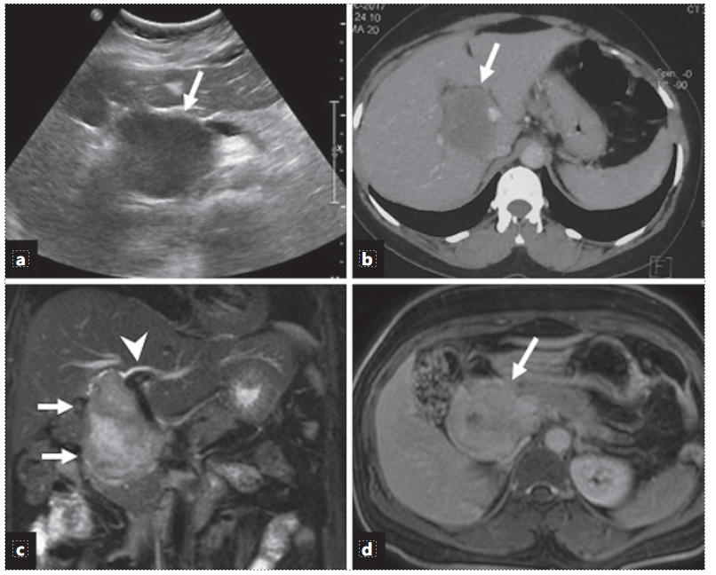
Fig. 1 a Ultrasound image shows a hypoechoic mass (arrow) at the porta hepatis. b Axial contrast-enhanced CT image shows a hypodense mass (arrow) at the porta hepatic splaying the portal veins. c Coronal T2-weighted MR image shows an oblong hyperintense mass (arrows) along the course of the CBD with mild intrahepatic bile duct dilatation (arrow-head). d Axial contrast-enhanced T1-weighted MR image (5 min delayed) shows late enhancement of the mass (arrow). 
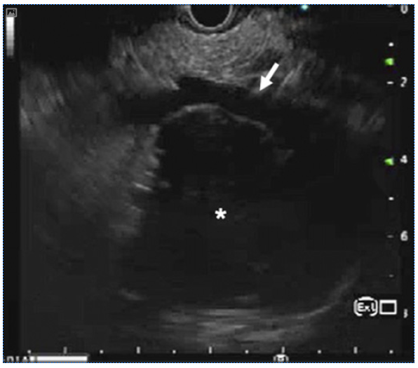
Fig. 2 Endoscopic ultrasound shows a hypoechoic mass (asterisk) at the porta hepatis abutting the portal vein (arrow). Image courtesy of: Dr Deepak Gunjan. 
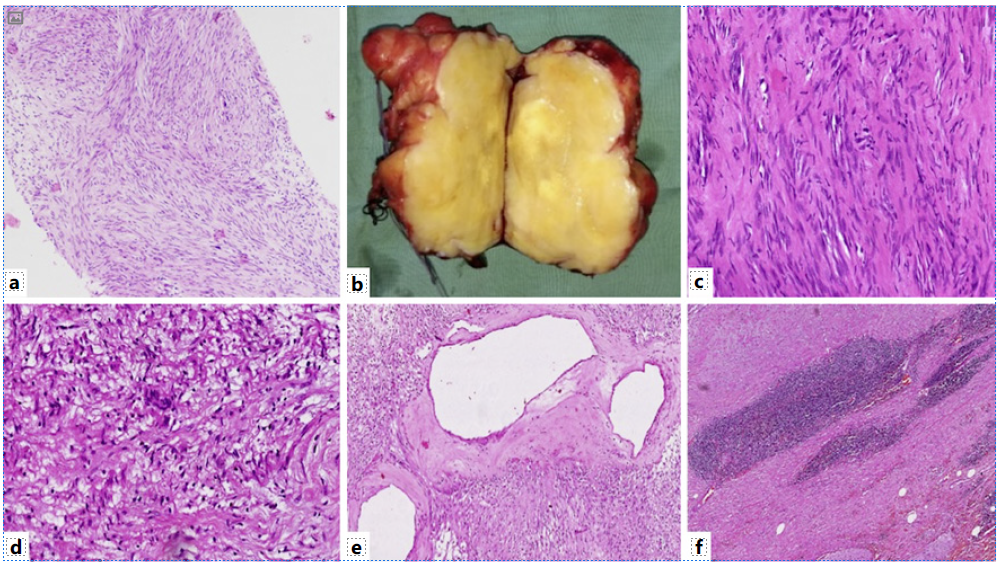
Fig. 3 a Biopsy section shows a benign spindle cell tumor (×100, H&E). b Gross examination shows nodular circumscribed yellow-white firm tumor. The tumor shows hypercellular areas with Verocay bodies (c) (×400, H&E) and hypocellular areas (d) (×400, H&E). Also, many dilated vessels with perivascular hyalinization (e) (×200, H&E) and lymphoid cuff at tumor periphery (f) (×200, H&E) are seen. 

Fig. 4 The tumor cells are immunopositive for vimentin (a), S100 (b), and SOX10 (c) while they are immunonegative for SMA (d), DOG1 (e), and show low Ki67 proliferation index (f) (×200). SMA, smooth muscle actin. 

Discussion

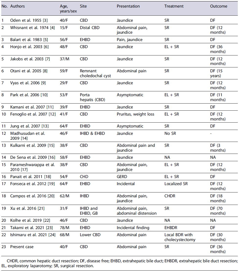
Schwannomas in porta hepatis and biliary tree are very rare and approximately 30 cases have been reported in literature till date. There are 22 reported cases of schwannoma in the biliary tract alone details of which have been summarized in Table 1 [3-24].

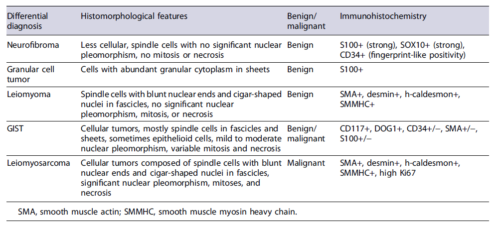
A significant female predominance with male-to-female ratio of 1:5.5 was seen in these patients. The mean age at presentation was 48.3 years (range 15-78 years). Abdominal pain and jaundice were the most common presenting symptoms in these patients (jaundice in 38%, abdominal pain in 19%, both pain and jaundice in 23% of patients, respectively). The most common location was CBD (57%). The preoperative clinico-radiological diagnoses were quite variable and comprised metastatic melanoma, gastrointestinal stromal tumor (GIST), lymphoma, adenocarcinoma, leiomyosarcoma. In all the cases, it was extremely difficult to correctly diagnose this tumor preoperatively, mainly due to the fact that tumors at this site can easily mimic bile duct adenocarcinoma and other malignancies such as lymphoma and IgG4-related diseases. Moreover, the location is difficult to approach for a minimally invasive technique such as fine needle aspirate. Surgical resection was carried out in most cases with unremarkable postoperative period [3-24]. In our case, a correct preoperative diagnosis on USG-guided biopsy helped in the adequate surgical management. Radiologically, contrast-enhanced MRI is better suited to visualize the extent and size of such soft tissue tumors and dilatation of bile duct radicles due to mass effect [25]. On MRI, schwannomas are hypointense on T1-weighted images and homogeneously hyperintense on T2-weighted images [26]. Usually, degenerative changes are uncommon in GI schwannomas, but if present can lead to error in diagnosis. Diagnosis in such cases can only be made on histology which requires excision of the mass. The most common differential diagnoses for schwannoma are other benign and malignant soft tissue tumors of the gastrointestinal tract such as neurofibroma, leiomyoma, GIST, and leiomyosarcoma, enumerated in Table 2.

Table 1 Details of previously reported cases of schwannoma in the biliary tract [3-24

Table 2 Discussion of common differential diagnoses of soft tissue masses in the biliary tract region 
Histologically, schwannomas show hypercellular areas (Antoni A) with Verocay bodies and hypocellular (Antoni B) areas. The tumor cells are predominantly spindle-shaped with hyperchromatic nuclei. Mitosis and necrosis are usually absent [10]. Schwannomas can show an array of degenerative changes such as hyalinization, calcification, emorrhage, myxoid change, cyst formation, focal bizarre nuclear atypia. Multiple variants are seen, namely, ancient, plexiform, cellular, epithelioid, microcystic or reticular, and melanotic or pigmented [14].

Immunohistochemistry plays an important role in distinguishing schwannoma from close differentials such as GIST, leiomyomas, or neurofibromas. Schwannomas are strongly immunopositive for S100 protein and SOX10. GISTs are immunopositive for c-KIT and DOG1, while leiomyoma and leiomyosarcoma will show strong and diffuse smooth muscle actin positivity. CD34 and calretinin might help in differentiating these from neurofibroma as both are immunopositive for S100. Neurofibroma shows moderate CD34 positivity. Fine et al. [27] in their study compared calretinin positivity in schwannoma and neurofibroma and 96% of schwannomas displayed strong calretinin staining compared to only 7% in neurofibroma. GI schwannomas show some histomorphological variations when compared to schwannomas at other regions in the form of predominance of hypercellular areas (Antoni A) and lack of nuclear palisading pattern as seen in conventional schwannoma [28]. Our case, although it showed both the areas, Lasota et al. [29] also reported lack of NF-2 gene alterations in the GI schwannomas suggesting it to be a morphologically and genetically distinct group of nerve sheath tumors. However, more data needs to be incorporated to arrive successfully at this conclusion. Being benign tumors, these have an excellent prognosis and complete surgical resection is the mainstay of treatment. Recurrence or malignant transformation is rarely seen [30].

Conclusion

CBD schwannomas are very rare and a surgeon’s nightmare until the histopathological examination is done. The present case adds to the rare list of schwan-nomas arising from CBD and also enumerates the di-agnostic challenges from a pathologist’s perspective.

Acknowledgment

The authors thank Dr. Deepak Gunjan (gastroenterologist) for contributing the EUS image.

References

1. Salvati M, Polli FM, Caroli E, Frati A, Missori P, Delfini R. Radiation-induced schwannomas of the nervous system. Report of five cases and review of the literature. J Neurosurg Sci. 2003;47(2):113-6; discussion 6. [ Links ]

2. Sanei B, Kefayat A, Samadi M, Goli P, Sanei MH, Khodadustan M. Gastric schwannoma: a case report and review of the literature for gastric submucosal masses distinction. Case Rep Med. 2018;2018:1230285. [ Links ]

3. Oden B. Neurinoma of the common bile duct; report of a case. Acta Chir Scand. 1955; 108(5):393-7. [ Links ]

4. Whisnant JD Jr, Bennett SE, Huffman SR, Weiss DL, Parker JC, Griffen WO Jr. Common bile duct obstruction by granular cell tumor (schwannoma). Am J Dig Dis. 1974;19(5):471-6. [ Links ]

5. Balart LA, Hines C Jr, Mitchell W. Granular cell schwannoma of the extrahepatic biliary system. Am J Gastroenterol. 1983;78(5):297-300. [ Links ]

6. Honjo Y, Kobayashi Y, Nakamura T, Takehira Y, Kitagawa M, Ikematsu Y, et al. Extrahepatic biliary schwannoma. Dig Dis Sci. 2003;48(11):2221-6. [ Links ]

7. Jakobs R, Albert J, Schilling D, Nuesse T, Riemann JF. Schwannoma of the common bile duct: a rare cause of obstructive jaundice. Endoscopy. 2003;35(8):695-7. [ Links ]

8. Otani T, Shioiri T, Mishima H, Ishihara A, Maeshiro T, Matsuo A, et al. Bile duct schwannoma developed in the remnant choledochal cyst-a case associated with total agenesis of the dorsal pancreas. Dig Liver Dis. 2005;37(9):705-8. [ Links ]

9. Vyas FL, Jesudason MR, Samuel R, Govil S, Jesudason SRB. Schwannoma of bile duct-a case report. Trop Gastroenterol. 2006;27:50-1. [ Links ]

10. Park MK, Lee KT, Choi YS, Shin DH, Lee JY, Lee JK, et al. A case of benign schwannoma in the porta hepatis. Korean J Gastroenterol. 2006;47(2):164-7. [ Links ]

11. Kamani F, Dorudinia A, Goravanchi F, Rahimi F. Extrahepatic bile duct neurilemmoma mimicking Klatskin tumor. Arch Iran Med. 2007;10(2):264-7. [ Links ]

12. Fenoglio L, Severini S, Cena P, Migliore E, Bracco C, Pomero F, et al. Common bile duct schwannoma: a case report and review of literature. World J Gastroenterol. 2007;13:1275-8. [ Links ]

13. Jung JH, Joo KR, Chae MJ, Jang JY, Lee SG, Dong SH, et al. Extrahepatic biliary schwannomas: a case report. J Korean Med Sci. 2007;22(3):549-52. [ Links ]

14. Madhusudhan KS, Srivastava DN, Dash NR, Gupta C, Gupta SD. Case report. Schwannoma of both intrahepatic and extrahepatic bile ducts: a rare case. Br J Radiol. 2009;82(982):e212-5. [ Links ]

15. Kulkarni N, Andrews SJ, Rao V, Rajagopal KV. Case report: benign porta hepatic schwannoma. Indian J Radiol Imaging. 2009;19(3):213-5. [ Links ]

16. De Sena G, Molino C, De Riitis MR, Candela S, Cifarelli V, Di Maio V, et al. Surgical management of schwannoma of biliary tract Chir Ital. 2009;61(1):119-21. [ Links ]

17. Parameshwarappa S, Rodrigues G, Kumar S, Patil B, Valliathan M. Schwannoma of common bile duct causing obstructive jaundice. Indian J Surg. 2010;72(Suppl 1):333-5. [ Links ]

18. Panait L, Learn P, Dimaio C, Klimstra D, Do KG, Schwarz T, et al. Resection of perihilar biliary schwannoma. Surg Oncol. 2011;20(4):e157-9. [ Links ]

19. Fonseca GM, Montagnini AL, Rocha Md S, Patzina RA, Bernardes MVAA, Cecconello I, et al. Biliary tract schwannoma: a rare cause of obstructive jaundice in a young patient. World J Gastroenterol. 2012;18(37):5305-8. [ Links ]

20. Marin Campos C, Garcia Sanz I, Munoz de Nova JL, Valdés de Anca A, Martín Pérez ME. Schwannoma of the biliary tract resembling cholangiocarcinoma: a case report and review. Ann R Coll Surg Engl. 2016;98(7): e143-6. [ Links ]

21. Xu SY, Guo H, Shen Y, Sun K, Xie HY, Zhou L, et al. Multiple schwannomas synchronously occurring in the porta hepatis, liver, and gallbladder: first case report. Medicine. 2016;95(33):e4378. [ Links ]

22. Kolhe KM, Amarapurkar A, Ingle M, Pandey V, Amonkar M, Khairnar H, et al. Jack in the duct: a case of common biliary duct schwannoma. ACG Case Rep J. 2019;6(10):e00223. [ Links ]

23. Takami K, Yamamoto K, Sakurai H, Kondo N, Shibata C, Katayose Y. Biliary schwannoma that required differentiation from bile duct cancer. Case Rep Gastroenterol. 2021;15(2):578-86. [ Links ]

24. Ishimaru N, Fujikawa H, Kobayashi Y. Successful preoperative diagnosis and minimally invasive surgery of bile duct schwannoma. Clin J Gastroenterol. 2021;14(1):336-40. [ Links ]

25. Bilgin M, Shaikh F, Semelka RC, Bilgin SS, Balci NC, Erdogan A. Magnetic resonance imaging of gallbladder and biliary system. Top Magn Reson Imaging. 2026 [ Links ]

26. Rha SE, Byun JY, Jung SE, Chun HJ, Lee HG, Lee JM. Neurogenic tumors in the abdomen:vtumor types and imaging characteristics. Radiographics. 2003;23(1):29-43. [ Links ]

27. Fine SW, McClain SA, Li M. Immunohistochemical staining for calretinin is useful for differentiating schwannomas from neurofibromas. Am J Clin Pathol. 2004;122(4):552-909;20(1):31-42. [ Links ]

28. Daimaru Y, Kido H, Hashimoto H, Enjoji M. Benign schwannoma of the gastrointestinal tract: a clinicopathologic and immunohistochemical study. Hum Pathol. 1988;19(3):257-64. [ Links ]

29. Lasota J, Wasag B, Dansonka-Mieszkowska A, Karcz D, Millward CL, Rys J, et al. Evaluation of NF2 and NF1 tumor suppressor genes in distinctive gastrointestinal nerve sheath tumors traditionally diagnosed as benign schwannomas: s study of 20 cases. Lab Invest. 2003;83(9):1361-71. [ Links ]

30. Yin SY, Zhai ZL, Ren KW, Yang YC, Wan DL, Liu XY, et al. Porta hepatic schwannoma: case report and a 30-year review of the literature yielding 15 cases. World J Surg Oncol. 2016;14:103. [ Links ]

Statement of Ethics Study approval from the institute’s Ethics Committee (AIIMS, New Delhi) was not required. A written informed consent was obtained from the participant for publication of the details of their medical case and any accompanying images.

Conflict of Interest Statement The authors have no conflicts of interest to declare.

Funding Sources No funding was received for this study.

Author Contributions Shilpi Thakur contributed to manuscript writing, editing, and data curation. Adarsh Barwad and Prasenjit Das contributed to review and editing. Nihar Ranjan Dash and Kumble S. Madhusudhan helped with clinical and radiological data curation. Rajni Yadav reviewed and edited the manuscript.

Data Availability Statement No data were generated during this study. Further inquiries can be directed to the corresponding author.

Received: March 29, 2023; Accepted: August 03, 2023

Correspondence to: Rajni Yadav, drrajniyadav@gmail.com

Creative Commons License This is an open-access article distributed under the terms of the Creative Commons Attribution License