SciELO - Scientific Electronic Library Online

 
vol.32 número2Carcinoma hepatocelular com envolvimento vascular e cardíaco num jovem com hepatite B não cirróticaSerologia Yersinia positiva e padrão do cólon em pedra de calçada: uma distração ou o principal culpado? índice de autoresíndice de assuntosPesquisa de artigos
Home Pagelista alfabética de periódicos  

Serviços Personalizados

Journal

Artigo

Indicadores

Links relacionados

  • Não possue artigos similaresSimilares em SciELO

Compartilhar


GE-Portuguese Journal of Gastroenterology

versão impressa ISSN 2341-4545

GE Port J Gastroenterol vol.32 no.2 Lisboa abr. 2025  Epub 15-Maio-2025

https://doi.org/10.1159/000539913 

Clinical Case Study

Aortoesophageal Fistula Mimicking Dieulafoy Disease: A Case Report

Fístula Aortoesofágica que mimetiza Doença de Dieulafoy: Descrição de um caso

Tatiana Pacheco1 

Pedro Costa-Moreira1 

Sara Monteiro1 

Joana Pinto1 

Luísa Barros Jorge Silva1 

1Gastroenterology Department, Centro Hospitalar do Tâmega e Sousa, Penafiel, Portugal


Abstract

Introduction:

Aortoesophageal fistula (AEF) is a rare and potentially fatal cause of upper gastrointestinal bleeding. The classic Chiari’s triad of symptoms and typical endoscopic findings are not present in all patients, making diagnosis challenging.

Case Presentation:

An 86-year-old man was admitted to the emergency room for melena and hematemesis with hemodynamic instability. He had a previous hospitalization for cardioembolic stroke complicated by hematemesis of unknown etiology after initiation of anti-coagulation (which was suspended), being discharged on aspirin. His medical history also included hypertension, diabetes, ischemic heart disease, and prostate cancer. On upper endoscopy, no lesions were found, despite the presence of a large non-mobilizable clot in the gastric fundus. He was admitted to the intensive care unit, and, on the next day, reassessment esophagogastroduodenoscopy was normal. On the eighth day of hospitalization, the patient presented with hemorrhagic shock due to new-onset hematemesis. Upper endoscopy revealed an esophageal 10-mm non-ulcerated mucosal depression with a visible vessel at 20 cm from the incisors, closed with 3 hemoclips. Thoracic CT angiography showed a brachiocephalic trunk aneurysm with aortoesophageal fistulization. He was deemed unsuitable for endovascular or surgical treatment. About 2 months later, the patient was admitted to the emergency room in cardiorespiratory arrest following an episode of hematemesis at home.

Discussion:

This report highlights the diagnostic and therapeutic complexity of AEF. Endoscopic treatment can be the main therapy in patients without indication for vascular intervention. The purpose was to palliate new bleeding episodes, maintaining a very poor prognosis.

Keywords: Esophageal fistula; Aortic diseases; Gastrointestinal bleeding; Computed tomography angiography; Aged 80 and over

Resumo

Introdução:

A fístula aortoesofágica (FAE) é uma causa rara e potencialmente fatal de hemorragia digestiva alta. A tríade clássica de sintomas de Chiari e os achados endoscópicos típicos não estão presentes em todos os doentes, o que torna o diagnóstico desafiante.

Apresentação do Caso: Um

homem de 86 anos foi admitido na sala de emergência por melenas e hematemeses com instabilidade hemodinâmica. Apresentou internamento recente por AVC isquémico cardioembólico complicado por hematemeses de etiologia indeterminada após início de hipocoagulação (que foi suspensa), tendo alta sob aspirina. Acresciam os antecedentes de hipertensão, diabetes, cardiopatia isquémica e neoplasia prostática. Na endoscopia digestiva alta (EDA) não foram encontradas lesões, apesar de coágulo gigante não mobilizável no fundo gástrico. Foi internado em Unidade de Cuidados Intensivos, tendo repetido EDA no dia seguinte que foi normal. No oitavo dia de internamento, apresentou-se com choque hemorrágico devido a novo episódio de hematemeses. A EDA revelou, aos 20 cm, uma depressão da mucosa não ulcerada de 10 mm com vaso no leito, encerrada com 3 hemoclips. O AngioTC torácico mostrou aneurisma do tronco braquicefálico com fistulização aortoesofágica. O doente não era candidato a tratamento endovascular ou cirúrgico. Cerca de dois meses depois, o doente foi admitido na sala de emergência em paragem cardiorrespiratória após episódio de hematemeses no domicílio.

Conclusão:

Este caso destaca a complexidade diagnóstica e terapêutica da FAE. O tratamento endoscópico pode constituir a terapêutica principal nos doentes sem indicação para intervenção vascular. O objetivo é a paliação de novos episódios de hemorragia, mantendo-se o péssimo prognóstico.

Palavras Chave: Fístula esofágica; Doenças da aorta; Hemorragia gastrointestinal; Angiografia por tomografia computadorizada; Idosos; 80 anos ou mais

Introduction

Aortoesophageal fistula (AEF) is a rare but catastrophic cause of upper gastrointestinal (GI) bleeding that occurs due to an abnormal communication between the aorta and the esophagus [1, 2]. The diagnosis should be suspected when upper GI bleeding occurs in the context of aortic disease, since the main etiology is secondary to aortic interventions, followed by native aortic aneurysm [2, 3]. Diagnosis is made based on clinical findings, upper endoscopy, and computed tomography angiography (CTA) [4-6]. The authors describe a rare case of an elderly patient who presented with intermittent bleeding and endoscopic findings suspicious of Dieulafoy disease, which were subsequently identified as an AEF.

Case Report

An 86-year-old Caucasian male was admitted to the emergency department (ED) due to melena and hematemesis with hemodynamic instability. His medical history includes arterial hypertension, diabetes, ischemic heart disease, and prostate cancer under surveillance. The patient had a hospitalization 2 months prior for an ischemic cardioembolic stroke complicated by hematemesis of unknown etiology after initiation of anti-coagulation, which prompted its cessation. He was discharged home on aspirin. After hospitalization, there were several visits to the ED due to melena, hematemesis, and symptoms related to anemia, requiring red blood cell transfusions and multiple upper endoscopies showing blood in the gastric lumen or no abnormal findings. Chronic medication also included bisoprolol, amlodipine, metformin, sitagliptin, esomeprazole, enzalutamide, and goserelin. He denied intake of alcohol or nonsteroidal anti-inflammatory drugs.

On admission, the patient was hypotensive, pale, and had cold extremities. His hemoglobin was measured at 6.1 g/dL; the remaining blood count was normal. There were no alterations in renal function, liver profile, and coagulation. He had hyperlactatemia (4.9 mmol/L) and troponin elevation (446 pg/mL) without chest pain or ischemic electrocardiographic changes. He received two units of packed red blood cells, fluid resuscitation, and intravenous esomeprazole, leading to hemodynamic stabilization in the ED. Upper endoscopy revealed a giant non-mobilizable clot in the gastric fundus, with no hemorrhagic lesions found throughout the exam. An abdominal CT angiogram showed no evidence of active GI bleeding. The patient was admitted to the intensive care unit, and next-day reassessment esophagogastroduodenoscopy (EGD) was normal. He remained clinically stable and without visible blood loss.

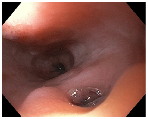
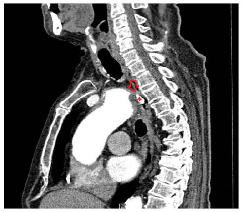
On the eighth day of hospitalization, the patient was readmitted to the intensive care unit in hemorrhagic shock due to new-onset hematemesis. He was intubated and treated with aggressive resuscitation and transfusion of red blood cells. EGD revealed an esophageal 10-mm non-ulcerated mucosal depression with a visible vessel at 20 cm from the incisors (shown in Fig. 1), closed with 3 hemoclips. Thoracic CT angiography showed a brachiocephalic trunk aneurysm with aortoesophageal fistulization (shown in Fig. 2). The vascular surgery department at our hospital and the cardiothoracic surgery department at a tertiary center were consulted, but the patient was deemed unsuitable for endovascular or surgical treatment given the anatomical characteristics of the fistula and his general condition. He subsequently recovered and was able to be discharged home. About 2 months later, the patient was admitted to the emergency room in cardiorespiratory arrest following an episode of hematemesis at home.

Fig. 1. Endoscopic image of AEF. 

Fig. 2. Endoscopic clip (*) identifying the level of AEF (arrow) on CTA sagittal view. 

Discussion

An AEF is an uncommon and potentially fatal cause of upper GI bleeding. As the name suggests, this condition arises from an abnormal communication between the aorta and esophagus, which allows high-pressure arterial blood to enter the esophagus [1, 2]. The middle portion of the esophagus is the most common site for aortoesophageal fistulization [3, 7]. This disease should be considered in patients who have a history of aortic intervention since most cases are secondary to vascular procedures. It can also originate from the native aorta, arising from thoracic aortic aneurysms, as in our patient, but also after ingestion of foreign bodies, or from tumor invasion [2, 3].

Although the first case of AEF was reported in 1818, the classic triad of symptoms was described by Chiari almost a hundred years later [8]. Patients may experience midthoracic pain, sentinel arterial hemorrhage, followed by a symptom-free interval, and finally a massive bleeding [7-9]. In a minority of cases, bleeding is intermittent, presumably due to the formation of a blood clot that temporarily seals the opening of the fistula [9, 10]. Our patient did not have known risk factors for the disease, such as previous aortic intervention, and presented with less typical symptoms, which may have contributed to a low index of clinical suspicion.

EGD plays an important role in the evaluation of upper GI bleeding; however, its sensitivity for AEF diagnosis is low. The diagnosis can easily go unnoticed in endoscopy if there is no active bleeding and because the typical endoscopic findings, such as submucosal tumor-like protrusion or pulsating arterial bleeding, are rarely found [3, 4, 10].

CTA is recommended as a first-line noninvasive examination in the suspicion of AEF and is diagnostic in most cases [4, 6]. In our case, the findings on EGD associated to intermittent bleeding raised the hypothesis of Dieulafoy lesion, which was not confirmed by CTA. In fact, the CTA was essential to establish the definitive diagnosis of AEF, as the clinical history and endoscopy findings were not suggestive. We emphasize the significance of performing a thoracoabdominal CTA when investigating the etiology of upper digestive bleeding. This is highlighted by the fact that the initial CTA was abdominal and unremarkable, while the subsequent thoracic CTA revealed the final diagnosis.

The management of AEF should involve a multidisciplinary approach and a therapeutic strategy individualized with consideration of both esophageal and aortic defects. A combination of surgery for the aorta (endovascular or open surgical repair) and esophagus (esophagectomy, esophageal stent, or repair) is usually adopted [2, 11]. In cases where correction of the aorta is not possible, as in our patient, the endoscopic treatment aims to palliate new episodes of bleeding, since the aortic defect and fistulous tract remain intact. The literature is scarce regarding the positioning of the different endoscopic interventions for palliation probably because cases of successful vascular treatment appear overwhelmingly more likely to be reported than cases of unfavorable clinical outcomes. Our therapeutic approach was chosen based on the location (20 cm of the incisors), the probable cause (Dieulafoy’s lesion), as well as the difficult access and stabilization of the endoscope that precluded other therapeutic options. We considered that a subsequent change in the endoscopic therapeutic strategy could precipitate a new episode of bleeding, and none of the endoscopic options allows definitive treatment of AEF.

Perhaps there may be a role for combining other endoscopic techniques in cases similar to ours, with only an indication for endoscopic treatment, such as the injection of sclerosants or tissue adhesives, if the diagnosis of AEF is known from the beginning.

In conclusion, the case highlights the diagnostic and therapeutic complexity of AEF. A low threshold for performing a CTA is recommended, particularly when there are frequent symptoms of upper digestive bleeding and clear signs observed during endoscopy. Endoscopic treatment not only allows hemostasis in the episode of acute bleeding but also may become the main therapy of AEF in patients without indication for vascular intervention for palliation of new bleeding episodes.

References

1. Hollander JE, Quick G. Aortoesophageal fistula: a comprehensive review of the literature. Am J Med. 1991;91(3):279-87. https://doi.org/10.1016/0002-9343(91)90129-l [ Links ]

2. Takeno S, Ishii H, Nanashima A, Nakamura K. Aortoesophageal fistula: review of trends in the last decade. Surg Today. 2020;50(12): 1551-9. https://doi.org/10.1007/s00595-019-01937-z [ Links ]

3. Li S, Gao F, Hu HO, Shi J, Zhang J. Risk factors for mortality in patients with aortoesophageal fistula related to aortic lesions. Gastroenterol Res Pract. 2020;2020: 4850287-11. https://doi.org/10.1155/2020/4850287 [ Links ]

4. Monteiro AS, Martins R, Martins da Cunha C, Moleiro J, Patrício H. Primary aortoesophageal fistula: is a high level of suspicion enough? Eur J Case Rep Intern Med. 2020; 7(8):001666. https://doi.org/10.12890/2020_001666 [ Links ]

5. Bogey WM, Thomas JH, Hermreck AS. Aortoesophageal fistula: report of a successfully managed case and review of the literature. J Vasc Surg. 1992;16(1):90-5. https://doi.org/10.1016/0741-5214(92)90423-6 [ Links ]

6. Hughes FM, Kavanagh D, Barry M, Owens A, MacErlaine DP, Malone DE. Aortoenteric fistula: a diagnostic dilemma. Abdom Imaging. 2007;32(3):398-402. https://doi.org/10.1007/s00261-006-9062-7 [ Links ]

7. Carter R, Mulder GA, Snyder EN, Brewer LA. Aortoesophageal fistula. Am J Sur. 1978; 136(1):26-30. https://doi.org/10.1016/0002-9610(78)90195-2 [ Links ]

8. Chiari H. Ueber Premdkorperverletzung des Oesophagus mit Aortenperforation. Ber Klin Wochenschr. 1914;51:7. [ Links ]

9. Glodean A, Grobholz R, El-Hag K, Ziaka M, Schmid JP. Midthoracic pain, sentinel arterial haemorrhage and exsanguination after a symptom-free interval (Chiari's Triad) is diagnostic of Arterio-Oesophageal Fistula: a lifethreatening cause of gastrointestinal bleeding. Eur J Case Rep Intern Med. 2021;8(3):002134. https://doi.org/10.12890/2021_002134 [ Links ]

10. Kayashima A, Mori H, Okuzawa A, Kubosawa Y, Hirai Y, Kinoshita S, et al. An esophageal ulcer associated with a thoracoabdominal aortic aneurysm. Case Rep Gastroenterol. 2019;13(1): 214-8. https://doi.org/10.1159/000500067 [ Links ]

11. Vitor S, Meireles L, Lopes J, Ribeiro LC, Velosa J. Secondary aortoesophageal fistula due to thoracic aortic stent graft: is there a role for endoscopic intervention? GE Port J Gastroenterol. 2015;22(3):128-9. https://doi.org/10.1016/j.jpge.2015.03.001 [ Links ]

Statement of Ethics Ethical approval was not required for this study in accordance with national guidelines. Informed consent was obtained from the patient’s relatives for the publication of the medical case and any accompanying images.

Conflict of Interest Statement The authors have no conflicts of interest to declare.

Funding Sources No funding was received.

Author Contributions Tatiana Pacheco: literature research, manuscript preparation, and drafting. Pedro Costa Moreira, Sara Monteiro, Joana Pinto, Luísa Barros, and Jorge Silva: critical revision of the manuscript. All authors have approved the final version of the manuscript.

Data Availability Statement All data generated or analyzed during this study are included in this article. Further inquiries can be directed to the corresponding author.

Received: March 29, 2024; Accepted: May 29, 2024

Correspondence to: Tatiana Pacheco, tatianasofiaapacheco@gmail.com

Creative Commons License This is an open-access article distributed under the terms of the Creative Commons Attribution License