


 Inglês (pdf)
 Artigo em XML
 Referências do artigo
 Enviar este artigo por email
 Citado por SciELO
  Similares em
SciELO
Similares em
SciELO 
 Permalink
A 58-year-old female, with a medical history of stage IIB Hodgkin lymphoma in remission for over 5 years post-chemoradiation, and treated invasive breast and renal carcinomas, was incidentally diagnosed with a cardiac gastric polyp on a follow-up computed tomog-raphy (CT). Upper endoscopy and endoscopic ultrasound (EUS) revealed a 50 mm ulcerated polypoid subepithelial lesion (Fig. 1) originating from a small stalk in the muscularis propria (MP). Biopsies and fine-needle biopsy were inconclusive but without epithelial neoplastic tissue. Subsequent staging showed no lymph nodes or distant metastasis. Given the high morbidity associated with surgery - total gastrectomy - the tumor’s small insertion in the MP, the patient’s lack of lymph node involvement or distant metastasis, and their preference for a less invasive approach, endoscopic submucosal dissection (ESD) was chosen as the optimal treatment option. En bloc ESD was performed without technical difficulties, revealing the lesion’s superficial origin in the MP (Fig. 2), which eliminated the need for perforation and subsequent defect closure. However, because of its dimension and shape, the specimen was fragmented during retrieval. Histological analysis confirmed gastric leiomyosarcoma with non-evaluable margins (Fig. 3). Following multidisciplinary discussion, surveillance was recommended. At 12-month follow-up, the patient remains with no signs of recurrence at CT and endoscopic evaluation.
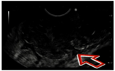
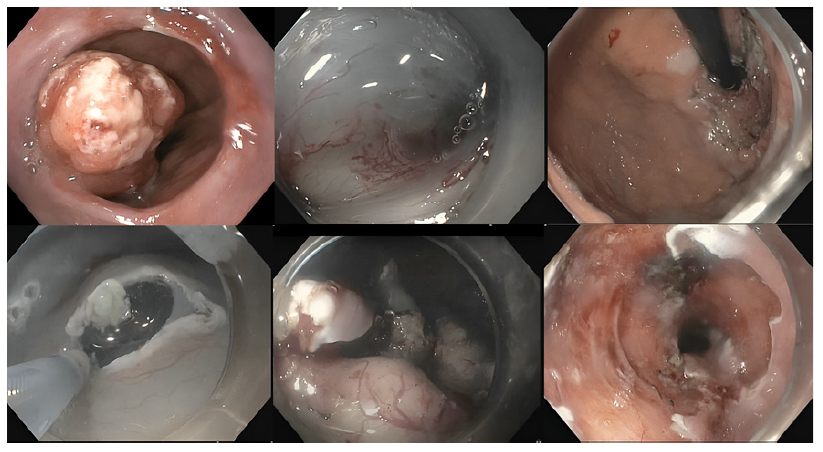
Fig. 2 Endoscopic submucosal dissection (ESD) procedure illustrating the initial lesion and key procedural steps, including the circumferential incision and submucosal dissection. This figure highlights the lesion at the start, the progression of the ESD, and the final appearance of the mucosal defect.
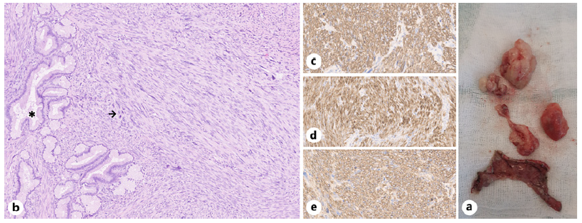
Fig. 3 Gastric leiomyosarcoma. a Retrieved specimen. b H&E showing normal gastric glands (*) and a fascicular spindle cell neoplasia (→) with marked cytological atypia and frequent mitosis. The neoplastic cells express smooth muscle immunohistochemical markers: actin (c), desmin (d), and caldesmon (e).
Primary leiomyosarcoma of the stomach is an extremely rare neoplasm [1] that can arise from the MP (more frequently) or the muscularis mucosa, with the latter presenting as intraluminal type and the former as infiltrative [2]. Ionizing radiation and Epstein-Barr virus have been suggested as associated risk factors [2].
Imaging diagnosis is often achieved using CT scans and EUS, which are valuable tools in evaluating these cases, specially the latter one as it enables the sampling of deeper tumor tissues with ultrasound-guided fine-needle aspiration. However, as demonstrated in our case, distinguishing leiomyosarcomas from other gastric tumors with these methods can be challenging. Endoscopic biopsies are also frequently ineffective due to the submucosal origin of the lesion. Smooth muscle tumors, including leiomyoma and leiomyosarcoma, typically show positivity for smooth muscle markers such as actin, desmin, and h-caldesmon, while they are generally negative for c-kit (CD-117) and CD-34 [3].
Surgical resection remains the standard treatment for smooth muscle tumors, as it allows for complete removal of the neoplasm and provides the best chance for curative outcomes [4]. However, there are limited reports in the literature exploring the use of ESD to treat these tumors [5]. ESD offers the advantage of en bloc resection, which is particularly beneficial for managing localized disease. In this case, although the specimen fragmented during retrieval - a potential disadvantage of this technique - the lesion itself was resected en bloc during the ESD, which is the most critical factor for achieving optimal clinical outcomes. While fragmentation may limit certain aspects of histological examination, the complete en bloc resection during ESD remains the primary determinant of clinical success. This approach can potentially minimize morbidity and mortality associated with more invasive surgical procedures, making ESD a viable alternative in selected cases in which the tumor is confined and surgical risks are high.

