What does this study add
We present an uncommon complication of secondary syphilis. Our goal is to raise awareness for this complication, so it’s included in the investigation of otherwise unexplained hepatitis and to search for hepatic alterations in patients diagnosed with syphilis.
Introduction
Syphilis, the infection caused by the spirochete Treponema pallidum, is a sexually and, less frequently, vertically transmitted disease. The untreated condition is polymorphic, progressing through active stages and latency. Early syphilis is contagious and occurs in the first 2 years of the infection, including primary, secondary, and early latent syphilis. Late syphilis (late latent and tertiary syphilis) is not contagious and has subtler manifestations1.
The primary lesion develops at the inoculation site as a firm, painless, ulcerated papule that spontaneously resolves. It is followed by dissemination and multiplication of the spirochete in several organs, a stage known as secondary syphilis. This is characterized by a broad spectrum of manifestations involving not only the skin, but also several internal organs. A latent phase follows, which is asymptomatic and can last for several years. Tertiary syphilis is rare nowadays, with severe involvement of several systems, such as the skin, bones, central nervous system, and heart1. Symptoms can be subtle and unspecific, a feature that has led to the historical denomination of “the great imitator”.
Hepatic disease is a possible but uncommon complication in the systemic spectrum of secondary syphilis2, feasibly caused by non-hepatotropic involvement of the liver by the bacteria3. There are less than 150 cases of this complication described until 20184. Herein, we present the case of a young adult man with syphilitic hepatitis.
Case presentation
A 34-year-old man presented to the emergency department with jaundice, malaise, and fatigue. He was otherwise healthy, with no history of new medications, recreational drugs, or alcohol consumption. He denied fever, abdominal pain, or anorexia. Laboratorial investigation revealed hyperbilirubinemia, elevated liver enzymes, and cholestasis (Table 1). Serologies for hepatitis viruses and human immunodeficiency virus were negative. He was admitted for further diagnostic investigation and monitoring.
Table 1 Values of liver enzymes before and after penicillin treatment
| Normal values | At admission | 10 days after treatment | |
|---|---|---|---|
| Total bilirubin (mg/dL) | < 1.20 | 12.84 | 3.52 |
| Direct bilirubin (mg/dL) | < 0.50 | 9.29 | 2.49 |
| AST (U/L) | < 34 | 254 | 84 |
| ALT (U/L) | < 55 | 354 | 158 |
| GGT (U/L) | < 64 | 1385 | 468 |
| ALP (U/L) | < 150 | 1408 | 704 |
AST: aspartate aminotransferase; ALT: alanine aminotransferase; GGT: gamma-glutamyl transpeptidase; ALP: alkaline phosphatase.
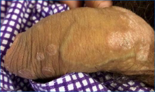
Dermatology was consulted regarding asymptomatic cutaneous lesions in the genital area with similar duration as the remaining symptoms. The patient had sexual intercourse exclusively with women (two different partners in the previous 6 months) and no history of previous sexually transmitted diseases was found. He denied genital or oral ulcers in the preceding months. There was no personal or family history of skin diseases. Examination disclosed psoriasiform erythematous and desquamative plaques and papules in the penis, with round shape and well-defined borders (Fig. 1), suggestive of secondary syphilis. A left inguinal adenopathy was palpable. Further laboratorial investigation revealed positive Treponema pallidum hemaglutination assay and Venereal Disease Research Laboratory, the latter with a 1:16 titer, supporting the clinical diagnosis. The diagnosis of secondary syphilis with probable hepatic involvement was considered and treatment with a single injection of intramuscular benzathine penicillin at dose of 2.4 million units was performed. Bilirubin, liver enzymes and cholestasis parameters progressively decreased following treatment (Table 1). Progressive improvement of jaundice and genital lesions was simultaneously observed.

Figure 1 Penile erythematous and desquamative plaques and papules, with round shape and well-defined borders, disclosing a psoriasiform appearance.
Additional laboratorial investigation was negative for hepatitis A, B, C, and E virus, cytomegalovirus, Epstein-Barr virus, and liver autoantibodies. Imaging studies, including abdominal ultrasonography and magnetic resonance cholangiopancreatography, were unremarkable. Liver biopsy was unspecific, with maintained hepatic architecture and mild inflammatory portal infiltration, with negative immunohistochemical staining for T. pallidum.
The patient was discharged 9 days after treatment, completely asymptomatic. He was referred to a Gastroenterology and Dermatovenereology outpatient clinic and had complete response to therapy, achieving a fully resolution of hepatic and skin alterations. The patient missed subsequent evaluations and was lost to follow-up.
Discussion
Liver damage in luetic infection occurs most frequently in early syphilis, particularly in secondary stage4, and syphilitic hepatitis is thought to occur in 0.2 - 2.7% of patients with secondary syphilis4-6. Mullick6 proposed four diagnostic criteria for syphilitic hepatitis in 2004: (1) abnormal liver enzyme levels; (2) serological evidence for syphilis in conjunction with an acute clinical presentation consistent with secondary syphilis; (3) exclusion of alternative causes of hepatic damage; and (4) improvements in liver enzyme levels after penicillin therapy. The presented case fulfilled all these criteria.
The pathogenesis of luetic hepatitis is still largely undetermined, but direct portal venous inoculation and immune-complex formation have been suggested7. However, given the paucity of spirochete direct recognition in the liver, direct hepatotoxicity induced by the bacteria is unlikely8,9.
Homosexual men in the fourth to fifth decades of life seem to be affected more often, in particular if infected with HIV4. The most frequent clinical findings include skin rash, anorexia, jaundice, and fever4. Laboratorial investigation exhibits a substantial increase in cholestasis parameters, like alkaline phosphatase (ALP) and gamma-glutamyl transpeptidase (GGT), with mild elevations in aminotransferases and total bilirubin4. Histologic features of syphilitic hepatitis include bile duct inflammatory infiltration, which correlates with ALP and GGT levels4. Spirochete identification with immunohistochemical staining is rare4, as in our case. Liver biopsy was performed in this patient as a part of the diagnostic approach of unexplained hepatitis, before the diagnosis of syphilitic involvement was made.
Penicillin remains the first treatment line, and the subsequent decrease of liver enzymes is one of the diagnostic criteria of syphilitic hepatitis6. Our patient was treated with the standard dose for secondary syphilis, and no iatrogenic reactions were observed.
Conclusion
The discrepancy between the estimated occurrence of syphilitic hepatitis and the number of published cases suggests that this condition is probably overlooked. The present case raises awareness for this complication, that should be included in the differential diagnosis of sexually active patients with abnormal liver enzymes of no obvious cause.


