




 Inglês (pdf)
 Artigo em XML
 Referências do artigo
 Tradução automática
 Enviar este artigo por email
 Citado por SciELO
  Similares em
SciELO
Similares em
SciELO 
 Permalink
Introduction
Hidradenitis suppurativa (HS), or acne inversa, is a chronic dermatological disease characterized by the inflammation of the hair follicle1. The lesions can range from inflammatory nodules to abscesses, mostly located in the axillary, inframammary, and anogenital regions1. The etiological complexity remains unclear, but it is associated with genetic predisposing factors, components of metabolic syndrome (dyslipidemia, peripheral insulin resistance, and hypertension), and smoking2. The most common sequelae are chronic pain, and extensive scarring, with an extensively debilitating outcome in terms of prognosis as well as social and psychological dimensions. It is recognized as the dermatological disease with the highest dermatology life quality index1,3.
The first clinical manifestations generally occur after puberty, affecting individuals aged between 20 and 40 years and it is more common in female gender4. The condition begins with occlusion of the hair follicle, resulting in dilation of the pilosebaceous structure and follicular hyperkeratosis, with sebaceous proliferation5. The deregulated inflammatory process then initiates, with the involvement of innate and adaptive immunity4-6.
The diagnosis of HS is based on the patient’s anamnesis and presentation of typical lesions, erythema, abscesses, nodules, tracts, and fistulas with local pain and scarring in more advanced stages5. Upon diagnosis, the disease is classified according to the Hurley score, which considers recurrent nodules and abscesses with minimal healing (Hurley 1), one or more sinus tracts and/or scars within a single body area (Hurley 2), and multiple or extensive sinus tracts and/or scars (Hurley 3)4. In addition, severity is classified according to the International Hidradenitis Suppurativa Severity (IHS-4) Score, which is divided into: slight (3 nodules or less), moderate (4-10 nodules), and severe (11 or more nodules), it uses a formula that sums up the number of nodules (multiplied by 1), abscesses (multiplied by 2), and draining fistulas (multiplied by 4)1,4,5.
The correct staging of lesions directly impacts the therapeutic approach and, despite being an essential tool in the primary analysis, the physical examination, even with the use of scales, becomes less sensitive and limited to verifying prodromes4,7. In light of the concern, specific ultrasound (US) metrics were developed, such as Sonographic Scoring of HS (SOS-HS), with the application of probes with high frequencies from 15 MHz to 70 MHz in the lesions, which allows early diagnosis and assessment of the severity of HS. Furthermore, the use of US has become more sensitive, being able to modify staging and assist in decision-making on previously established therapeutic approaches1,5,8-10.
The late diagnosis of HS is one of the factors that hinders treatment and contributes to the rapid progression and severity of the disease, which challenges the instruction of the correct therapy and the maintenance of follow-up in care units, in a disease that is already highly complex5,8. Besides the US high-frequency probes, the vascular assessment of lesions with Doppler can lead to early diagnosis and correct management, as well as identifying the early signs of tract, fistulas, and abscess formation, characteristics of disease severity5,8,11.
In relation to the therapeutic response by Hidradenitis Suppurativa Clinical Response (HiSCR) score, the use of US optimized its accuracy, as demonstrated in the study by Caposiena et al. (2019), with an 8-week follow-up of patients using oral clindamycin and rifampicin. It was also demonstrated that there is a correlation between ultrasonographic and histopathological findings, observed through Doppler intensity and neutrophilic infiltration besides the regular accuracy between the fistula diameter in US and histopathological evaluations12.
Therefore, given the holistic impact of the US in HS approach, this study aims to conduct a systematic review to assess the effectiveness of the classification metrics used in HS, as well as the comparative evidence between clinical and imaging staging methods, with a particular focus on the use of high-frequency US.
Methods
This systematic review was conducted in accordance with the Preferred Reporting Items for Systematic Reviews and Meta-Analyses (PRISMA) and the International Prospective Register of Systematic Review (PROSPERO), with registration number no. CRD4202346559413.
The PICO strategy was used to develop the research question, which is an acronym for patients, interventions, comparisons, and outcomes. Thus, we have patients with HS lesions (P), diagnostic evaluation (I), comparison between physical examination and the use of US (C), and diagnostic accuracy (O). The main research result is that patients with HS lesions can have greater accuracy in the diagnostic assessment when US is performed instead of a physical examination14.
Based on the research question, data were collected from scientific databases such as SCIELO, LILACS, MEDLINE, PUBMED, IBECS, and WPRIM. The descriptors approved in DECS/MESH are (hidradenitis suppurativa), AND (ultrasonography), AND (ultrasound), and (diagnosis). The defined inclusion criteria consisted of publications from the last 10 years, in Portuguese, English, and Spanish; scientific documents of article quality; randomized, quantitative, and qualitative studies; cohort studies; and multicenter and retrospective studies.
The exclusion criteria included narrative reviews, duplicate documents, book chapters, opinion articles, letters to the editor, and works that did not correspond to the scope of the research. Furthermore, studies that did not use the US as the primary radiological diagnostic methods were also excluded.
The MS Excel and MS Word platforms (Microsoft Inc., USA) were used to select the results and identify duplicates. Subsequently, the titles and abstracts were assessed and read in full by three independent reviewers. Any doubts or conflicts of information between the authors were resolved through a discussion with a fourth author in a consensus meeting.
The information found in each article was distributed in a table created by the authors, which included the following: author, title, year, language, journal, research location, country, number of research participants, objective, US frequency, US location, and conclusion.
To assess the risk of bias among the selected studies, the bias of randomized clinical trials was assessed using the Cochrane Collaboration Tool, which analyzes selection bias, performance bias, detection bias, attrition bias, reporting bias, and other biases. In addition, other sources of bias were considered using the conflict of interest statement14.
Results
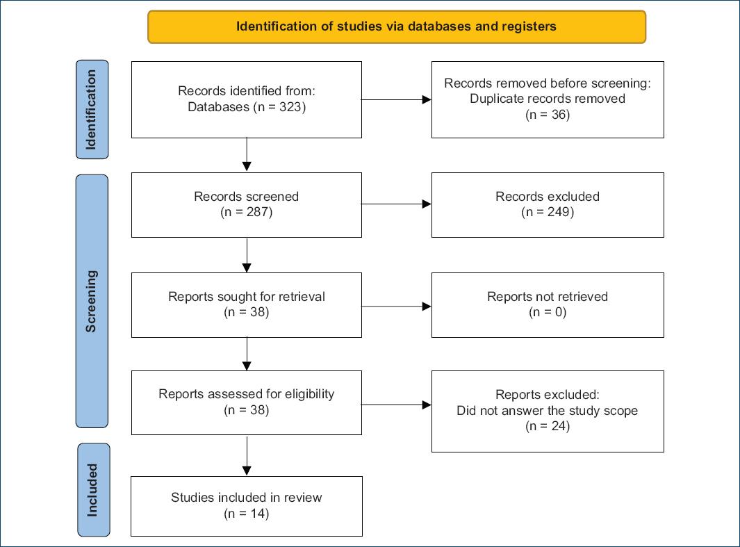
A total of 323 articles were found during the database search. At the end of the evaluation, 14 articles were eligible for inclusion in this systematic review, as shown in figure 1.
Fourteen articles were included in this study, including seven retrospective studies, four cross-sectional observational studies, a longitudinal study, an open study, and a collaborative initiative. All data were collected using the PUBMED database. Regarding the US frequency specifications, the main ones were 15-22 MHz and 10-22 MHz; the other frequencies used were included in various 6-22 MHz and 70 MHz. Approximately 1,485 HS lesions were properly included in the analysis of the studies, where the body regions most researched were the axillary and pelvic regions, covering the roots of the thighs, perineum, inguinal, and genitals; other areas of focus included the chest and inframammary, occipitocervical and periumbilical regions8,11,15,16.
In the set of nine articles analyzed, it was observed that the introduction of a combination of physical examination and imaging assessment resulted in significant changes in the classification of injuries and patient management. In four of these studies, patients presented a more severe classification after undergoing the examination, whereas in three articles, a comparison with physical examination revealed subclinical lesions. The impact of early diagnosis using US on female patients of different ages was observed in one of the articles included. Yet another study advocates both US for staging and as a guide for managing the treatment of lesions with Guided Galvanic10,17-21.
Notably, only one study correlated the use of Power Doppler for lesion characterization. All studies based their evaluations on the SOS-HS classification compared with clinical semiology scores, such as IHS4 and Hurley. One article addressed the issue of fistulous tracts, highlighting the gluteal region as the most susceptible to this type of lesion, and another study investigated the association between the presence of lymph node enlargement and HS7,22-24.
Selected articles are listed in table 1.
Table 1 Summary of the studies on the utility of ultrasonography in hidradenitis suppurativa
| Title | Author/year | Study design | Lesions sample | US probe frequency | Study objective | Conclusion |
|---|---|---|---|---|---|---|
| Sonographic Evaluation of HS with Smartphone-Linked Portable US | Weigelt, 202118 | Retrospective study | 33 | 10 MHz | Assess the ability of a novel smartphone-linked pUS device to identify key sonographic lesions of HS. | Demonstration of the ability of US to identify the key sonographic HS lesions. US is a simple and affordable way to integrate into clinical and research settings, with clear potential benefits to patients. |
| US as a diagnostic and management tool in HS patients: a multicentre study | Martorell, 201922 | Cross-sectional multicentre study | 143 | 18 MHz | Determine the usefulness of US as a characterizing diagnostic tool in patients with HS compared with physical examination and assess the potential need for disease management modification. | Support US as an essential tool in the diagnosis and management of HS, as physical examination significantly underestimates severity and staging. US can decrease the performance of late, blinded, or multiple invasive procedures, with consequent scarring, and positively impact the quality of life. |
| US In-Depth Characterization and Staging of HS | Wortsman, 201321 | Retrospective study | 142 | 7-18 MHz | Systematically evaluate the addition of US to the clinical assessment of a large series of HS patients and explore the relevance of sonographic examination and scoring. | Clinical examination can underestimate the severity and disease involvement of HS. The addition of US to clinical evaluation allows the uncovering of subclinical pathology and better assessment of the extent of the lesions. |
| Ultrasonography in the pathway to an optimal standard of care of HS: the Italian US Working Group experience | Lacarrubba, 201916 | Retrospective study | 434 | 14-20 MHz | Compare clinical HS-PGA with US HS-PGA in the evaluation of HS patients in a large cohort of patients. | Confirmation that isolated clinical grading may underestimate the real disease severity and US can be an essential non-invasive imaging tool for a more accurate diagnosis, staging, treatment planning, and monitoring, and should be included as an optimal standard of HS care. |
| Pain and inflammation in HS correspond to morphological changes identified by high-frequency US | Zarchi, 201523 | Cross-sectional observational study | 20 | 6-18 and 10-22 MHz | Investigate correlation and validity of simple patient- and investigator-assessed inflammation-related items with high-frequency US morphological changes in HS. | Patient assessments of flare activity and pain and investigator assessment of erythema are strongly associated with morphological changes identified by US and might be strong indicators of the degree of inflammation in HS. |
| Power Doppler (PD) US assessment of vascularization in hidradenitis suppurativa lesions | Caposiena Caro, 201715 | Retrospective study | 198 | 10-18 MHz | Describe the PD signal of HS nodules, abscesses, and fistulas. | PD may categorize the vasculature component, relevant to assess disease severity. PD changes in vascularization might support prognosis and anticipate the need for more aggressive therapies achieve a more complete knowledge and proper disease management. |
| Assessment of Inter-rater reliability of clinical hidradenitis suppurativa outcome measures using ultrasonography | Lyons, 20228 | Longitudinal observational study | 20 | 22 MHz | Evaluate inter-rater reliability in HS disease severity assessment using clinical and US techniques. | US can improve inter-rater reliability for assessing HS activity and severity. Clinical grading often underestimates the true extent of disease. US + clinical examination decreases variation in staging and severity between providers, supplying appropriate treatment recommendations and evaluating treatment response. |
| Color Doppler US assessment of morphology and types of fistulous tracts in HS | Wortsman, 201625 | Retrospective analysis | 96 | 18 MHz | Categorize fistulous tracts in HS. | Color Doppler US may categorize fistulous tracts relevant for assessing disease severity, support the prognosis (reversibility of physiopathological changes), perhaps predict a future need for or response to more aggressive treatments, and achieve more anatomically oriented, early, and precise HS management. |
| Doppler US-based non-invasive biomarkers in HS: evaluation of analytical and clinical validity | Grand, 202119 | Cross-sectional observational study | 65 | 10-22 MHz | Evaluate the analytical and clinical validity of sonographic epidermal thickness, PD, and dermal tunnel diameter in HS. | Sonographic epidermal thickness and dermal tunnel diameter have acceptable levels of analytical validity in assessing HS lesions compared with histology. PD signs demonstrate both acceptable levels of clinical and analytical validity. Need to assess the response of these biomarkers to therapeutic interventions and their utility as diagnostic and predictive biomarkers for directing and optimizing patient care. |
| US evaluation as a complementary test in HS: proposal of a standardized report | Martorell, 201724 | Collaborative initiative | NA | NA | Develop a consensus US report that could summarize the relevant anatomical characteristics and staging of patients considering the experience of radiologists and dermatologists working on HS imaging. | A standardized sonographic report on HS approved by the consensus of an international group of radiologists and dermatologists as a first attempt to include US as the first elective medical test for staging and monitoring patients. |
| Advanced evaluation of HS with ultra-high frequency US: a promising tool for the diagnosis and monitoring of disease progression | Oranges, 202010 | Retrospective study | 116 | 7-18 MHz and 48-70 MHz | Compare high-frequency US (HFUS) with ultra-high frequency US (UHFUS) in the analysis of main HS lesions and evaluate the potential role of UHFUS in clinical practice. | UHFUS is a very promising diagnostic method: identifies non-observable findings (early diagnosis), contributes to understand the evolution from early to advanced stages, allows more effective monitoring, and can perform a detailed pre-surgical mapping in patients likely to undergo surgery. |
| Seventy-MHz US detection of early signs linked to the severity, patterns of keratin fragmentation, and mechanisms of generation of collections and tunnels in HS | Wortsman et al., 202011 | Cross-sectional study | 139 | 15-18 MHz and 71 MHz | Test the capability of 70-MHz US for detecting initial US HS signs linked to severity. | US can detect early HS signs significantly linked to severity and 2 types of keratin fragmentation that could support the generation and perpetuation of the fluid collections and tunnels. US can help prompt diagnosis and management, development and testing of medications, and measure of treatment outcomes. |
| Clinical and ultrasonographic characterization of hidradenitis suppurativa in female patients: impact of early recognition of the disease | Di Cesare et al., 20237 | Retrospective study | 53 (females only) | 16-18 MHz | Investigate clinical and ultrasonographic features to improve earlier disease recognition, provide clinical features to be investigated, also in a multidisciplinary team, and lead to timely and effective treatment protocols. | The Doppler signal could be a useful tool for introducing adjuvant systemic treatment to reduce the activity and dimensions of nodules abscesses, and tunnels before surgery and to identify predictive failure markers of biological therapies. |
| Safety and effectiveness of percutaneous US-guided galvanic current in tunnels of patients with HS: A pilot study | Soto-Moreno et al., 202417 | Open study (one-way repeated measures design over time) | 26 | 15-18 MHz | 1 - Evaluate the effectiveness and safety of percutaneous US-guided glucocorticoid (GC) administration in inflamed and/or draining tunnels. 2 - Evaluate US reduction of GC-treated lesions and duration of remission in complete responding lesions at week 4. | US parameters of depth and volume could potentially serve as predictors of response to GC (single administration) that appear to be an effective therapeutic alternative in inflammatory and draining HS tunnels. |
US: ultrasound; HS: hidradenitis suppurativa.
Discussion
Current clinical staging classifies the scales into qualitative analysis, such as the Hurley score, and quantitative analysis, to which the Sartorius and HS Physician Global Assessment (HS-PGA) are applied. To evaluate the response to therapy, the HiSCR is consolidated as a metric to analyze its effectiveness24.
In HS cases, US can detect subclinical anatomical findings, which can significantly modify the basic description of the lesions and their staging, leading to a change from conservative to surgical treatment or vice versa. According to Wortsman (2013), its application altered the treatment in 82% of the evaluated patients; specifically, the clinical approach was altered to surgical in 24% of the cases8,21.
Semiological limitations include palpation techniques, the inconsistency of which can lead to no differentiation between fistulas and drainage abscesses, the impossibility of distinguishing between nodules and fistulous tracts, as well as the stratification of the erythematous extension22. The basic descriptions of the clinical findings may differ from those presented by US, such as the evaluation of a nodule, which may clinically represent a pseudocyst, liquid collection, or fistulous tract. Meanwhile, in US, a collection does not necessarily imply the presence of purulent material, and some cultures may even be negative22.
Ultrasonography also allows a deeper investigation of the skin layers and tissue anatomy, as well as detecting vascularization patterns and its surrounding areas with Doppler, while monitoring the progression of the disease20,22. For HS classification through US, three or more of the following criteria are required: increased in the size of hair follicles, dermis layer changes, presence of pseudocysts, liquid collections, and fistulas. Moreover, staging can be established using US imaging with an exclusive metric, such as SOS-HS, which is mainly based on subclinical findings that cannot be detected on clinical examination. The criteria included are the number of lesions and their locations, the type of abscess detected (whether it has a fistulous tract, drainage, or scarring) along with dermal alterations and vascularization patterns20. It enables the description of fistulas and collections, anechoic or hypoechoic pseudocyst nodules, hair follicle enlargement, or changes in thickness and echogenicity, allowing the mapping of the disease and its activity25.
The multicenter study by Martorell (2019), which compared the use of US with physical examination for evaluation of HS, concluded that a physical exam alone significantly underestimated the severity of the case22. For this assessment, a linear transducer with a frequency of 18 MHz was used to examine areas such as the axilla, breast, periumbilical, inguinal, buttocks, thighs, and perineum. After the US examination, most of the cases had their severity classification modified, resulting in adjustments or changes in treatment. Modifications in drug dosage were observed in 96 patients compared to 78 patients after the clinical examination. In addition, the number of patients who remained on unchanged treatment decreased from 63 to 45 (p < 0.01)22.
Based on the results, a predictive factor Hurley classification was established when comparing the two types of examination. A statistically significant difference was found in relation to age. Patients who required a change after US examination were, on average, 6.5 years younger (95% CI, 0.95-12.16; p < 0.05). The average number of nodules found during the clinical examination was 4.4 (SD, 5.9), which was significantly higher than the number found during the US examination: 1.4 (SD, 1.9; p < 0.01). On the other hand, the average number of abscesses detected during the US examination was double that detected during the physical examination (0.7 [SD, 14 vs. 1.5] [SD, 1.9; p < 0.01]). In addition, an increase in the number of fluid collections identified through US compared to physical examination was observed22.
Moreover, the pathogenesis of HS itself leads to poor validation of semiology, since the anatomical changes occur in deep layers of the dermis, compromising the application of clinical scores25. In contrast, extensive lesions with high inflammatory activity and erythema may benefit from clinical assessments11,25. Worstman et al. (2020) and Oranges et al. (2022) used 70 MHz US high-frequency, which showed anatomical changes such as keratin fragmentation patterns and pathophysiological mechanisms linked to abscesses and collections. It was possible to identify the bridge-connecting sign of the band between the base of adjacent hair follicles, and the sword sign, defined as a fragment of the hair shaft extruding through a dilated hair follicle. These findings would not be identifiable by clinical examination, as they represent specific and deep structural changes10,11,24.
Furthermore, fistulous tracts are critical indicators of the advanced stage of HS25. The US imaging detects subclinical signs and its progression precisely, optimizing clinical analysis, according to Wortsman et al. (2016)25.
In terms of clinical correlation with US findings, the analysis of Zarchi et al. (2015) illustrated that the inflammatory signs reported by patients, such as pain, erythema, and increased local sensitivity, are proportional to the diameter of the nodule identified, corroborating with clinical care approach23.
Lyons et al. (2022) performed a retrospective study based on a clinical examination of the HS lesions, using three score systems: Hurley, Sartorius, and HiSCR. Subsequently, the analysis was initiated using US with 22 MHz probes. In the pre-US period, the clinical methods showed low assertiveness, except for the Hurley score. Post-US application revealed greater sensitivity in the Sartorius and HiSCR metrics, attributable to their ability to distinguish fistulas, nodules, sinus tracts, abscesses, inflammation, and scars. Despite the excellent characterization of the Hurley score, it does not measure the current activity of the disease, such as the patient’s pain and discomfort. Furthermore, individuals with the same Hurley staging may present different clinical conditions, with other qualification measures required8.
As well as exposed, Lyons et al. (2022) and Grand et al. (2021) demonstrated a comparison between the application of both methods in a sample of lesions. The dermal thickness and diameter of the pilosebaceous tunnels allowed staging with greater accuracy through US analysis, with clinical semiology reporting low or no validity in view of the lesions. The authors highlight adversities that can, in fact, limit the semiological analysis, such as the rapidly evolving temporal clinical condition, slow histological change due to acute inflammation, and the score applied, IHS4. In addition to evaluating the disease classification, US improved longitudinal support regarding prognosis and therapeutic response19,24.
In the Italian retrospective study conducted by Lacarrubba et al. (2019), the US frequency of 14-20 MHz was used. A direct comparison was carried out between the clinical and US assessments, which revealed a significantly higher proportion of patients classified as having moderate to very severe HS by US. A total of 117 patients (26.96%) were categorized in a more severe category by US assessment (HS-PGA) compared with clinical assessment. The staging of the disease was changed to a more severe stage in 44.7% of patients previously diagnosed with Hurley stage 1, based on clinical evaluation. Specifically, on clinical assessment, 26.6% of the patients were diagnosed with Hurley stage 1, 49% with stage 2, and 24.5% with stage 3. In contrast, US classified 14.7% of patients as Hurley stage 1, 55.9% as stage 2, and 29.4% as stage 316.
These results indicate that relying solely on clinical classification to assess the severity of HS can lead to an underestimation of its true severity. Unlike the previous study by Wortsman et al. (2013), which employed different clinical and US scores (Hurley vs. SOS-HS), the Italian study compared two similar clinical and US scores (clinical HS-PGA vs. HS-PGA US), thus reducing possible biases16.
A retrospective study by Weigelt in 2021 showed that the use of portable US at a frequency of 10 MHz can effectively identify the main lesions associated with HS. It allows a successful integration of this imaging test into routine clinical and research environments. In the following study, these echographic lesions were identified in 10 out of 16 patients (62.5%). Among these findings, five liquid collections were suggestive of an abscess (31.25%), three sinus tracts/fistulas (18.75%) and one pseudocyst (6.25%). Moreover, subclinical lesions were identified in 18.75% of cases, allowing for timely interventions that had a clear positive impact on the clinical outcomes of these patients18. This study highlights the importance of portable 10 MHz US as an affordable and effective alternative for detecting and assessing HS lesions, allowing for more appropriate treatment and improving patients’ quality of life7,18. Unfortunately, US can be inaccessible in clinical practice in some places due to the high cost of the equipment18.
The application of power to Doppler demonstrates the expansion of diagnosis to staging and more invasive procedures, as reported by Caposiena et al. (2018). In this study, a correlation was observed between the presence of a positive signal on the power Doppler image and the size of the lesion. Besides, patients in stages 2 or 3 of the HS Evaluation System had a higher rate of sensitivity on the power Doppler. Even so, no correlations were found between vascularization and patient characteristics such as age, gender, duration of the disease, location of the lesions, and lifestyle habits such as smoking. Nevertheless, this tool can help with the introduction of adjuvant systemic treatments and detect negative predictive values in regard to biological therapies15,17.
Besides, obesity plays a significant role in the severity of HS at the expense of increased skin folds and friction, which results in sweat retention and increased sweating. The condition progresses with increased levels of inflammatory interleukins, tumor necrosis factor-alpha, and C-reactive protein. These factors mainly explain the high power Doppler sensitivity in this population7,15.
By applying more recent therapies, such as galvanic current, as described by Soto-Moreno (2024), the authors obtained a positive predictive value of the therapeutic response, which was confirmed with the use of US, with an improved analysis of the volume and depth parameters. The aid of technology can provide supporting evidence of the benefits of new treatments17.
The use of US in longitudinal analysis, however, does not have a medical consensus, as shown by Martorell et al. (2017) based on the application of a questionnaire form to 14 professionals, including radiologists (57%) and dermatologists (43%), from nine different countries. The recommendation for the use of US in axillary and perianal lesions was unanimous among professionals, mainly because of the high prevalence of subclinical lesions in these regions, despite other divergences described24.
The limitations of the US include the risk of cross-infection, pain, and device hygiene, as reported by DiCesare et al, (2023). In parallel, there are also technological factors, such as the skill of the operator and the performance of the device, with high frequencies often being more advantageous7,23.
Conclusion
In conclusion, this study establishes that the US is an indispensable, non-invasive imaging tool for the accurate diagnosis, classification, treatment planning, and longitudinal approach of HS. Although neither clinical nor imaging methods (US) are sufficiently sensitive to quantify lesion progression due to external interference factors, the correct application of US enhances the accuracy of staging. It can identify more severe cases of HS compared to clinical assessment alone, due to a sharper description of clinical findings such as nodules, abscesses, and fistulas, along with the detection of subclinical conditions. Furthermore, US optimizes clinical score accuracy and correlates with the histology and pathophysiology of the disease, supporting a holistic analysis of HS and its complications.
Despite the clinical gains made by the US, the device is unable to replace human hands, and the clinical reasoning needed to manage these patients. Furthermore, the dermatologist’s central role as a conductor is essential for therapeutic success, given the technical and academic expertise developed during medical formation.

