




 Inglês (pdf)
 Artigo em XML
 Referências do artigo
 Tradução automática
 Enviar este artigo por email
 Citado por SciELO
  Similares em
SciELO
Similares em
SciELO 
 Permalink
Introduction
Alopecia areata (AA) is one of the most common forms of nonscarring alopecia encountered by dermatologists. Though classified as nonscarring, there have been reports elucidating progression of the disease to scarring, with eventual permanent hair loss1. Furthermore, the classical peribulbar lymphocytic infiltrate, which is considered the hallmark histological feature of AA, is not present in all scalp biopsy specimens. The frequency this and other histological changes are observed depends on the disease stage when the biopsy is performed. Thus, it becomes pivotal for the dermatopathologist to thoroughly scrutinize the slide, in order to establish a correct diagnosis2. Besides, as AA is usually diagnosed on clinical grounds and biopsies seldom performed, careful evaluation with a detailed exploration and interpretation of histological specimens in AA still remains an unmet need. In the last three decades, use of transverse sections in evaluating scalp biopsy specimens has richly enhanced the study of the histological profile of AA. This technique enables inspection of numerous follicles in one section, making visualization of various features in a more systematic way3. Nevertheless, the significance of vertical sections cannot be undermined, and some authors suggest combination of both horizontally and vertically sectioned scalp biopsies to maximize the diagnostic yield in most alopecia cases, including AA4,5. However, in Sperling’s report, vertical sections in the diagnosis of AA were rarely necessary,6 thereby raising an inquiry regarding the ideal approach for scalp biopsies in AA. As of now, a definitive answer to this question is lacking, allowing flexibility in the choice for the diagnostic approach based on the clinical setting where one works.
Given the paucity of studies among Indian patients regarding the histological pattern of AA, this study was performed with the aim to evaluate the microscopic features of AA, using both vertical and horizontal biopsy sections.
Methods
This was a prospective, descriptive study carried out over a 1-year period, after obtaining permission from the institutional ethical clearance committee [Registration number KMC/IEC/Dept. Res./016/2021-24 (DVL)].
A total of 30 patients with a clinically unequivocal diagnosis of AA were enrolled in the study after their informed consent. In each case, two 4 mm punch biopsies were performed, one specimen being sent for vertical, and the other for horizontal sectioning. Both biopsies were taken from the advancing edge of an area of active hair loss. Specimens were fixed in 10% formalin and stained with hematoxylin and eosin. Horizontal sections were performed with particular emphasis to include the infundibular and isthmic levels, so that maximum findings were obtained. Thus, in each patient, two horizontal sections were evaluated, one at the isthmic level and the other at the infundibular portion.
The histopathological features studied included
– Number of anagen and nonanagen (catagen and telogen) follicles in each section.
– Evidence of “nanogen” (dystrophic) follicles, i.e., miniaturized follicles difficult to categorize as anagen, catagen, or telogen.
– Number of terminal and vellus hairs.
– Inflammatory infiltrate surrounding the hair bulb.
– Predominant cells in the inflammatory infiltrate.
– Pigment incontinence.
– Follicular streamers (stelae), i.e., residual fibrovascular tracts representing the impermanent lower third of the hair follicle below the bulge region.
– Follicular miniaturization.
– Dilated infundibular openings.
Results
Our patient group included 18 females and 12 males with the patchy subtype of AA, aged 7-45 years with duration of AA between 8 months and 2 years. None of the patients had positive family history of AA.
On histopathology, nonanagen hair follicles represented 70-80% of terminal hair follicles in 12 (40%) patients, and 33-69% in the remaining 18 (60%) patients. The mean ratio of anagen and nonanagen follicles was 1:1.7. Among nonanagen follicles, catagen follicles were recognized by a deep red/pink trichilemmal keratinization around the hair shaft, along with apoptosis in the outer root sheath (Fig. 1), and telogen follicles as structures resembling an asterisk or daisy shape composed of basaloid cells with minimal or no central keratinization (Fig. 1).
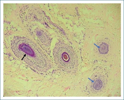
Figure 1 Horizontal section showing a catagen follicle (black arrow), recognized by a deep red/pink trichilemmal keratinization around the hair shaft, and two telogen follicles (blue arrows), composed of basaloid cells with no central keratinization [H&E 100×].
“Nanogen” (dystrophic) follicles were identified in three (10%) patients. In these follicles, there was decreased thickness of the epithelial component with fusion of the internal and external root sheaths, and detritus of amorphous keratin was observed in place of the existing hair shaft (Fig. 2).
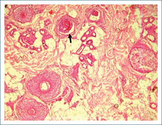
Figure 2 Horizontal section depicting a “Nanogen” follicle (black arrow), identified by decreased thickness of the epithelial component with fusion of internal and external root sheaths and detritus of amorphous keratin in place of the existing hair shaft [H&E 100×].
Miniaturized hair follicles constituted more than 50% in 18 (60%) of the specimens (Fig. 3).
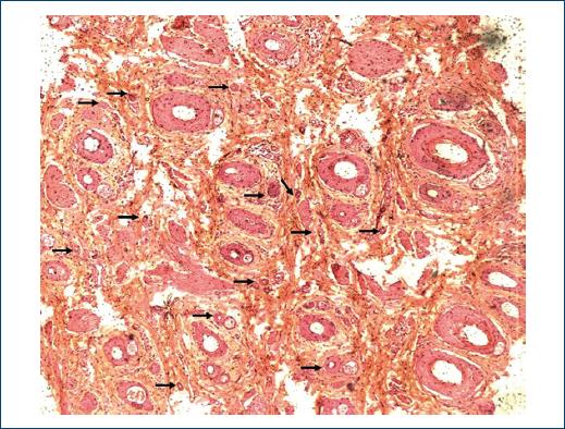
Figure 3 Transverse section in which are present numerous miniaturized hair follicles (black arrows; [H&E 40×]).
The classical lymphocytic peribulbar infiltrate resembling a “swarm of bees” was not identified in any of our study subjects. Sparse infiltrates of lymphocytes were identified in 18 (60%) patients, with the remaining 12 (40%) patients delineating no evidence of inflammatory infiltrates.
Pigment incontinence was observed in six (20%) patients, with clumped melanin pigment aggregates present within follicular streamers (Fig. 4).
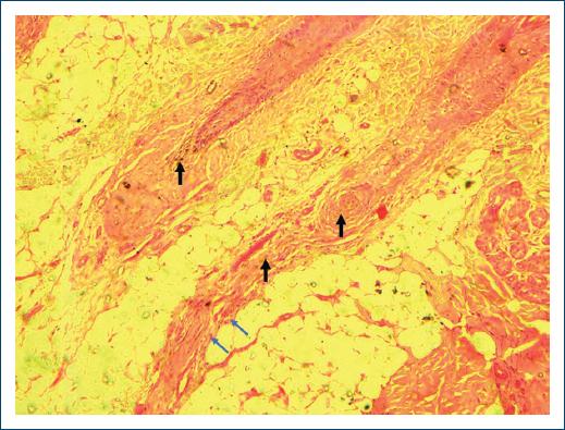
Figure 4 Vertical section demonstrating pigment incontinence (black arrows) and follicular streamers characterized by concentrically arranged connective tissue consisting of collagen fibers, fibroblasts with intermingled blood vessels and lymphocytes (blue arrows; [H&E 100×]).
Follicular streamers were identified in eight biopsy specimens (26.7%). They appeared as concentrically arranged connective tissue composed of collagen fibers and fibroblasts, intermingled within blood vessels and sparse infiltrates of lymphocytes (Fig. 4). This was the only finding better appreciated in vertical sectioning.
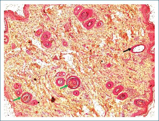
Dilated follicular infundibula occurred in 15 (50%) specimens (Fig. 5).
Discussion
Histopathological interpretation of scalp biopsies from alopecia is often arduous. Particularly in AA, likelihood of missing the diagnosis is high, if the classical “swarm of bees” pattern is absent. Furthermore, microscopic changes of AA vary with the stage of the disease, with prognostic and therapeutic implications.
In the histological profile of our study, the “swarm of bees” peribulbar lymphocytic pattern was not identified in any of the biopsy specimens. Additionally, we observed that longitudinal sections by themselves were insufficient in making a final diagnosis of AA, and horizontal sections were mandatory for a decisive conclusion. This contrasted with the findings by Chaitra et al.7 and Garcia and Poletti8, who observed peribulbar lymphocytic infiltrates in all their study subjects. Peckham et al.9 and Genedy et al.10 on the other hand identified peribulbar lymphocytes in 84 and 70% of their cases, respectively; and Yoon et al.11 reported similar findings in 48.1% of the specimens examined. This wide variation (from 0% to 100%) raises the question of interobserver variability and/or sampling bias (number of step sections). For example, Chaitra et al.7 performed 15 sections to conclude the presence of peribulbar lymphocytes in all cases, although the exact number of slides which delineated this finding was not clearly specified.
The striking absence of peribulbar lymphocytes in our series needs contemplation. Of course, long-standing disease definitely serves as a valid exposition for some patients, but the reason for their conspicuous absence in the advancing border of active lesions requires further elucidation. Although, a definitive explanation for this disparity remains elusive at this juncture, we suggest the possibility of an unidentified environmental factor, along with genetic polymorphisms to be responsible for this observation, whose confirmation requires molecular and genetic analysis.
A major disadvantage of vertical sections in evaluating AA (or other nonscarring alopecias) is the presence of few hairs in the biopsy specimen. Despite including full skin thickness, from the horny layer up to the subcutis, the chances of obtaining a diagnostic follicle are considerably low in the first section, making serial step sections mandatory for a final diagnosis, as well as to reduce the chances of any form of sampling error12,13. Besides, to get the desired diagnostic follicle, the number of sections required can even go as high as 30-116 (mean 53) which may not be a practical proposal14. Bathish et al.15 reported the need for serial vertical sections in 54% of their cases of noncicatricial alopecias to establish a confirmatory diagnosis, whereas in the remaining 46%, a conclusive diagnosis was forthcoming in the initial vertical section examined.
In our study, serial vertical step sections were not performed, because a straightforward conclusion was reached when examining the horizontal cuts. In line with our findings, transversal sections become important as they include all follicular units needed for examination, enabling apparent qualitative and quantitative material for the diagnosis16-18. Besides, it is suggested that when a single biopsy is obtained, transverse sections are better suited for diagnosing noncicatricial alopecias, while vertical sections are more informative for the scarring alopecias, specifically for lichen planopilaris19.
Peribulbar lymphocytic infiltrates and an increased number of nonanagen follicles are highly characteristic of AA, and their combined presence provides a plausible diagnostic clue toward AA in acute episodes2,20.
In the subacute stage of AA, however, decreased anagen and increased telogen and catagen hairs represent the hallmark finding; with reduced terminal hairs and increased miniaturized hairs typifying the chronic stage of the disease2. In these scenarios, telogen effluvium (TE) and androgenetic alopecia (AGA) constitute the most likely differentials. Even so, in TE, unlike AA and AGA, miniaturized hair follicles are rarely encountered; serving as a valuable pointer in its diagnosis. On the other hand, in AGA, telogen percentages and vellus hair counts almost equal that of chronic AA, making it very difficult to distinguish the two conditions histologically. In this circumstance, the presence of dilated follicular infundibula and sparse inflammatory infiltrates of eosinophils and mast cells serve as important indicators in favor of AA2. The formation of dilated follicular infundibula occurs secondary to stasis of sebum in the follicular infundibulum of the thinned hair shafts, with subsequent dilation that closely resembles a “swiss cheese”21. This microscopic pattern corresponds to yellow dots observed on dermoscopy, and if a biopsy is performed from these sites, it is unlikely to miss this finding. However, in the absence of dermoscopic correlation, chances of obtaining the adequate specimen may be variable. These yellow dots correspond to the dilated follicular infundibula of the vellus-like anagen (also referred to as “nanogen”) and telogen follicles that characterize the chronic phase of AA22.
The most consistent finding documented in our study was the elevated levels of nonanagen follicles, which was in agreement with the interpretation of Whiting23 and Peckham et al.9 who reported that the absence of inflammatory infiltrates, the presence of catagen and telogen hairs, and reversal of anagen: nonanagen ratio constitute the most common microscopic findings in AA. Miniaturized hairs were another frequent observation. This finding is of diagnostic significance as it enables differentiation from trichotillomania where increased telogen/catagen percentages are identified without any evidence of follicular miniaturization24. Moreover, in trichotillomania, although pigment casts are seen within the follicular canal, they are not associated with an infiltrate of eosinophils or lymphocytes or melanin in fibrous tracts as in AA, which can help distinguishing the two conditions histologically9. In the follicular stella examined, the presence of lymphocytes enabled distinction from trichotillomania, but eosinophils were not detected in our studied sections. In conclusion, a thorough examination of biopsies labeled clinically as AA is essential to rule out its histological mimickers, in particular trichotillomania.
Conclusion
Although clinical diagnosis of AA is uncomplicated, recognizing the microscopic details requires prowess. Based on our study, transverse sections become imperative for long-standing AA (> 1 year), where the classical “swarm of bees” pattern is frequently absent. In these cases, vertical sections are usually insufficient in arriving at a specific diagnosis, thus making evaluation of horizontal sections (both qualitative and quantitative) essential for obtaining a precise diagnostic conclusion.

