




 Inglês (pdf)
 Artigo em XML
 Referências do artigo
 Tradução automática
 Enviar este artigo por email
 Citado por SciELO
  Similares em
SciELO
Similares em
SciELO 
 Permalink
Introduction
Basal cell carcinoma (BCC) is the most prevalent and incident type among malignant skin neoplasms, although it is not highly lethal. It originates in epidermal cells, arising from keratinocytes or cutaneous appendages such as pilosebaceous follicles and eccrine sweat gland ducts. The most widely accepted hypothesis is its origin from pluripotent immature epithelial cells that have lost the ability to differentiate and keratinize1.
The highest incidence in fair-skinned patients with clinical signs of chronic sun exposure and its preference for sun-exposed areas are striking characteristics in epidemiology, supporting the relationship with cellular DNA damage caused by ultraviolet radiation (UVR). Skin affected by chronic UVR experiences prolonged cellular proliferation, and direct DNA damage is propagated during genetic material replication.
Sun exposure from childhood to old age has a cumulative effect, increasing the chances of developing BCC. Therefore, it is more commonly found in elderly individuals with prolonged sun exposure throughout their lives. Its course is generally slow, remaining small for several years, but in some subtypes like sclerodermiform, it can grow rapidly, infiltrating adjacent tissues2.
However, it is acknowledged that up to a third of BCC cases occur in covered areas such as the axillae, groin, buttocks, perianal, genital, and pubic regions, the cause of which remains uncertain and suggests pathogenic mechanisms different from those related to UVR-induced DNA damage. Recent discoveries regarding BCC pathogenicity highlight the involvement of specific genetic alterations, particularly in young patients with sporadic lesions2,3.
The present case with an atypical location underscores the importance of dermatological surveillance in unusual topographical areas and proposes other pathogenic mechanisms in the development of BCC.
Case report
Male patient, 43 years old, referred to the dermatologist by his urologist, with a history of a lesion on the right scrotal region that had been progressively growing for 2 years, associated with mild local itching. On physical examination, we observed a slightly erythematous plaque with darkened spots, well-defined borders, and central ulceration on the right side of the scrotal region, measuring approximately 1.5 cm in diameter (Fig. 1). Dermoscopy revealed a lesion with a pink background with clefts, telangiectasia, and blue-gray blue-globules and chrysalises (Fig. 2).
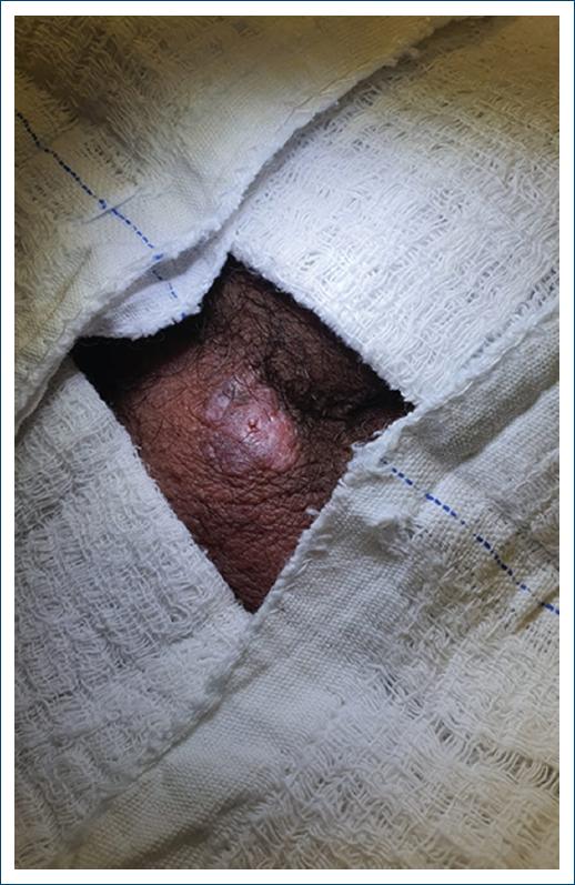
Figure 1 Nodular lesion measuring 1.5 cm, well-defined, reddish in color, with a shiny surface, central ulceration, and the presence of telangiectasias.
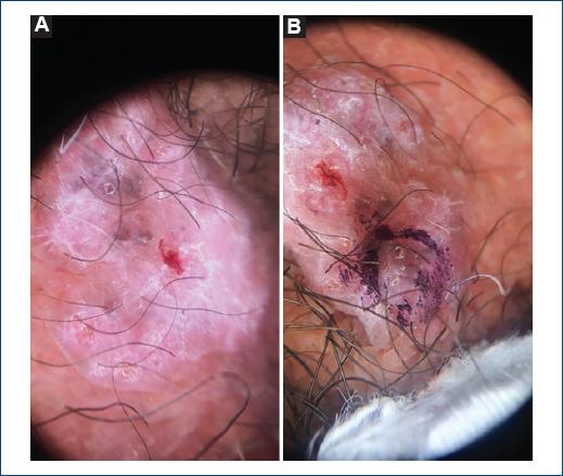
Figure 2 A and B: dermoscopy of the lesion, showing blue-gray globules, telangiectasias, and chrysalises on a pink background.
Histopathological analysis of a skin biopsy revealed the presence of rounded clusters of basaloid cells extending from the lower portion of the epidermis and invading the papillary dermis, with the formation of a peripheral palisade and retraction fissure between epithelial clusters and the stroma. There was focal cystic change and the presence of melanin, with a fibromyxoid-like stroma (Fig. 3).
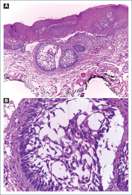
Figure 3 A: proliferation of basaloid cells with peripheral palisading, fibromyxoid stroma, and lack of connections with the epidermis. Stained with hematoxylin and eosin (H&E), magnification ×40. B: basaloid cells with peripheral palisading and fibromyxoid stroma. Stained with hematoxylin and eosin (H&E), magnification ×100.
Once the diagnosis of BCC was confirmed, the patient was referred back to the urologist and underwent excision of the lesion with safety margins. Surgical reconstruction was performed with a simple approximation of the edges.
At present, the patient is under annual follow-up with the dermatology team, with no recurrence of no new malignant lesions reported for 2 years and 4 months.
Discussion
Although the prevalence of BCC increases with sunlight exposure, the distribution of lesions is not always correlated with areas of maximum sun exposure, such as the head and neck4. The reasons for the occurrence of BCC in non-sun-exposed areas remain controversial in the literature.
Some studies associate its emergence with continuous contact with substances such as tar derivatives, mineral oils, petroleum, solvents, and lanolin oils. In addition, non-occupational agents such as radiotherapy, arsenic, prolonged fungal infections, and chronic trauma to the scrotum have been implicated in its pathophysiology. Genetic syndromes also represent plausible risk factors, such as albinism, xeroderma pigmentosum, basal cell nevus syndrome, Rombo syndrome, and Bazex syndrome5.
Recent findings on BCC pathogenicity highlight the involvement of specific genetic alterations, especially in young patients with sporadic lesions. Chromosome 9, particularly the PTCH1 gene, plays a crucial role in BCC development. Studies indicate that up to 40% of sporadic tumors exhibit microsatellite alterations on this chromosome, resulting in PTCH1 gene inactivation and uncontrolled cellular proliferation2,3.
Furthermore, two new genes have been identified in basal cell tumorigenesis: DENND1A and BANP/SMAR1. DENND1A, located on chromosome 9, although understudied, suggests a relevant role in basal carcinogenesis, while BANP/SMAR1, a tumor suppressor that acts on the p53 pathway, influences cyclin D1 transcription and inhibits transforming growth factor-beta signaling. Alterations in BANP/SMAR1 have been associated with carcinogenesis, including in other cancer types such as breast cancer. These findings underscore the importance of understanding the genetic basis of BCC to develop new therapeutic approaches and prevention strategies, especially in young patients with genetic predispositions1,6.
BCCs on the scrotum represent < 0.5% of all cases7,8. The estimated annual incidence is 1/1,000,000 inhabitants, constituting < 5% of all tumors in this region. In nearly 10% of reported cases, a more aggressive potential is suggested, with a higher risk of metastasis compared to other locations. Its invasive behavior can reach the anal canal, and external anal sphincter, and, more rarely, progress to metastasis in regional lymph nodes7,8.
The scrotal region is considered atypical for BCC, being underestimated in its frequency and clinical relevance. Neglecting clinical reasoning regarding nodular-ulcerated lesions in unusual topographies significantly impacts patient prognosis.
Conclusion
Delays in seeking medical attention and late definitive diagnosis were frequently observed in most reported cases, highlighting the need to raise awareness among health-care professionals about the possibility of BCC in genital and perineal areas, as well as the increased risk of metastasis associated with tumors in these regions. This study underscores the importance of a vigilant and early approach in identifying and managing BCC, especially in atypical locations, to enhance clinical outcomes and patient prognosis.

