




 Inglês (pdf)
 Artigo em XML
 Referências do artigo
 Tradução automática
 Enviar este artigo por email
 Citado por SciELO
  Similares em
SciELO
Similares em
SciELO 
 Permalink
Introduction
Blue nevi are benign melanocytic lesions formed by accumulation and differentiation of melanocytic cells in the dermis, caused by failure of adequate melanocytic migration from the neural crest into the dermal–epidermal junction. They are characterized by a blue-gray color, secondary to the deep location of the melanin pigment1. These lesions are usually solitary, but can sometimes be grouped in an agminated pattern, first described by Upshaw in 19472. Agminated blue nevi are formed by a cluster of bluish-pigmented flat or raised lesions in a well-defined area of ≤ 10 cm3. They are quite rare, with only a few cases reported in the literature. Because of their infrequency, exact behavior and prognosis are still uncertain. We report the case of a patient with an extensive agminated blue nevus and review the literature on this subject.
Case report
A 64-year-old female patient was referred to the dermatology clinic for evaluation of an extensive pigmented lesion on her posterior left arm. Her medical history was notable for systemic lupus erythematosus (SLE) since her early twenties, currently using azathioprine 100 mg daily, prednisolone 5 mg daily, and methotrexate 10 mg weekly PO. The patient indicated the lesion was present since birth; however, she noticed progressive growth in size since puberty and new pigmented papules inside the main lesion. Family history of skin cancer was negative.
On physical examination, the patient had an 8 × 7 cm bluish plaque on her posterior left arm filled by multiple isolated maculopapular lesions of blue and dark brown colors, with different sizes and forms. (Fig. 1). Dermatoscopy revealed light brown patches with round dark brown and blue structures with a homogeneous pattern (Fig. 2).
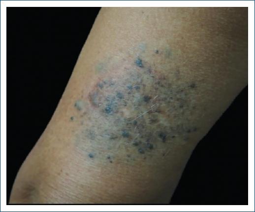
Figure 1 Plaque on posterior left arm filled by multiple maculopapular lesions of blue and dark brown colors, with different sizes and forms.
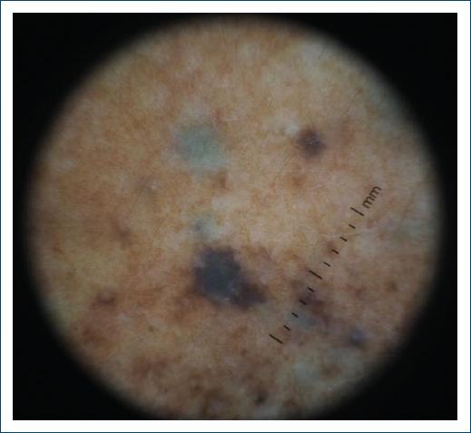
Figure 2 Dermatoscopy shows light brown patches and round dark brown and blue structures with a homogeneous pattern.
An elliptical incisional biopsy was performed. Histopathologic examination revealed intradermal islands of spindle cells with little pigment and a focal nodular downgrowth into the hypodermis mixed with melanophages. No atypia, mitoses, or necrosis was found. (Fig. 3). Clinical and histopathologic findings were consistent with the diagnosis of an agminated blue nevus with common and cellular blue nevus components.
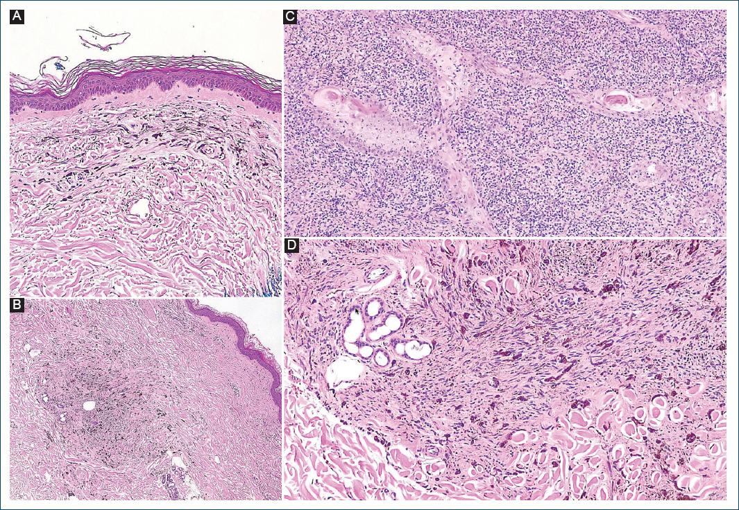
Figure 3 Histopathology with superficial (A) and deep (B, C, D) intradermal ovoid and dendritic cells with little pigment, both isolated and clustered within islands. These cells show no atypia, mitosis, or necrosis.
The patient is currently being followed up in the dermatology clinic for the last three years, with periodic checkups and evaluation of the lesion. Besides a 1 cm growth, the lesion has not undergone other significant changes.
Conclusion
There are three types of solitary blue nevi: common, cellular, and combined. Unusual variants of grouped blue nevi have also been described4. Agminated blue nevi represent a rare entity with an unclear incidence and pathogenesis. Cutaneous trauma was originally proposed as a predisposing factor after a case report of a cluster of blue nevi appearing after a severe sunburn5; however, since then, most reported cases have had no associated cutaneous injury. In contrast to isolated blue nevi, agminated forms appear to be more commonly congenital or manifesting at earlier ages in life4. They have no predilection for specific body sites and have been documented on the face, trunk, extremities, and genitalia3,4.
Somatic mutations in GNAQ and GNA11 are frequent in blue nevi, appearing in more than > 80% of cases5. These mutations produce a constitutive activity of heterotrimeric G proteins, which permanently activate the Ras signaling pathway, implicated in the regulation of cell proliferation. A case report in a patient with an agminated blue nevus and genetic profiling revealed a mutation in GNAQ4, indicating these mutations may also be prevalent in this variant.
Dermoscopic findings appear to be similar to those of common blue nevi. A review of the reported cases demonstrated a structureless homogeneous pattern to be the prevailing finding3. One case report also described linear pigmented structures appearing as “darker sulci”5. Our patient’s dermoscopy revealed consistent findings, with dark brown and bluish structures with a homogeneous pattern.
Differential diagnosis of this entity includes agminated intradermal Spitz nevus combined with speckled lentiginous nevus, malignant blue nevi, and even melanoma, reason why histopathologic examination is usually recommended6. Microscopic findings in agminated blue nevi consist of proliferation of melanocytic cells in the upper and deeper dermis. Most reported cases showed elements consistent with common blue nevi, with dermal pigmented spindle-shaped dendritic melanocytes and a branching network of dendritic processes. A few cases have also documented a cellular blue nevi-type component, exhibiting a biphasic appearance with classic blue nevi features and cellular areas composed of spindled to oval melanocytes with clear or finely pigmented cytoplasm3,5, such as in our case.
Recent data suggest that blue nevi tend to remain stable throughout the years7. There are some cases however that tend to have an expanding nature, documented mostly on the cellular blue nevi subtype. Progression to melanoma is also more common in cellular blue nevi8. Even though the exact nature and prognosis of agminated blue nevi have not been defined, malignant melanoma arising in these lesions has been documented9. A case report of a changing agminated blue nevi in a patient with dermatomyositis has been documented, suggesting a potential role for immunosuppression in this progression10. This is comparable to our patient’s immunosuppressive treatment and the behavior of her lesion.
Agminated blue nevi represent a rare subtype of blue nevi. Most of the few reported cases have exhibited a benign course; however, there is no validated evidence of the benign course. We report this case of an agminated blue nevus in a patient with systemic lupus erythematosus and immunosuppressive treatment. Because of ongoing changes within the lesion and uncertainty of behavior in this pathology, our patient remains in follow-up with periodic examinations.

