




 Inglês (pdf)
 Artigo em XML
 Referências do artigo
 Tradução automática
 Enviar este artigo por email
 Citado por SciELO
  Similares em
SciELO
Similares em
SciELO 
 Permalink
Introduction
Granular cell tumor (GCT) or granulosa cell myoblastoma, described by Abrikossoff in 1926, is a rare benign neoplasm that probably originates from Schwann cells1. It is believed to occur due to the altered metabolism of these cells, a theory reinforced by the constant presence of the S-100 protein in immunohistochemistry (IHC). It affects predominantly individuals between the third and sixth decades of life (30-60 years), predominantly females (1.8-2.9:1)2, with phototypes V to VI, being a rare diagnosis in the pediatric population. It represents only 0.5% of all soft tissue tumors3.
In the general population, GCT most frequently affects the tongue and oral cavity (30–50%)4 and can also affect other locations. In children, it most commonly affects the oral mucosa and extremities. The lesions normally appear as solitary, slow-growing, asymptomatic, or painful nodules with a firm consistency. The clinical differential diagnoses are fibroma, hidradenoma, and lipoma4. Multiple nodules in the same person have been reported (5-16%), associated with congenital syndromes.
Around 1-2% of cases progress to malignancy with a poor prognosis. There are reports of regional or distant metastases with multiple organs involved4. The clinical parameters for malignancy include size greater than 4 mm, invasion of adjacent tissues, and rapid progression4. Malignant tumors are clinically larger and often located in subcutaneous tissue.
The histological examination demonstrates pseudoepitheliomatous hyperplasia. In the dermis, there are large polygonal or round cells with small central vesicular nuclei and abundant eosinophilic granular cytoplasm. These granules are lysosomes or components of the Golgi complex. Positive Periodic Acid-Schiff (PAS) granules and resistant diastasis indicate the presence of myelin. The cells may present ovoid-pustular Milian bodies, specific to the disease, but found in few cases, normally in older lesions (they represent the accumulation of granules inside the lysosomes in cells that have already lost mitochondria and the endoplasmic reticulum). Perineural and perivascular involvement are common and are not markers of a worse prognosis. The criteria for malignancy include the presence of necrosis, elongated and thin cells, large vesicular nuclei, increased mitotic activity, nuclear pleomorphism, and increased nucleus-cytoplasm ratio4. Tumors that present three or more of these histological characteristics are considered malignant2. Finally, immunohistochemical findings are not specific, with positivity for S100 protein, CD8, vimentin, myelin basic protein, and neuron-specific enolase being more common.
Complete excision of the tumor, with safety margins, is recommended due to local recurrence4. For cases with atypical histological characteristics, lymph node dissection is recommended4. Mohs micrographic surgery (MMS) can be an effective tool in invasive tumors or tumors that occur in areas of functional or aesthetic impairment (where maximum tissue preservation is required)4. During MMS, special markers may be required, such as S100, as perineural tumor invasion is difficult to detect by conventional hematoxylin-eosin staining.
The prognosis of GCT is good. Cases of recurrence are generally related to incomplete excision of the tumor (15% of cases). Progression to malignancy is rare (less than 2% of the cases)4. This dermatosis must be followed due to recurrence or development of greater aggression.
Case report
Male patient, 9 years old, phototype IV, complaining of the periungual hardening nodule on the tip of the fifth finger of the right hand presenting slow growth, local pain, and occasional itching for 2 years.
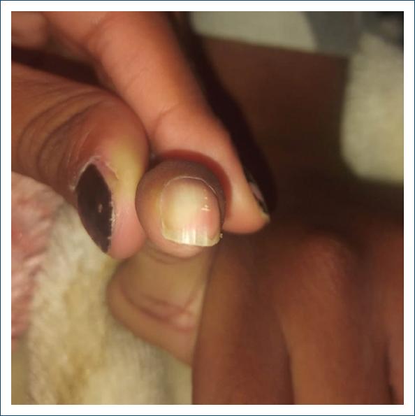
Dermatological examination revealed a normochromic, hardened nodule on the distal phalanx of the fifth right finger with slight nail changes (Fig. 1).
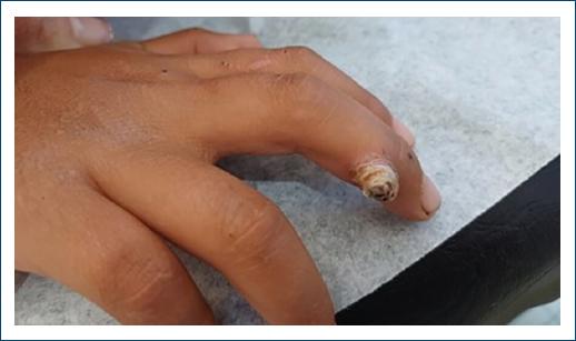
Figure 1 Clinical image showing a nodule on the tip of the fifth finger with minor deformity of the nail plate.
A spindle biopsy was performed, which demonstrated an epidermis without atypia and, in the dermis, a proliferation of cells with a large, frankly acidophilic cytoplasm and vesicular, central nuclei, without atypia (Fig. 2). There were no signs of malignancy.
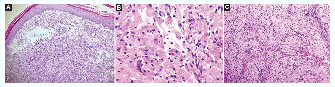
Figure 2 Histopathology showing large polygonal or round cells with small central vesicular nuclei and abundant eosinophilic granular cytoplasm (H&E).
Immunohistochemical examination was positive for S-100 protein, CD68, inhibin, and SOX-10 and negative for the markers Ki67 and epithelial membrane antigen (EMA).
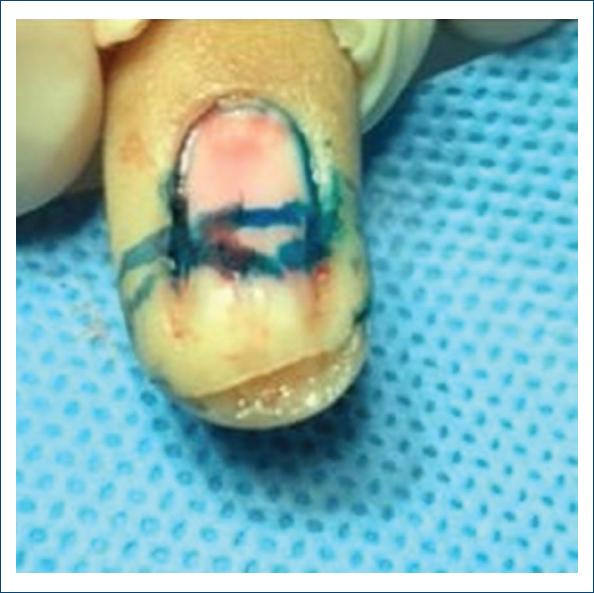
Given the lack of definition of the tumor margins, with the aim of preserving tissue in a noble area and the high degree of clinical infiltration, the Mohs technique was chosen (Fig. 3).
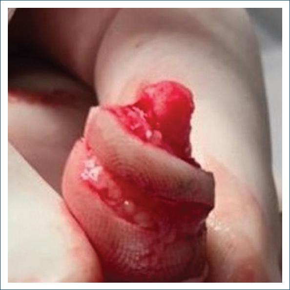
Resection was performed in two phases, with lateral and deep involvement in the first phase without signs of malignant transformation. There was no involvement of the nail matrix, only the cutaneous portion of the distal half of the distal phalanx of the affected finger (Fig. 4). Finally, the reconstruction of the wound was made with a skin graft from the contralateral arm. The 1-year clinical follow-up of the patient was carried out without signs of recurrence after surgery and a good aesthetic result (Fig. 5).
Discussion
The presented case of a GCT brings particular aspects regarding age, gender, and location2, parameters that are not within the typical epidemiological profile of this disease.
There are 14 case reports of GCT in children, aged between 5 and 12 years. Locations included extremities, palms, soles, back, and anterior thorax3. In two cases, the Mohs technique was performed without signs of recurrence, even though in one of them a small margin was revealed.
GCT with nail involvement is very rare and only seven cases have been described in the literature (one in the index finger and six in the hallux). Nail injuries, which are often painful, can occur depending on the structure affected. If proximal, a longitudinal groove may appear on the nail plate. If distal, it generally presents as a warty appearance1. Dupin et al. described a case with distal subungual hyperkeratosis in the hallux that recurred after the first surgical approach, requiring expansion in a second approach. Furthermore, there is a report of MMS performed in a patient with extensive involvement of the labia majora of the vagina, with the aim of sparing tissue4.
Due to the success provided by this technique to this child, it is possible to use this technique for other tumors with a high recurrence rate, malignant potential, or involvement of noble and deep structures. The two main objectives of MMS are resolution of the injury and tissue preservation.
Finally, MMS was chosen in the case reported due to its location, difficult delimitation, and risk of malignancy and recurrence. As it was a child, the preference for the graft was due to its practicality in a less time-consuming surgery. It was believed that there would be a need for some repair afterward, but it was not necessary, and the patient evolved with practically no nail dystrophy, just a slight increase in the convexity of the nail plate, with no impact on his quality of life.
Abrikossoff’s tumor, despite its rarity, should be considered as a differential diagnosis for both the dermatologist and pathologist. Even though it has some specific histological characteristics, IHC is necessary for a diagnostic conclusion. Its treatment must be assertive, as it presents uncertain behavior regarding potential malignancy.

