




 Inglês (pdf)
 Artigo em XML
 Referências do artigo
 Tradução automática
 Enviar este artigo por email
 Citado por SciELO
  Similares em
SciELO
Similares em
SciELO 
 Permalink
Introduction
Stevens-Johnson syndrome (SJS) and toxic epidermal necrolysis (NET) belong to the same disease spectrum of a severe and acute drug reaction, with epidermal necrolysis and detachment, skin blistering, cutaneous and mucosal erosions, and, commonly, systemic involvement1-3. The distinction between SJS and TEN depends on the percentage of detached skin surface (up to 10% in SJS, more than 30% in TEN, and between 10 and 30% in SJS/TN overlap)1,2. Annual incidence varies between 1.2 and 9.2 cases/million people for SJS, and 0.4-1.9/million for TEN4,5, with a mean mortality of 30%1,3.
Epidermal loss may affect response to infections, termo-regulation, and hydroelectrolytic balance. Despite different pathophysiologies and the need for timely suspension of the possible culprit and use of systemic immunomodulatory or immunosuppressant drugs, skin involvement may be viewed as a severe superficial but extensive burn4,5. A cornerstone of treatment lies in the proper care of denuded skin, to improve its protective activity, reduce pain, accelerate re-epithelialization, and, therefore, reduce time to complete healing and decrease the main complications, namely the risk of infection and sepsis, the predominant cause of death. An appropriate choice of the dressing and its correct use are essential, although this aspect is often overlooked1,3.
The main objective of the current systematic review is to assess the most frequently used local skin cleansing measures for SJS/TEN and, especially, wound dressings, the way they were used, and their advantages.
Methods
For this systematic review, searches were performed in Cochrane Library, Embase, MEDLINE, and PubMed databases, with the following search terms: (SJS OR NET) AND (Wounds OR Dressings). Articles published in the last 11 years were selected (from 01 January 2013 to 31 December 2024). Inclusion criteria were: articles on the subject of SJS/TEN and dressings; articles that described dressings used; and articles published in English, Spanish, or Portuguese (Fig. 1). Meta-analysis, systematic review, cohort, and case-control articles were included in the current review. Exclusion criteria adopted were: articles focusing on treatment of SJS/TEN solely on mucosal membranes (as opposed to skin), and articles that did not directly address SJS/TEN.
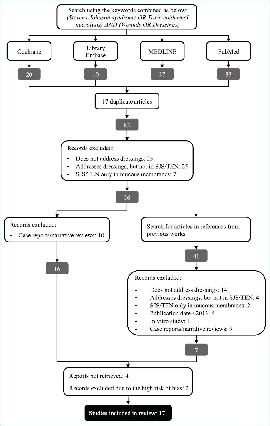
Three independent researchers carried out searches and all articles filtered in the four databases were then initially selected based on their titcles identifying whether they meet the inclusion criteria. No automation tools were used in this review. After the exclusion of duplicate studies, abstracts were critically appraised by three independent reviewers, to choose which articles would be fully read. Reference lists of selected articles were also evaluated, looking for other eligible works. Selected articles were then assessed regarding the risk of bias, using Joanna Briggs Institute (JBI) Critical Appraisal Tools6, by two independent researchers. When these two researchers assigned different gradings for the same article, a third researcher proceeded to evaluate that article, and the two most similar evaluations were considered. A different version of the JBI Critical Appraisal Tool was used according to the category of each study: cross-sectional, case-control, cohort, review, or systematic review. This tool is constituted by 6-11 questions (which vary for each type of study) assessing the risk of bias. For each single article, the obtained score was divided by the maximum possible score to establish its grade (for instance, if six out of eight questions were answered positively for a study, its final score was 0.75). The study was then classified as high (score < 0.50), moderate (score 0.51-0.70), or low (score > 0.71) risk of bias. Articles deemed as with a high risk of bias were then excluded from this review.
Three independent authors extracted general data for each study (title, publication year, study type, name of first authors, name of journal), as well as data on study participants (mean age of patients, gender, comorbidities, nutritional status, mortality, affected body surface area, systemic treatments used, and pain assessment) and on dressings (what was used for cleansing, debridement, topical therapy/dressings and their fixation; frequency of dressing changes and time until re-epithelialization). When some of the data was not identified, it was considered “not applicable” or “not available.” All data were collected in a descriptive way, given the high frequency of incomplete information.
Extracted data were aggregated into different topics, in the structure of a narrative synthesis, focusing mainly on dressings. Results obtained from studies were also aggregated and shown as tables.
The current review has passed through PROS PERO system7 and is registered under number CRD42023483491. Moreover, it followed PRISMA guidelines8 for its construction.
Results
Upon preparation of this systematic review, 100 articles were initially identified, out of which 17 were duplicates, and 67 did not meet the inclusion criteria. The remaining 16 articles had their reference lists searched through to find other eligible studies (41 additional articles). However, 34 out of the additional articles did not meet the inclusion criteria, and only the remaining seven were added to the 16 originals, totaling 23 articles. Out of these, four were excluded because they were not available in full, even after trying to contact authors and editors through email. Two further manuscripts were excluded because of assigned grades lower than 0.50 in JBI Critical Appraisal Tools. Ultimately, there were 17 remaining articles to be included in this systematic review (Fig. 2).
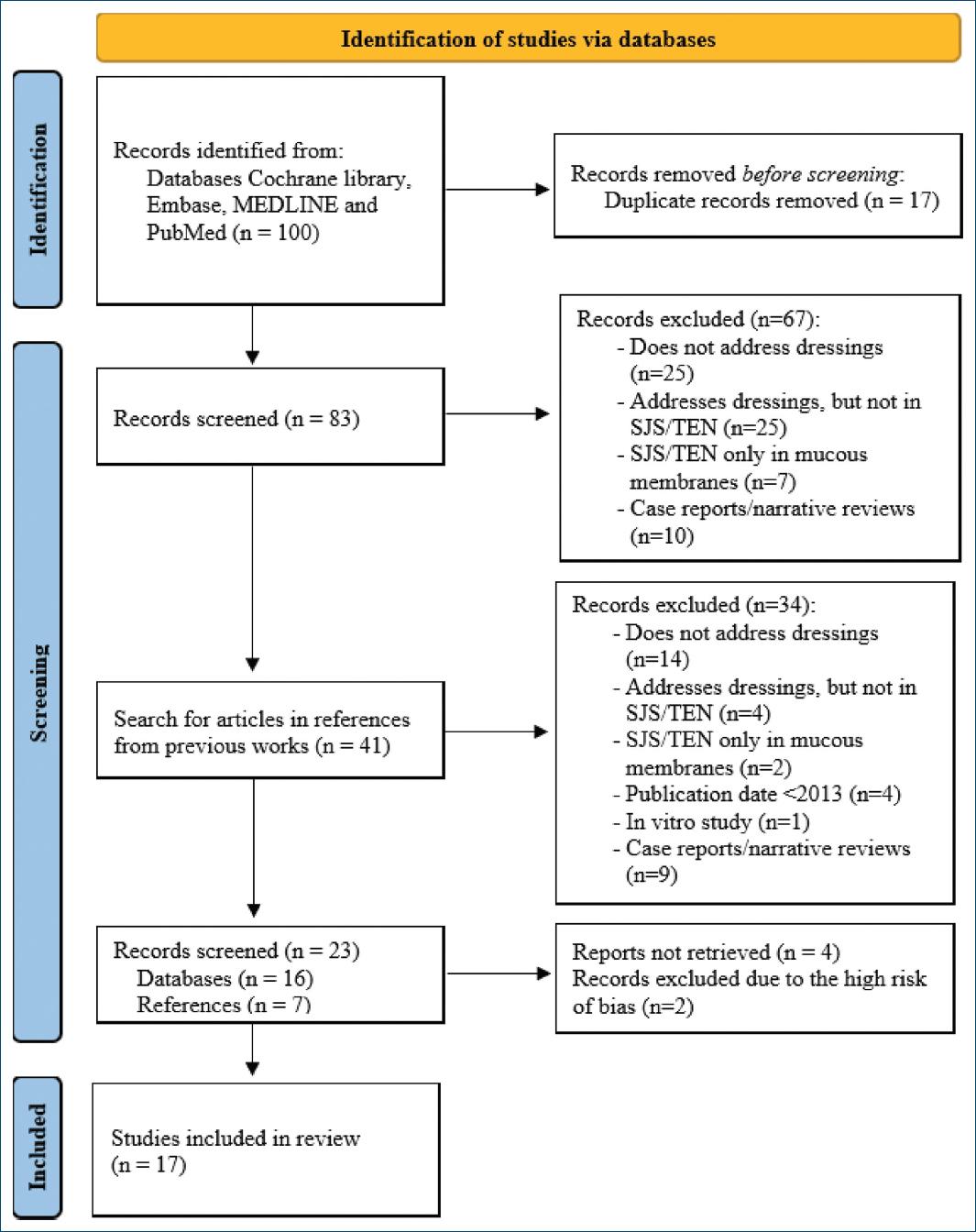
The 17 articles included five case-control studies, three systematic reviews, six expert consensus, two literature reviews, and one meta-analysis. General data for each study, including demographic/epidemiological data and systemic treatments used or recommended are displayed in table 1 and henceforth described.
Table 1 General data of studies included in a systematic review on dressings in SJS/TEN
| Reference | Study design | Mean age (years) | Participants, by gender | Comorbidities (n) | Nutritional status | Mean BSA (%) | Systemic treatment (% of individuals/studies) | Mortality (%) |
|---|---|---|---|---|---|---|---|---|
| Schwartz et al.17 | Expert consensus | NA | NA | NA | NA | NA | No level of evidence for any systemic treatment | NA |
| Mahar et al.9 | Systematic review | 46.8 | 291F/218M | Nd | Nd | All > 30% | IVIG (40%); SCS (30%); fluid replacement with albumin (35%) | 30 |
| Huang et al.10 | Case-control | 59.5 | 10F/10M | Nd | Nd | 55.2% (30-85%) | Nd | 5 |
| Dodiuk-Gad et al.18 | Multicentric – Expert consensus | NA | NA | NA | NA | NA | Most used IVIG, SCS, ciclosporin, anti-TNF. | NA |
| Young et al.11 | Case-control | 52 | 19 F / 5 M | SAH (18), smoking (7), hyperlipidemia (6), CAD (3), CHF (4), DM (6), seizures (4) | Nd | 63% | IVIG (79.17%), SCS (29.17%) | 12.5 |
| Creamer et al.16 | Guidelines | NA | NA | NA | Daily intake of 20-25 kcal/kg (early catabolic phase); 25-30 kcal/kg (recovery anabolic phase) | NA | Mainly supportive care + causative drug withdrawal | NA |
| Curtis et al.24 | Expert consensus | NA | NA | NA | NA | NA | IVIG, ciclosporin | NA |
| Wolkenstein and Wilson19 | Expert consensus | NA | NA | NA | NA | NA | No consensus. Most used IVIG, ciclosporin | NA |
| Cartotto20 | Literature review | NA | NA | NA | NA | NA | No consensus. Most evidence support IVIG, ciclosporin | NA |
| Rogers et al.12 | Case-control | 59.5 | 26F/16 M | Nd | Nd | 30% (10%-40%) | IVIG, ciclosporin, etanercept | Nd |
| Paggiaro et al.13 | Systematic review | 37.3 | 14 F/6 M | Nd | Nd | 73.9% (45%-90%) | IVIG (75%) | 10 |
| Castillo et al.4 | Literature review | NA | NA | NA | NA | NA | SCS, IVIG | 10.8 |
| Richard et al.14 | Multicentric – Expert consensus | NA | NA | NA | NA | NA | IVIG (41.9%), SCS (9.7%), ciclosporin (6.5%), plasmapheresis (3.2%) | NA |
| Jaller et al.23 | Systematic review | NA | NA | NA | NA | NA | Nd | NA |
| Lee et al.15 | Meta analysis | 51.8 | 100 F/127 M | NA | NA | NA | IVIG, ciclosporin | 29.1 |
| Dastagir et al.21 | Case-control | 66.3 | NA | Nd | Nd | NA | Nd | 19 |
n: number of individuals; NA: not applicable; Nd: not described; BSA: body surface area; F: female; M: male; IVIG: intravenous immunoglobulin; SCS: systemic corticosteroids; TNF: tumor necrosis factor; G-CSF: granulocyte colony-stimulating factor; CRP: C-reactive protein; DM: diabetes mellitus; SAH: systemic arterial hypertension; CAD: coronary arterial disease; CHF: congestive heart failure; SLE: systemic lupus erythematosus; CKD: chronic kidney disease; AF: atrial fibrillation atrial; MM: multiple myeloma; HCV: hepatitis C virus; COPD: chronic obstructive pulmonary disease.
General data
Out of the 17 articles included in this review, seven described the age and sex of participants (54.6% women and a mean age of 53.03 years)9-13. Only six articles reported the average percentage of body surface affected (BSA) in the participants, with an overall average of 52.51% BSA across these articles.
Most often mentioned comorbidities in studies were systemic arterial hypertension, epilepsy, and diabetes mellitus corroborating anticonvulsant drugs as common culprits in the pathogenesis of SJS/TEN11.
In agreement with the lack of consensus on the best systemic treatment for SJS/TEN the majority of patients in these studies received either IVIG4,11-15 or systemic corticosteroids9,11,14, followed by ciclosporin4,12,14,15. Nutritional status, which is of utmost importance in this disease, was not commonly addressed. Nutritional therapy after hospital admission with oral or enteral feeding was frequently used, with one study reporting a daily caloric goal of 20-25 kcal/kg during the early catabolic phase and 25-30 kcal/kg during the recovery anabolic phase16.
The next topics encompass each of the steps involved in wound management and dressing: cleansing, debridement, topical therapy, dressings, and their fixation, frequency of changes, time for re-epithelialization, pain assessment, and mortality. Data regarding employed or recommended dressings are displayed in table 2.
Table 2 Specific data on most used dressings for SJS/TEN in included studies
| Reference | Study design | Materials/techniques used for dressings | Frequency of dressing changes | Time until re-epitheliazation | Pain scale | |||
|---|---|---|---|---|---|---|---|---|
| Cleansing | Debridement | Topical therapy (% of use/citations) | Fixation | |||||
| Schwartz et al.17 | Expert consensus | Nd | Nd | Paraffin gauzes, porcine xenografts, human allografts, biosynthetic skin substitutes with porcine collagen | Nd | Nd | Nd | Nd |
| Mahar et al.9 | Systematic review | Chlorhexidine (1) | Routine active debridement (9/20 studies) | Aseptic dressings (5%), silver nitrate-impregnated (10%), synthetic skin substitutes (20%), porcine xenograft (10%), absorbent gauze (5%), cadaveric/porcine graft (5%), emulsifying ointment/ aqueous cream (5%), and silver sulfadiazine (10%) | Nd | Cadaveric or porcine graft changed daily (1) | Nd | Nd |
| Huang et al.10 | Case–control | Nd | Nd | Silver-containing hydrofiber and petrolatum gauze versus silver sulfadiazine | Nd | Case versus control twice weekly versus daily | Case versus control group: 16.75 versus 17.50 days (not significant) | Hydrofiber group; lower pain scores |
| Dodiuk-Gad et al.18 | Multicentre – Expert consensus | Nd | Nd | Silver-impregnated dressings, synthetic dressings, topical antimicrobials, bioactive skin substitutes | Nd | Nd | Nd | Nd |
| Young et al.11 | Case–control | Nd | Hydrotherapy + sedation | Porcine xenograft versus silver-impregnated dressing | Nd | Nd | Nd | Pain score 2.8 versus 6 |
| Creamer et al.16 | Guidelines | Warmed sterile water, saline, or chlorhexidine | if infected or necrotic areas hydrosurgery or antimicrobials (iodopovidone or chlorhexidine) | Greasy emollient (50:50 white soft paraffin/liquid paraffin) + topical antimicrobial + non-adherent dressing. Detached areas: non-adherent dressings + secondary foam or synthetic membranes or allograft/xenograft | Nd | Nd | Nd | Nd |
| Curtis et al.24 | Expert consensus | Nd | Most no debridement | Petrolatum gauze; silver-containing non-adherent dressings | Nd | Nd | Nd | Nd |
| Wolkenstein andWilson19 | Expert consensus | Nd | Nd | Detached skin as biologic dressing. Caution with silver-impregnated dressings in large areas | Nd | Nd | Nd | Nd |
| Cartotto20 | Literature review | Nd | Routine removal of remaining epidermis | Porcine xenograft or cadaveric allograft or biosynthetic skin substitute + antimicrobial secondary dressing | Fixation staples | Nd | 12-14 days | Nd |
| Rogers et al.12 | Case–control | Soap and chlorhexidine upon admission | Mechanic debridement upon admission | Biosynthetic membrane with porcine collagen +, followed by antimicrobial mesh with nanocrystalline silver versus petrolatum ointment or silver-based dressing or greasy tulle gauze | Nd | silver-containing screen 48-72 h. Control group: Nd. | 13 versus 12 days | Nd |
| Paggiaro et al.13 | Systematic review | Nd | Nd | Xenograft / cadaveric allograft / amniotic membrane | Nd | Nd | Nd | Nd |
| Castillo et al.4 | Narrative review | Nd | Nd | Biosynthetic dressings versus silver-impregnated fibers | Nd | weekly on average | 14.16 ± 9.42 days | Biosynthetic dressings improved comfort (5/22 studies) |
| Richard et al.14 | Multicentre Expert consensus | Diluted chlorhexidine (51.6%), water (22.6%), saline (12.9%), other (12.9%) | Nd | Topical antibiotics (58.1%), whirlpool baths (12.9%), topical corticosteroids (6.5%), silver foam (48.4%), biologic skin substitutes (45.2%), nanocrystalline silver mesh (32.3%), petrolatum gauze (29%), silver sulfadiazine (12.9%), and non-adherent gauze with PHMB (6.5%) | Nd | Daily (41.9%), every 3 days (22.6%), 2 days (6.5%), weekly (6,5%) | Nd | Nd |
| Jaller et al.23 | Systematic review | Warm sterile water, saline, or diluted chlorhexidine (1:5000) | No consensus on surgical debridement | High-potency topical corticosteroids in non-detached skin. Topical antimicrobials or silver-impregnated dressings on denuded areas, biosynthetic skin substitutes, cryopreserved cadaveric allografts, or porcine xenografts | Nd | Depending on dressing from daily to every 7-14 days | 12.5 days (biosynthetic skin with collagen versus 16 days for debridement | Nd |
| Lee et al.15 | Meta analysis | Nd | Nd | Nd | Nd | Nd | 17 (debridement) versus 14 days (dressing) | Nd |
| Dastagir et al.21 | Case–control | Nd | Surgical | Nd | Nd | Nd | Nd | Nd |
| Enescu et al.22 | Case–control | Nd | Hydrotherapeutic blister debridement (maximum of 10% BSA) | Suprathel® versus polyhexanide gel | Nd | No exchange versus daily | Nd | Nd |
Nd: not described; PHMB: polyhexamethylene biguanide; BSA: body surface area.
Cleansing
Cleansing consists of the removal of debris, foreign bodies, and sources of infection through applying solutions such as water, saline, and antibacterial solutions, among others2. Out of the 17 articles included in this review, 12 (70.6%) did not mention wound cleansing4,10,11,13,15,17,18-22. Among the others, all five reported or recommended cleansing with chlorhexidine9,12,14,16,23, whereas two also recommended distilled water14,16, and three saline solution14,16,23.
Debridement
Debridement, comprising strategies intended for the removal of devitalized necrotic tissues that could impair healing and cause secondary infections, is not a consensual therapy, as shown in a systematic review concluded in 202023. Out of the 17 selected articles for the current review, 10 (58.8%) did not detail if debridement was performed4,10,13,14,16-19,21,22. In total, 6 (35.3%) studies opted for mechanical or surgical debridement, mostly in an operating room or under sedation upon arrival in the reference center, manually or with surgical gauzes and brushes12,20,22. Other types of debridement have been cited such as in hydrotherapy rooms (with or without sedation) or hydrosurgery16. An expert consensus published in 2016 in the Journal of the American Academy of Dermatology24, found that 67.7% of experts did not perform debridement, and another systematic review concerning burn centers published in 20149, found that only 13 out of 20 selected studies mentioned debridement, with 9 (69.3%) in favor of routine active debridement and 4 (30.7%) indicating not do debride detached epidermis. A 2024 meta-analysis evaluating surgical debridement compared to the use of a bandage alone found no difference in mortality, but there was a significantly shorter re-epithelialization time in the group without debridement15.
Topical therapies
Topical therapy represents the dressing itself or topical pharmacologic agents applied directly on the wound surface. Dressings described for SJS/TEN varied from petrolatum-impregnated gauzes to silver-containing dressings, silicone foams, collagen dressings (not specifying the subtype of collagen), porcine xenografts, among others23. Most studies report silver-based dressings (mostly silver-impregnated meshes), followed by biosynthetic membranes, and by xenografts or allografts.
Topical therapies were variable among studies, with no consensus on which is the best. In a systematic review made in 202023, high-potency topical corticosteroids were recommended in areas of erythematous skin not yet detached, and topical antimicrobials (not designating which one) or silver-impregnated dressings (also not designating which one) in areas of detached skin. Other mentioned dressings were biosynthetic skin substitutes with porcine collagen and cryopreserved cadaveric allografts or porcine xenografts for areas with epidermal detachment. For detached areas, British guidelines16 recommend a first layer of dressing with a non-adhering material, such as PHMB-impregnated non-adhering gauze or silicone mesh, and a foam or other absorbing dressing as a second layer. Another systematic review, published in 20149, indicates silver nitrate-impregnated dressings as the most used, followed by synthetic skin substitutes, porcine xenograft, absorbing gauze, cadaveric allograft, emollient ointments and aqueous creams (not specified).
In 6 (35.3%) selected studies, silver-based topical therapies are mentioned as the treatment of choice, varying from silver ointments/creams to silver-impregnated foams, gauzes, or meshes4,10,11,16,18,24. In 5 (29.4%) there are no further details on the chosen silver-based dressing11,16,18,24, and in 2 (11.8%) studies silver-impregnated hydrofiber foam was chosen4,10. A 2016 expert consensus19 advised caution when using silver-impregnated dressings over large areas, due to the risk of increased silver absorption on detached epidermis with systemic complications in debilitated patients.
Biosynthetic dressings with a combination of collagen mesh and silicone were frequently cited in 4 (23.5%) recent studies, as they require no changes, reduce pain, and increase comfort, allowing a similar, or even shorter, re-epithelialization time when compared to traditional topical therapies12,16,17,20.
Six (35.3%) articles mentioned the use of allografts and porcine xenografts, which are used as skin substitutes on non-infected detached epidermis, and generally do not need frequent changes11,16,17,20. A further option is the use of the patient’s own detached epidermis as a biological dressing, a possibility mentioned by 2 (11.8%) articles: the UK guidelines and an expert consensus16,19.
Petrolatum-based products are still often employed, as mentioned in 4 (23.5%) studies, but more commonly as emollients in areas where the skin is not yet detached16,17,20,24. Nevertheless, petrolatum is being progressively replaced with previously mentioned therapies. The application of petrolatum-impregnated gauzes (as a means to keep the wound moist, facilitate dressing change, and reduce pain) has not been shown to be statistically significantly beneficial over other topical therapies10.
Fixation
Dressing fixation in SJS/TEN is an important step and should ideally be non-adhering and not cause damage to the skin23, as the skin is fragile, and inadequate fixation may worsen the area of epidermal detachment and pain. The primary dressing, i.e., the one that is in direct contact with the wound, may or may not need to be covered by a secondary dressing. Despite its importance, out of the 17 articles in this review, 14 (82.3%) did not mention fixation at all4,9,10,12-24, or described the use of gauzes for fixation11,21.
Dressing change frequency
The frequency of wound dress changing was variable for each dressing. Biosynthetic skin substitutes, allografts, and xenografts usually do not require changing12,20. Some studies9,21 performed daily changes, notably if there was facial epidermal detachment. However, patient discomfort is reportedly greater, when cleansing and dressing changes are more frequent. Silver-impregnated dressings were left in place for a longer interval, with changes varying from every 2 to 7 days4,10. In a 2018 expert consensus14, the ideal change interval was also diverse among experts, with changes daily, every 2 or 3 days, or weekly, respectively, in 41.9%, 6.5%, 22.6%, and 6.5% weekly, and 22.6% in alternative periods.
Re-epithelialization
Time until total re-epithelialization was not mentioned in 11 (64.7%) of the selected studies9,13,16-19,21,22,24. On average, the time until re-epithelialization was lower than 2 weeks, regardless of the topical therapy used4,12,20. A 2020 systematic review23 reported re-epithelialization time to be shorter when using a biosynthetic skin substitute with porcine collagen, with an average of 12.5 days, compared to 16 days when using paraffin gauzes with daily changes.
Mortality
Few articles associate the choice of topical therapy for SJS/TEN with mortality rates. Among selected studies, 11 (64.7%) did not mention mortality12,14,16-20,22-24. In the remainder, mortality was < 30% of cases, with no reduced mortality reported for any specific dressing4,9,10,11,13,15,21.
Pain assessment
Thirteen (76.5%) studies did not mention whether a pain scale was used9,12-14,16-20,23,24, and few studies related it to the wound dressing choice. A pain Visual Analog Scale was used to compare groups with distinct silver-based therapies in a 2014 case–control study10. The group using silver hydrofiber scored significantly (p = 0.02) better on this scale (5.75/10, standard-deviation 1.39) than the group using silver sulfadiazine (7.42/10, standard-deviation 1.31), probably as a result of decreased need of manipulation and dressing changes in the silver hydrofiber group. A 2016 case–control study11 compared patients with porcine xenografts to patients with silver-impregnated dressings and found pain to be considerably lower for the first group (2.8 vs. 6/10 points). Another systematic review published in 20184 found, in five out of 22 articles that cite pain assessment, that biosynthetic dressings result in better comfort, although without altering healing time.
Discussion
The percentage of epidermal detachment in SJS/TEN has a direct relationship with mortality and worse prognosis, therefore a fundamental part of care in SJS/TEN includes local wound management and use of proper dressings23. An ideal dressing should be comfortable for the patient and protect denuded skin from secondary infections, hypothermia, or fluid loss, reduce pain, avoid expansion of detachment, and promote re-epithelization1. Dressing change may be performed in an operative setting, under general anesthesia, considering the extreme pain1. It is recommended that care of SJS/TEN patients be done, whenever possible, in specialized centers or burn units, but there are no specific guidelines for dressing application and there is a great deal of variability in topical therapies used in all steps of wound care in these conditions14.
The current systematic review highlights that there is still no established consensus on wound management in patients with SJS/TEN, especially when regarding the choice of proper dressing. A range of different dressings have been used, such as silver-based dressings, biosynthetic membranes, xenografts, allografts, and the patient’s own detached skin (which may be kept in place and used as a biologic dressing). High variability in care reflects the lack of standardized guidelines and differentiation of treatment according to each center and the patient’s individual characteristics.
Silver-impregnated dressings are commonly employed due to antimicrobial properties, but attention should be given to risks of systemic absorption and adverse effects, notably if used in extensive areas19. Biosynthetic dressings and xenografts have shown advantages regarding patient comfort, pain reduction, and need for less frequent dressing changes, and may favor re-epithelization and decrease infection risk, but scientific evidence is still scarce and unable to infer superiority for any specific class of dressing11-13,16,17,20.
This review highlights priorities for future research in this area. There appears to be little information regarding the importance of topical treatments in SJS/TEN. Consequently, choosing the best dressing for these diseases remains a challenge. There are several options in medical literature for each step of wound management but it is not clear what initial measures should be taken by doctors and healthcare teams when admitting patients with SJS/TEN, or what subsequent measures and dressings should be performed. There was likewise no conclusion on the best way of wound cleansing and/or fixation, and whether debridement should or not be performed.
In view of this, there is a clear need for clinical trials to directly compare different available topical therapies to establish protocols that optimize clinical outcomes, decrease mortality, and improve the quality of life in patients with SJS/TEN. Until then, an individualized and multidisciplinary approach remains the most suitable means of managing these severe and potentially lethal diseases.
Our study has some strengths – it provides an updated review of an important subject for which there is a scarcity of data published in medical literature. This study, however, also has several limitations. An important one is the overall small number of studies included. Furthermore, there is a lack of studies with a higher level of evidence–for instance, there was no randomized clinical trial available that would fulfill the inclusion criteria. Another limitation is the heterogeneity of articles–different designs such as case-control studies and guidelines, for instance, do not allow direct comparison among selected studies.
Conclusion
Regarding topical therapies in the management of SJS/TEN patients, the most used dressings were silver-containing ones, followed by synthetic membranes and allografts or xenografts. However, despite miscellaneous therapeutic options, there is no consensus on an ideal protocol for SJS/TEN patients. More powerful study designs are needed to methodologically assess which are the best topical therapies in practice. In this way, considering that wound management is essential for proper healing and reduction of morbidity and mortality in SJS/TEN, further research on this subject is highly needed.

