




 Inglês (pdf)
 Artigo em XML
 Referências do artigo
 Tradução automática
 Enviar este artigo por email
 Citado por SciELO
  Similares em
SciELO
Similares em
SciELO 
 Permalink
Introduction
Takayasu’s arteritis (TA) is a vasculitis characterized by granulomatous inflammation, fibrosis and thickening of the wall of large and medium-sized vessels, such as the aorta and its branches1. The prevalence of this disease is higher in young women, especially in Asia and the Middle East2. Rarely, the microcirculation may also be involved, with ocular (retinopathy, uveitis, scleritis), cardiac (myocarditis) and cutaneous manifestations2. The prevalence of the latter is estimated to be around 2.8% to 28% of TA patients3–7. From a clinical and histological point of view, cutaneous manifestations can be divided into specific for TA–acute inflammatory nodules, erythema nodosum-like subcutaneous nodules, erythema induratum, pyoderma gangrenosum- like ulcers, livedo reticularis, purpuric and necrotic lesions–and nonspecific–urticaria-like lesions, erythematous macules and papules, eczematiform lesions, among others3,8–11. More than one type of lesion may be present in the same patient11. This report represents a case of small cutaneous vessel vasculitis manifesting as erythema nodosum-like subcutaneous nodules in a patient with active TA disease activity.
Case report
We report the case of a 29-year-old woman, with no previously known comorbidities. She had been diagnosed with Takayasu’s arteritis 4 years prior to her Dermatology evaluation. The diagnosis was made when she developed pericarditis, differential blood pressure between the upper limbs higher than 10 mmHg, and an elevation of inflammation parameters. The diagnosis was confirmed with an angiography CT scan, which showed reduction of the caliber of the supra-aortic branches of the thoracic and the abdominal aorta.
Several immunosuppressive drugs were administered in order to control disease activity, with favorable response to an induction dose of prednisolone (1 mg/kg/day). Corticosteroid tapering and switching to corticoid-sparing drugs was attempted, but the patient was intolerant to some drugs, namely adalimumab, azathioprine and methotrexate and others failed to reduce the inflammatory activity, namely tocilizumab and infliximab. Oral prednisolone was used as maintenance therapy, however clinical criteria of active disease (Indian Takayasu Clinical Score 5) and analytical criteria (sedimentation velocity of 64 mm/h) were sustained.
The patient was referred to a Dermatology appointment due to a skin condition characterized by centimetric, erythematous, painful (particularly in the morning) and non ulcerated nodules on the soles and inner edges of both feet (Fig. 1). Lesions had subsided partially after increasing the dose of prednisolone, but had not resolved completely. Laboratory tests with blood and urine samples, specifically regarding auto-immune vasculitis and cryoglobulinemia, were negative.
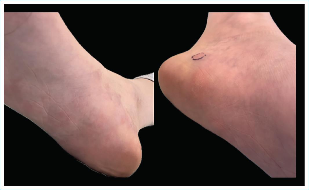
Figure 1 Erythematous nodules on the sole and inner edges of both feet. The place where the skin biopsy was performed is highlighted with a blue circle.
Therefore, a skin biopsy of one of the nodules of the inner edge of the left foot was performed. Dermatopathology findings were compatible with leukocytoclastic vasculitis and adjacent granulomatous panniculitis (Fig. 2). It was concluded that the small vessel inflammation occurred in the context of TA, manifesting as erythema nodosum-like subcutaneous painful nodules. For the small vessel vasculitis, the patient was started on colchicine 1 mg per day and maintained a corticotherapy regimen of 15 mg of prednisolone per day with improvement of the cutaneous lesions. Even though systemic inflammation is not controlled with this dose of systemic corticosteroids, the patient refuses other biologic treatments at the moment.
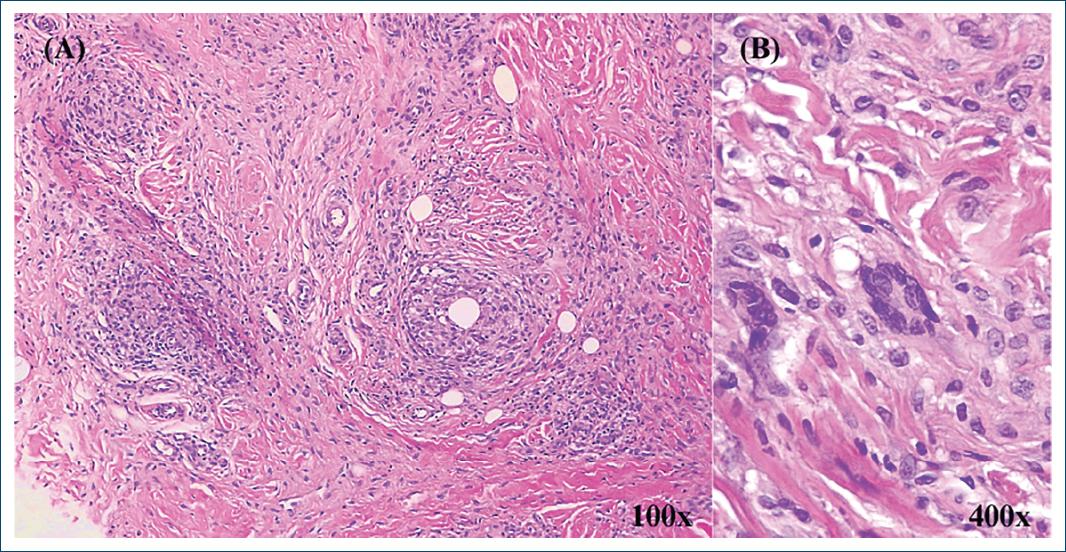
Figure 2 Histopathologic findings (H&E stain). A: scattered perivascular neutrophils in the deep dermis and the subcutaneous fat, as well as a lymphohistiocytic infiltrate along the lobules and septae of fat tissue, with multinucleated giant cells. B: higher magnification showing multinucleated giant cells in the infiltrate.
Discussion
The first skin manifestations in TA were described in 1985 by Mousa et al.12, who associated the nodular skin lesions with the systemic vasculitis in a patient with TA. Overall, dermatologic manifestations in patients with TA may result from the occlusion of large vessels –presenting as Raynaud’s phenomenon and digital gangrene–or from the inflammation of small cutaneous vessels3,9. The latter may manifest as erythema nodosum, ulcerated nodules, pyoderma gangrenosum-like lesions, among others3,8–11. In North America and Europe, acute inflammatory nodules and erythema nodosum are the most prevalent skin manifestations3, while pyoderma gangrenosum has been described more frequently in the TA Japanese population and predominantly affects the upper arms9. Less frequently, findings compatible with cutaneous lupus erythematosus with no systemic evidence of lupus have been reported3.
Histologically, granulomatous or necrotizing vasculitis of small and medium vessels in the dermis and hypodermis, accompanied or not by septal and/or lobular panniculitis have been previously described3,5,7,9,11. Granulomatous vasculitis can be found in various types of vessels in these patients, from large to small sized arteries, capillaries and venules3. Panniculitis is, in some cases, the only finding on the skin biopsy; in others, like ours, it is interpreted as a continuum of the inflammatory changes of the vasculitis found in the deep dermis into the hypodermis, particularly in erythema nodosum and subcutaneous ulcerated nodules3.
Establishing a relation between cutaneous findings and TA may be doubtful. Francès et al.3 suggested three arguments to sustain this association, based on the findings described in the literature, the presence of granulomatous necrotizing vasculitis of small cutaneous vessels (similar to findings on the walls of large vessels), and the chronological association between skin findings and disease activity3. The association is established when other etiologies have been excluded, usually by performing tests that screen for other granulomatous diseases (chest radiography, tuberculin test, anti-streptococcal antibodies, yersinia titers)3,9.
Disease progression of TA can be divided into an initial inflammatory systemic phase and a subsequent occlusive phase with manifestations caused by the stenosis of large and medium sized arteries13. This evolution may not be linear, with overlap and/or inversion of the natural history of TA13. The appearance of cutaneous manifestations, including painful erythema nodosum-like nodules, may accompany an initial period of constitutional systemic symptoms, preceding the vessel inflammation phase of the vasculitis (sometimes several years), or manifest simultaneously with a worsening of the large vessel occlusion3,5,11. In Francès C, et al’s3 cohort, skin lesions were present in all disease phases, however acute and subacute nodules were more prevalent in early stages of the vasculitis, while pyoderma gangrenosum, purpuric and necrotic lesions accompanied the occlusive stage. Moreover, cutaneous lesions in TA do not appear to correlate with disease severity or location of the large vessel occlusions5.
The painful, acute, subcutaneous nodules found in our patient are concordant with the erythema nodosum-like manifestations described in literature. Histologically, these lesions may initially be indistinguishable from classic erythema nodosum; however, later in disease progression, small cutaneous vessel vasculitis with or without panniculitis may develop and contribute to the differential diagnosis8.
The inflammatory process of small and large vessels may correlate due to anatomical similarities between skin vessels and the vasa vasorum on the outer layers of the walls of large vessels7,9. Also, evidence suggests that both forms of inflammation respond favorably to systemic corticotherapy and other immunosuppressants, along with systemic inflammation2,5,7,8,10,11. Therefore, cutaneous manifestations of small vessel inflammation are considered markers of disease activity3,9,11. In our patient, vasculitis was not under control with the current therapy so this skin condition corroborates the existence of inflammatory activity and may be useful as a marker of response and/or flare under future therapeutic options, due to the simultaneous involvement of small, medium and large vessels.
We conclude that an early correlation between cutaneous findings and constitutional and cardiovascular symptoms of TA may lead to earlier diagnosis and a more prompt control of inflammation.

