




 Inglês (pdf)
 Artigo em XML
 Referências do artigo
 Tradução automática
 Enviar este artigo por email
 Citado por SciELO
  Similares em
SciELO
Similares em
SciELO 
 Permalink
Introduction
Immunoglobulin G4-related disease (IgG4-RD) is a rare systemic inflammatory condition characterized by tissue infiltration by IgG4-expressing plasma cells and progressive fibrosis, firstly identified in the 21st century. It occurs more frequently in middle-aged to elderly males and can synchronous or metachronously involve multiple organs1,2. Although the pathophysiology of IgG4-RD is not fully uncovered, studies suggest a predominance of the T helper (Th) type 2 immune response, with interleukin (IL)-4 playing a central role in the production of IgG43. The disease clinical heterogeneity makes its diagnosis frequently challeging, with dermatologic manifestations appearing in a minority of cases1. Systemic corticosteroids are the first-line treatment; however, refractoriness and relapse on discontinuation are common4,5. Herein, we report a case illustrating the efficacy of dupilumab, a monoclonal antibody blocking IL-4 and IL-13 receptors, in treating cutaneous IgG4-RD, underscoring its potential role for patients who are resistant to corticosteroids.
Case report
A 59-year-old man was referred to our dermatology department due to a disseminated and extremely pruritic dermatosis with 7 years of evolution. In addition, he referred asthenia and frequent episodes of diarrhea. Fecal occult blood test was negative and thoracic, abdominal and pelvic computed tomoghrapy (CT) showed pulmonary emphysema, nodular thyroid, and prostatic calcifications. Systemic steroids provided relief, but the disease quickly relapsed on its tapering. Patient medical history was remarkable for allergic rhinitis, uncontrolled non-insulin dependent diabetes mellitus, dyslipidemia, and an episode of unilateral proptosis and diplopia 4 years before. At that time, as radiological examination demonstrated thickening of the left extraocular muscles, a diagnosis of inflammatory myositis was assumed, and the patient was treated with oral corticosteroids, resulting in clinical and imaging regression.
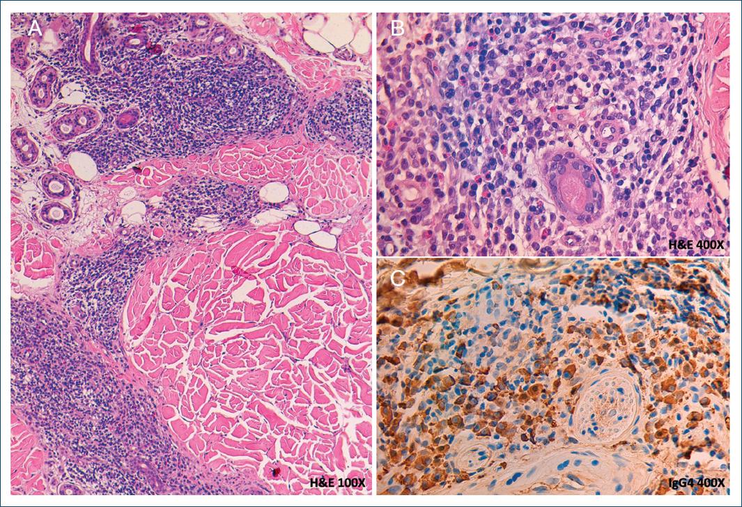
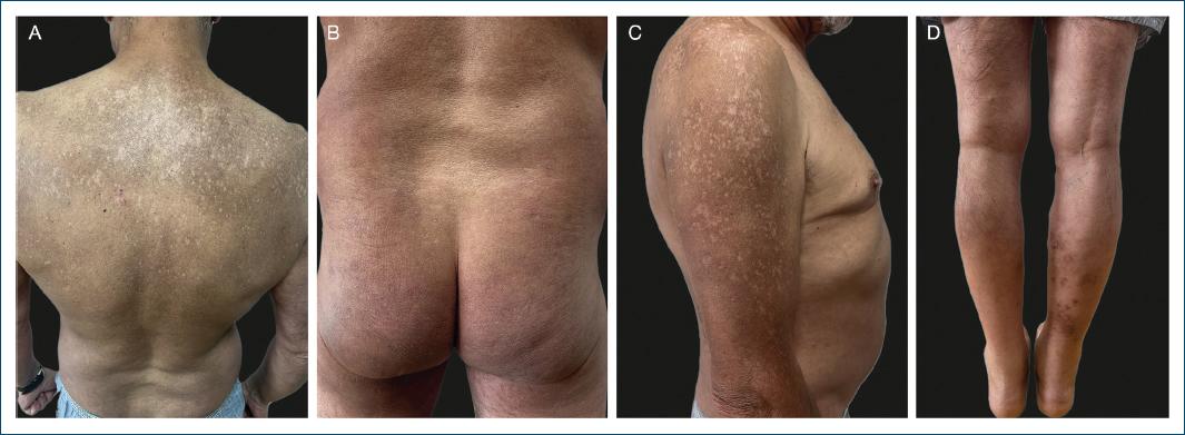
Dermatological examination showed a symmetric and extensive eruption affecting the face, trunk, buttocks, and upper and lower limbs, characterized by erythematous-brownish papules and papulonodules, some with crusts, amidst with excoriations and hyper- and hypopigmented macules and patches (Fig. 1). Bilateral mobile and painless cervical lymphadenopathies were palpable. Cutaneous lesions caused a debilitating pruritus (itch numeric rating scale 8/10) that markedly interfered with patient’s quality of life (dermatology life quality index [DLQI] 11). The diagnoses of eczema and nodular prurigo were considered, and a skin biopsy was performed, revealing a dense perivascular and perifollicular lymphoplasmacytic infiltrate, rich in IgG4-positive cells (IgG4+/IgG+ > 40%) and eosinophils (Fig. 2). Laboratory tests showed IgG4 levels of 1570 mg/dL and immunoglobulin E (IgE) of 4563 KUI/L. The diagnosis of IgG4-RD was hence evoked, and complementary study revealed overlapping findings on body CT and colloid goiter on thyroid aspiration cytology. Treatment with dupilumab (300 mg sc every 2 weeks with a loading dose of 600 mg) was initiated as monotherapy. After 6 months, sparse cutaneous lesions were observerd and lower serum IgG4 and IgE levels (1450 mg/dL and 601 KUI/L, respectively) were noted. After 1 year of therapy, dermatosis resolution (Fig. 3), and a significant improvement of asthenia, gastrointestinal symptoms and quality of life (DLQI 0) were observed, accompanied by a noticeable decrease in serum IgG4 and IgE levels (724 mg/dL and 228 KUI/L, respectively).
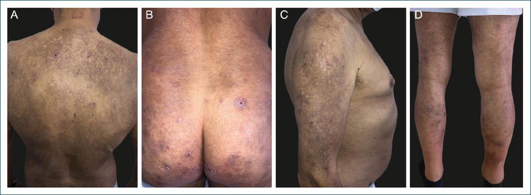
Figure 1 Baseline clinical presentation, with erythematous-brownish papules and papulonodules, excoriations and hyper- and hypopigmented macules and patches, mainly located on the A: trunk, B: buttocks and C and D: upper and lower limbs.
Discussion
IgG4-RD is an immune mediated condition characterized by tumefactive lesions and progressive fibrosing of affected tissues1,6. Its true incidence is unknown and potentially underestimated, due to lack of awareness. Although the immunopathogenesis of IgG4-RD is not entirely understood, a Th2 immune reaction is proeminent, with IL-4, IL-5, and IL-10 playing a role in stimulating the expression of IgG4 and IgE and eosinophilia. IL-4 is the key cytokine, inducing IgG4 class-switch mediated by Th follicular cells. IL-13 promotes mitochondrial dysfunction, cellular senescence, and fibrosis, but its role in the fibrosis of IgG4-RD is still under debate2,7. Up to 31% of patients with IgG4-RD have an atopic background, suffering from allergic rhinitis, atopic dermatitis, or asthma1,3.
IgG4-DR can virtually affect any organ and usually follows a subacute course with relatively mild symptoms8. Extracutaneous presentations include tumor-like enlargement, inflammation, and fibrosis of tissue such as lacrimal and salivary glands, orbits, gallbladder, pancreas, thyroid, lungs, kidneys, and lymph nodes4. Asthenia and weight loss are common unspecific symptoms, while organ-specific manifestations such as diarrhea, respiratory symptoms, or xeropthalmia vary widely. Erythematous and pruritic nodules, papules, and plaques are the most common cutaneous lesions, mainly appearing on the head-and-neck regions, but also on the trunk and extremities. Purpura and prurigo nodularis-like lesions have also been reported9,10. A Japanese study including 80 patients showed that skin lesions were identified in only 6.3% of IgG4-RD cases11. Cutaneous lesions are usually associated with systemic involvement, particularly of the orbit, lacrimal, and salivary glands4. Due to its clinical heterogeneity, the differential diagnosis of IgG4-RD is broad, depending on the clinical presentation and specific sites of involvement9.
Histopathology of affected organs shows dense lymphoplasmacytic infiltrates rich in IgG4+ plasma cells with frequent eosinophilic infiltration, storiform fibrosis, and obliterative phlebitis, the latter two seldom observed in skin biopsies1,4,11. Most patients present elevated serum IgG4 levels, which tends to correlate with the extent of organ involvement, hypergammaglobulinemia, elevated serum IgE (60%), and mild-to-moderate peripheral eosinophilia (34%)1. Comprehensive diagnostic criteria include clinically characteristic swelling or masses in single or multiple organs, serum IgG4 levels > 135 mg/dL, and histological features, namely, a ratio of IgG4+/IgG+ plasma cells > 40% on biopsy, that nevertheless is not specific as isolated and finding1,4.
Prevention of irreversible fibrosis and organ function impairment is the major treatment goal. Systemic corticosteroids are the first-line therapy; however, relapses on discontinuation occur in up to 53% of cases5.
Dupilumab, an IL-4 and IL-13 inhibitor, is approved for the treatment of type 2 inflammatory diseases such as atopic dermatitis, chronic prurigo, asthma, and chronic rhinosinusitis and nasal polyposis. Limited studies have demonstrated its safety and promising role in the treatment of IgG4-RD, mainly focusing on outcomes of asthma and rhinosinusitis2,7,12,13. Kanda M et al. reported a case series of four patients diagnosed with IgG4-related disease, predominantly affecting the submandibular and lingual glands, paranasal sinuses, and lungs, who were treated with dupilumab2. Similarly to our case, the two patients who underwent monotherapy with dupilumab took 3-6 months to experience a therapeutic effect.
Recent studies have focused on alternative therapies to corticosteroids, driven by a progressively deeper understanding of IgG4-RD pathophysiology. In vitro testing has shown that T cells from patients with IgG4-RD exhibit aberrant activation, associated with reduced expression of the inhibitory molecule CTLA4. However, in a case series of three patients diagnosed with IgG4-RD, the response to abatacept, a recombinant fusion protein of CTLA4, was inconsistent14. Considering the role of Janus kinases (JAK)/STAT pathway in the intracellular signaling of cytokines such as IL-4 and IL-13, JAK inhibitors may potentially mitigate chronic inflammation and tissue fibrosis in IgG4-RD. In fact, tofacitinib at 5 mg/day as monotherapy showed promising results in two patients with IgG4-RD, leading to partial or complete clinical response15.
Studies focusing on the role of IL-13 inhibitors for the treatment of this disease are lacking. Current evidence points to a significant but problably not dominant role of IL-13 in the pathophysiology of IgG4-RD, which could justify the lack of studies with this biological agents.
Conclusion
This report highlights the efficacy of dupilumab in the treatment of cutaneous IgG4-RD, a debilitating and potentially difficult-to-treat rare condition. We further underline the role of dermatologists in the diagnostic workup of this disease, to be considered in the presence of a persistent pruritic dermatosis with diverse systemic manifestations.

