




 Inglês (pdf)
 Artigo em XML
 Referências do artigo
 Tradução automática
 Enviar este artigo por email
 Citado por SciELO
  Similares em
SciELO
Similares em
SciELO 
 Permalink
Introduction
Generalized pustular psoriasis (GPP) is a rare and aggressive subtype of psoriasis, characterized by a widespread eruption of sterile, subcorneal pustuleson a background of erythema, often associated with systemic involvement manifested as fever and leukocytosis1. Possible complications include sepsis and hepatic, respiratory, or renal impairment2.
GPP can develop in patients with or without a prior history of psoriasis. While its pathogenesis is not fully understood, various precipitating factors have been reported, such as infections or drug exposure3.
In the setting of a generalized cutaneous eruption followingdrug exposure, a comprehensive differential diagnosis is essential to distinguish among severe drug- induced dermatoses, such as Stevens-Johnson Syndrome (SJS)/Toxic Epidermal Necrolysis, Drug Rash with Eosinophilia and Systemic Symptoms Acute Generalized Exanthematous Pustulosis (AGEP), or GPP. In some cases, the initial presentation of the eruption is nonspecific, and a definitive diagnosis can only be established later, making clinicopathologic correlation essential within the diagnostic algorithm4.
The recommended first-line therapies for GPP in adults include acitretin, methotrexate, cyclosporine, and biologic agents, although there are few randomized controlled trials available to guide therapy3.
Dapsone is a sulfone antibiotic that has been proven useful in some dermatological conditions including dermatitis herpetiformis, pyoderma gangrenosum and bullous diseases. Due to its antineutrophilic activity, dapsone can also be an effective treatment for pustular disorders, such as GPP. There is a number of case reports documenting its efficacy in the management of treatment-resistant GPP5.
We present the case of a 37-year-old woman with an acute flare of generalized pustular psoriasis induced by terbinafine, resistant to conventional therapy and successfully treated with dapsone.
Clinical case
A 37-year-old woman presented to the Emergency Department (ED) with an erythematous generalized rash that had been rapidly progressing over the past four days. The patient had no personal or family history of skin disorders and no known history of drug allergies. One week before the eruption, she had been started on terbinafine 250 mg once daily for tinea unguium. On the first day of the rash, she sought the care of her primary care doctor and was started on deflazacort 30 mg once daily. On admission, the patient looked uncomfortable, with mild pruritus, but was apyrexial and there was no peripheral lymphadenopathy.
During the physical examination, poorly defined erythematous-violaceous patches were seen on the trunk (Fig. 1) and limbs, affecting over 50% of the body surface area (BSA). On the lower abdomen and thighs, these patches had coalesced and presented scarce, nonfollicular, pinhead pustules (Fig. 2). In some areas, there was limited epidermal detachment, with positive Nikolsky sign, affecting less than 10% of the BSA. The remainder of the patient’s skin was clear, including the face, scalp, palms, soles, and mucous membranes.Upon examination of the nails, xanthonychia and distal onycholysis of the toenails on both feet were observed. The patient underwent evaluation by Ophthalmology and Otorhinolaryngology in the ED, both of which confirmed the absence of ophthalmologic or mucosal involvement.
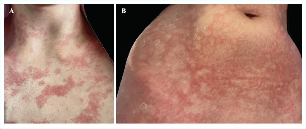
Figure 1 Poorly defined erythematous–violaceous patches on the trunk of the patient, starting from the neck (A) and coalescing in the lower abdomen (B).
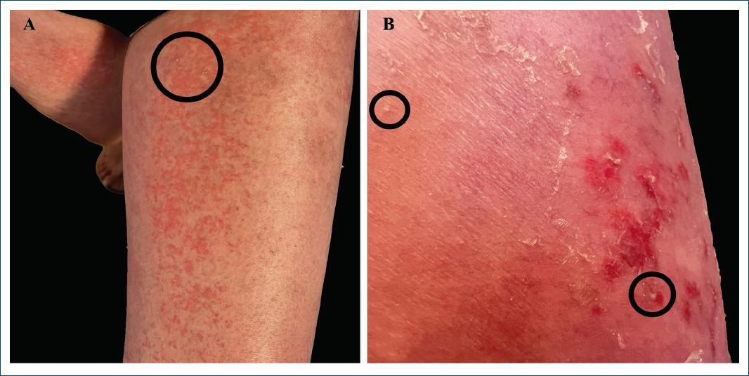
Figure 2 Erythematous coalescing patches on the thighs (A) of the patient presenting with some epidermal detachment (B) and scarse, nonfollicular, pinhead pustules (black circles).
Routine biochemical analyses, comprising renal and liver tests, were normal. Full blood count revealed a hemoglobin level of 13.5 g⁄dL (normal range 12.0-16.0 g⁄dL), with leukocytosis 17.2 × 10⁹⁄L (normal range 3.6-11.0 × 10⁹⁄L) and neutrophilia 15.03 × 10⁹⁄L (normal range 1.30-8.80 × 10⁹⁄L). Blood cultures were negative, and the chest X-ray was normal.
The initial differential diagnosis included terbinafine- induced SJS and AGEP. Hence, the patient was hospitalized for observation and initiated prednisolone 1 mg/kg/day.
A 4-mm punch biopsy was taken from the patient’s thigh. Histopathological examination revealed subcorneal pustules (Fig. 3). The epidermis exhibited orthokeratosis, with sparse permeation of neutrophils in the underlying spinous layer (Fig. 4). There was no evidence of spongiosis or acantholysis, and keratinocyte apoptosis was minimal. The dermis containeda superficial perivascular lymphohistiocytic infiltrate, accompanied by frequent neutrophils that extend into the interstitium of the superficial reticular dermis. There were no observable eosinophils in the dermis nor in the epidermis, and there was also no vacuolar interface change in the epidermis. Direct immunofluorescence microscopy studies were negative. These findings were more suggestive of the diagnosis of pustular psoriasis than of AGEP, its closest histological mimic.
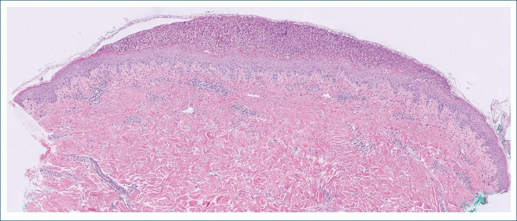
Figure 3 Histopathology of a thigh lesion skin biopsy revealed a subcorneal macropustule.The dermis contained a superficial perivascular lymphohistiocytic infiltrate accompanied by multiple neutrophils that extend into the interstitium of the superficial reticular dermis. Hematoxylin and eosin, 50×.
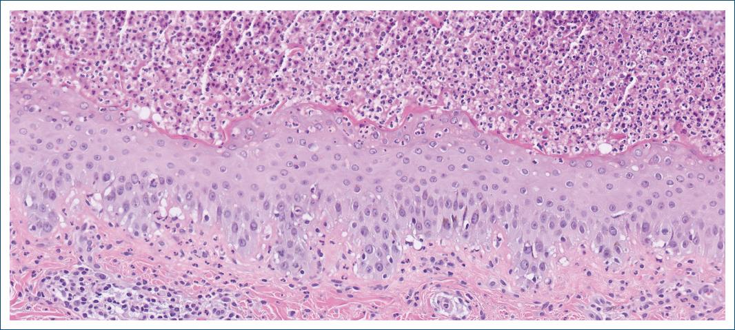
Figure 4 Histopathology of a thigh lesion skin biopsy revealed permeation of neutrophils in the spinous layer. There was no evidence of spongiosis or acantholysis, and keratinocyte apoptosis was minimal. Hematoxylin and eosin, 200×.
After two weeks of systemic corticosteroid therapy, no clinical improvement was noted. Additionally, during the hospitalization, the patient gradually developed few thick silvery scaly plaques on her scalp. Thus, the clinical diagnosis was nowfelt to be more in keeping with terbinafine-induced GPP. Cyclosporine 3 mg/kg/day was started, as well as slow corticosteroid tapering, but there was no response after four days of therapy. After excluding glucose-6-phosphate dehydrogenase (G6PD) deficiency, the patient was switched to dapsone 100 mg daily, and within 2 days the eruption was almost cleared. The patient was discharged on the third day, maintaining dapsone on an outpatient basis, and a complete clearance was seen after 2 weeks.
One month later, the patient made the decision to stop the treatment on her own, and 2 days later the eruptionrelapsed. Dapsone was reintroduced, leading to a new resolution of the rash within just 3 days. At the 3-month follow-up, the patient’s skin remained clear. Dapsone was tapered to 50 mg daily and, after another month, 50 mg every other day. Throughout this period, no adverse effects were observed, and regular laboratory tests, including complete blood counts, showed no abnormalities. Dapsone was discontinued after 5 months of therapy, and the patient remained in remission at the 8-month follow-up.
Discussion
As with many medications, oral terbinafine carries the potential foradverse skin reactions, varying from mild maculopapular reactions to severe drug eruptions, such as GPP6.
The challenge demonstrated in this case lies in differentiating between SJS, AGEP, and GPP in the presence of a nonspecific drug-induced rash exhibiting characteristics of all three conditions upon admission.
Initially, the patient presented with some areas of epidermal detachment, and the possibility of an early SJS had to be considered given the severity and possible complications of this condition. Thus, systemic corticosteroid therapy was instituted along with close monitoring of the eruption’s progression. However, the lack of mucosal lesions and clinical stability prompted us to reconsider the diagnosis.
Considering the presence of sterile pustules, albeit sparse, along with leukocytosis and neutrophilia, the possibility of AGEP was also evaluated. However, AGEP typically resolves within a few weeks following the cessation of the causative drug, whereas our patient did not show any improvement while on systemic prednisolone over the course of two weeks. Moreover, the leukocytosis and neutrophilia could be interpreted in the context of prior use of deflazacort.
As it is described in the literature, a GPP diagnosis often becomes more evident asdisease progresses7, and indeed, our patient developed typical psoriatic plaques on her scalp during hospitalization. Additionally, the skin biopsy results further reinforced the diagnosis of GPP. Although AGEP and GPP demonstrate considerable histopathologic overlap, both presenting in the pattern of a pustular dermatosis, this case revealed an absence of dermal eosinophils, vacuolar interface change, and eosinophilic spongiosis, all of which can be used as criteria8 to help favoring GPP. Finally, the toenail alterations upon admission could already be a sign of psoriasis. It is, in fact, common to misdiagnose nail changes as onychomycosis when they may be attributed to other underlying conditions. Thus, performing a mycological examination is crucial for an accurate diagnosis.
Among the available first-line therapies for GPP, cyclosporine was initiated due to its rapid onset of action, with clinical improvements documented in the literature occurring within the first days of therapy4. However, our patient did not show any sign of improvement over the course of 4 days of cyclosporine. Following this lack of response, we decided to switch to oral dapsone based on multiple case reports demonstrating its efficacy in treatment-resistant GPP5. Dapsone is a readily available drug associated with fewer side effects and not as immunosuppressive as other conventional agents9. Given that the patient was of child bearing age, acitretin was not a viable option.
In this case, a significant response to dapsone was observed within a few days of therapy. This was further corroborated when the patient discontinued dapsone, leading to recurrence of the skin lesions, and achieved resolution once again within just 3 days upon resuming the medication.
Dapsone’s efficacy in GPP and other sterile pustular dermatoses can be explained by its mechanism of action targeting neutrophils, which involves theinhibitionof the myeloperoxidase system, as well as the suppression of neutrophil adhesion and chemotaxis5,10. Side effects of oral dapsone include methemoglobinemia, hemolytic anemia, neutropenia, and agranulocytosis. A G6PD deficiency should always be ruled prior to starting dapsone and regular monitoring with a complete blood count should be performed. Less commonly, dapsone may also cause hepatitis, renal toxicity and hypersensitivity reactions9.
The clinical course of GPP can be unstable with frequent flares, either precipitated by re-exposure to known triggers or occurring without an identifiable cause2. Regarding our patient, although dapsone was discontinued after 5 months, continued patient monitoring is essential and re-exposure to terbinafine should be avoided. In the event of a recurrence, dapsone could be restarted considering the excellent response.
In conclusion, this case highlights the challenge in differentiating between SJS, AGEP, and GPP when presented with a drug-induced generalized, nonspecific, pustular skin eruption. Additionally, it supports the use of dapsone as an effective, readily available, and safe therapeutic option for treating GPP.

