




 Inglês (pdf)
 Artigo em XML
 Referências do artigo
 Tradução automática
 Enviar este artigo por email
 Citado por SciELO
  Similares em
SciELO
Similares em
SciELO 
 Permalink
Introduction
The nail apparatus performs essential functions for the organism, such as mechanical protection, assistance in fine tactile discrimination, object grasping, and contribute to the esthetics. Loss of nail integrity compromises various daily activities, directly affecting quality of life1.
Diagnostic gaps in nail disorders in clinical practice are particularly attributable to insufficient familiarity with diagnostic methods, such as dermoscopy, dermatopathology of the nail, microbiological cultures, and, especially, nail biopsy. In dermoscopy, for example, nail signs are magnified and, together with physical examination, may enable diagnosis2. Proper use of this tool –as well as the others– requires thorough knowledge of nail anatomy, since the presentation of pathologic changes depends the affected area of the nail apparatus.
Among the nail diseases encountered in office practice, 50% are fungal, while the other 50% include neoplastic, inflammatory, traumatic, and systemic-related conditions3. The fungal nail disease, termed onychomycosis, employs direct microscopic examination (DME) as its primary diagnostic test, due to its low cost and the provision of immediate information that can be crucial for determining appropriate patient therapy4.
Nail neoplasms include both malignant and benign tumors, which are often mistaken for onychomycosis or benign nail pigmentation5. Nail biopsy is critically important in this context, being at times both diagnostic and therapeutic. Its performance depends on understanding the surgical anatomy of the nail unit, proper anesthesia and hemostasis, and targeting an abnormality for which histopathology can yield a definitive diagnosis.
Indications and techniques for nail biopsy vary according to the site and type of pathology. Nail bed biopsies can be performed easily with minimal scarring and are most commonly used to diagnose tumors, infectious and inflammatory nail disorders6. The principal indication for nail biopsy is to establish or exclude the diagnosis of melanoma6.
Diagnosing nail conditions can sometimes be a challenging task, not only because of the large number of cutaneous and systemic diseases involving the nails, but also due to their shared clinical features. Therefore, understanding the peculiarities of each pathology, as well as the anatomy and physiology of the nail apparatus, is essential for a more correct diagnosis and for delivering more targeted and effective treatment.
Anatomy and physiology of the nail
The nail unit is one of the principal skin appendages, functioning as a mechanical and sensory protective organ7. Nail anatomy comprises five components: the nail plate, commonly known as the nail; the matrix, which produces the nail plate and lies beneath the proximal nail fold, and can be visible on the thumbs and great toes through the nail plate as the lunula, the proximal, distal, and lateral nail folds; and the nail bed, which supports the nail plate. Beneath the nail bed lie connective tissue and the phalanx with its ligaments that attach the nail to the underlying joint structures8.
The nail plate is a modified form of the stratum corneum and is curved along both the longitudinal and transverse axes. This curvature allows anchorage into the proximal and lateral nail folds, ensuring stability. The nail folds assist in securing the plate to the nail bed; when diminished, there is a tendency for onycholysis (nail detachment), and when thickened, pathological ingrowth may occur9. The proximal nail fold, together with the cuticle and the plate, protects the matrix from radiation and chemical irritants that could impair its function10.
Maturation and differentiation of matrix keratinocytes proceed along a distally oriented, oblique axis. Thus, keratinization of distal matrix cells forms the ventral aspect of the nail plate, while keratinization of proximal matrix cells forms the dorsal aspect11. Nail plate abnormalities generally result from pathologies affecting the matrix or from space-occupying lesions beneath the nail fold11.
Fingernail growth takes approximately 6 months to completely replace the nail plate, whereas toenail growth takes about 12 months12. Nail growth rate decreases with age and may be partially or fully halted by systemic diseases, trauma, or certain medications12.
The objective of this study is to describe the main nail pathologies in terms of their clinical and etiological characteristics, as well as to establish objective criteria and the main diagnostic tools for recognizing these most prevalent nail conditions.
Methods
This study is a structured narrative literature review, guided by the principles of Preferred Reporting Items for Systematic Reviews and Meta-Analyses rather than by the full protocol of a systematic review, which employed the terms “nail diseases,” “nail disorders,” “diagnosis,” “nail neoplasm,” “melanonychia,” “pigmentation disorders,” “nail surgery” and “histopathology” in combination with the Boolean operators “and” and “or.” Priority was given to articles addressing the diagnosis of nail conditions, including original articles, literature reviews, meta-analyses, guidelines and consensus statements, and case reports focusing on nail diseases diagnosed by clinical, dermatoscopic or histopathological methods. Inclusion criteria comprised articles published in the PubMed database between 2000 and 2024, in portuguese, spanish or english, and relevant to the research objectives. Exclusion criteria included studies without an available abstract, published outside the specified period, or not focused on the diagnosis of nail diseases.
Results
A total of 9526 articles were retrieved from PubMed for analysis. Of these, 3253 articles were excluded due to platform filtering based on language and publication period, and 6263 articles were excluded for not meeting the inclusion criteria or for not being related to the research objective (Fig. 1).
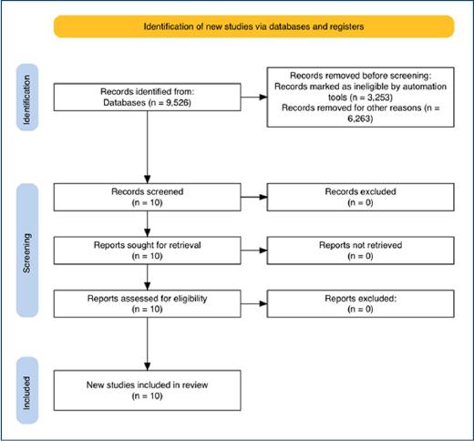
Figure 1 Flow diagram of the literature review according to Preferred Reporting Items for Systematic Reviews and Meta-Analyses guidelines.
After exclusions, the remaining articles consisted of one prospective study and nine review articles, which were thoroughly analyzed for this research. In this regard, the article analyses adhered to criteria of methodological consistency, scientific evidence, and practical applicability, and were categorized by the type of nail condition addressed (inflammatory, neoplastic, infectious, traumatic), the diagnostic method utilized, and the clinical relevance of the findings (Table 1).
Table 1 Articles selected for review
| Title of article | Authors/year | Content | Category |
|---|---|---|---|
| “Optimal diagnosis and management of common nail disorders” | Lee and Lipner, 2022 | Etiology, clinical presentation, diagnosis, and treatment of infectious and inflammatory nail diseases | Infectious, traumatic, and inflammatory |
| “Dermoscopy in the Evaluation of Nail Disorders” | Starace et al. 2021 | Clinical presentation, diagnosis, and dermoscopy of nail lesions | Infectious, neoplastic, and traumatic |
| “Role of tangential biopsy in the diagnosis of nail psoriasis” | Bertanha et al. 2024 | Clinical presentation and biopsy-based diagnosis of inflammatory nail diseases, such as nail psoriasis | Inflammatory |
| “Nail neoplasms” | Park et al. 2017 | Clinical presentation, diagnosis, and histopathology of benign and malignant nail neoplasms | Neoplastic |
| “Nail Biopsy: A User’s Manual” | Grover and Bansal, 2018 | Techniques and types of nail biopsy for the diagnosis of nail diseases | Infectious and neoplastic |
| “Diagnosis and Management of Malignant Epithelial Nail Unit Tumors” | Iorizzo et al. 2024 | Clinical presentation, diagnosis, dermoscopy, treatment, and prognosis of malignant epithelial tumors of the nail | Neoplastic |
| “Differential diagnosis of pigmented nail lesions” | Bertanha et al. 2024 | Clinical presentation, dermoscopy, and diagnosis of pigmented nail disorders. | Infectious, neoplastic, and traumatic |
| “Histopathology of the nail unit” | Fernandez-Flores et al. 2014 | Clinical presentation, histopathology, and diagnosis of nail unit diseases | Infectious, neoplastic, and inflammatory |
| “Nail surgery: General principles, fundamental techniques, and practical applications” | Queirós et al. 2022 | Techniques and types of nail surgery for the treatment of nail lesions | Infectious, neoplastic, and traumatic |
| “Diagnosis of Melanonychia” | Starace et al. 2021 | Clinical presentation, dermoscopy, histopathology, and diagnosis of pigmented nail disorders | Infectious and neoplastic |
The selection of nail diseases was based on the clinical significance of the results and the volume of information available in each included article; two nail conditions were discussed within each category (Table 2).
Table 2 Diagnostic tools to assist in each nail condition
| Condition | Dermoscopy | Microscopy | Fungal culture | Nail biopsy |
|---|---|---|---|---|
| Nail psoriasis | X | - | - | X |
| Nail lichen planus | X | - | - | X |
| Onychomycosis | X | X | X | - |
| Paronychia | - | - | - | - |
| Glomus tumor | X | - | - | X |
| Onychopapilloma | X | - | - | X |
| Subungual melanoma | X | - | - | X |
| Squamous cell carcinoma | X | - | - | X |
| Onychocryptosis | - | - | - | - |
| Subungual hematoma | X | - | - | - |
Discussion
Inflammatory nail disorders
Nail psoriasis
The diagnosis of nail psoriasis can be based solely on cutaneous and/or osteoarticular signs and symptoms if a confirmed diagnosis of psoriasis vulgaris exists13. In the absence of these signs, diagnosis can be complex and must rely on complementary examinations. Hyperkeratosis, onychorrhexis, nail plate discoloration, and nail plate thickening generally resemble onychomycosis, which occurs in up to 60% of patients13.
Nail unit involvement is characterized by pitting, rough and brittle nail plates, and pronounced transverse (Beau’s) lines14-16. Signs of nail bed involvement include distal onycholysis with a proximal yellow-orange margin and serrated border, splinter hemorrhages, oil-drop (salmon) patch, subungual hyperkeratosis, and red or black hemorrhagic spots14-16.
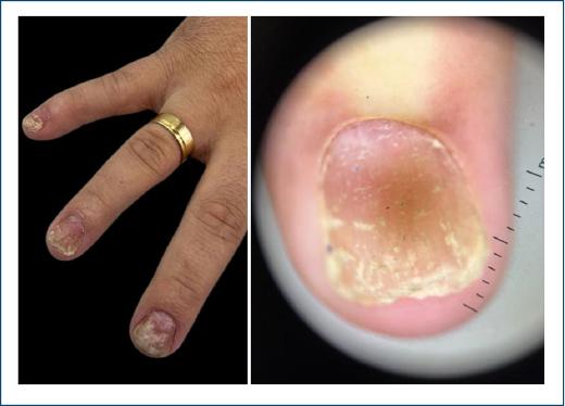
Nail pitting, distal onycholysis (separation of the nail plate from the underlying nail bed), oil-drop (salmon) patches, and splinter hemorrhages are key dermatoscopic features of nail psoriasis16,17. The formation of depressions, the characteristic nail pitting, is the most frequent finding and typically appears irregular on dermatoscopy, representing an indicative signal of nail psoriasis involvement (Fig. 2). This feature arises from parakeratosis of the nail matrix, leading to extrusion of parakeratotic cells as they emerge from beneath the proximal nail folds, thereby creating indentations on the nail surface14-16.
The biopsy technique ideally indicated for the diagnosis of nail psoriasis is a punch biopsy without prior nail plate avulsion, as it preserves the morphology of the superficial tissue. However, it is difficult to introduce the punch through the nail plate while maintaining adhesion of the plate to the nail bed/matrix during instrument rotation without shear18. To reduce technical difficulty, the digit is soaked in warm water for a few minutes or the nail plate may be thinned by gentle abrasion19. Delay in performing the procedure and in specimen fixation can damage elements of the nail bed/matrix and compromise diagnostic accuracy20.
In this regard, tangential excisional biopsy has gained prominence in the diagnosis of inflammatory nail diseases. In the tangential technique, the nail plate is carefully removed with a scalpel blade, preserving epithelial integrity and enabling artifact-free histopathological processing18. When bed and matrix alterations are present, a nail bed specimen is always preferable, both for technical ease and for a lower risk of scarring.
In a recent study by Bertanha et al. on tangential biopsy in nail psoriasis, good clinic-histopathological correlation was obtained using the following criteria: a mandatory criterion, dilated dermal papillary vessels, and at least three secondary criteria, namely, absence of spongiosis, presence of parakeratosis, psoriasiform epidermal hyperplasia, focal thinning of the granular layer and suprapapillary epidermis, and intra- or subcorneal neutrophilic exudate18.
Nail lichen planus (LP)
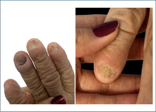
Nail involvement occurs in 10% of patients with LP. Early recognition is crucial, as the disease can inflict extensive damage to the nail matrix, resulting in anonychia and proximal pterygium21. Clinical features of nail LP include brittle, roughened nail plates, a red or mottled lunula, onychorrhexis (vertical ridging of the nail plate), and longitudinal striations22,23 (Fig. 3).
Onychoscopy may aid in detecting early LP changes22,23. Nail-bed involvement is manifested by plate fragmentation, chromonychia (discoloration of the nail plate or subungual tissue), splinter hemorrhages, onycholysis, subungual hyperkeratosis, and longitudinal grooves22,23.
Twenty-nail dystrophy, or trachyonychia, is regarded as a variant of nail LP; its uniform appearance and the absence of proximal pterygium distinguish it from classic nail LP21. The idiopathic atrophic form presents acutely and typically progresses to diffuse nail destruction within a few months21.
The diagnosis of nail LP requires biopsy with clinic-histopathological correlation. Key histopathological features include hyperkeratosis, hypergranulosis, a band-like inflammatory infiltrate, basal cell degeneration, and pigment incontinence24. In rare instances, subepidermal blister formation with prominent aggregates of colloid bodies may be observed, corresponding to the uncommon bullous variant of nail LP24.
Infectious nail disorders
Onychomycosis
Onychomycosis is the most prevalent nail disease and can sometimes be confused with a variety of benign and malignant nail conditions2,25. This nail disorder represents all fungal infections of the nails and may involve the nail bed, plate, and matrix. It is the most prevalent nail disorder, affecting toenails more frequently than fingernails due to slower growth, reduced blood supply, and prolonged exposure to dark, humid environments25,26.
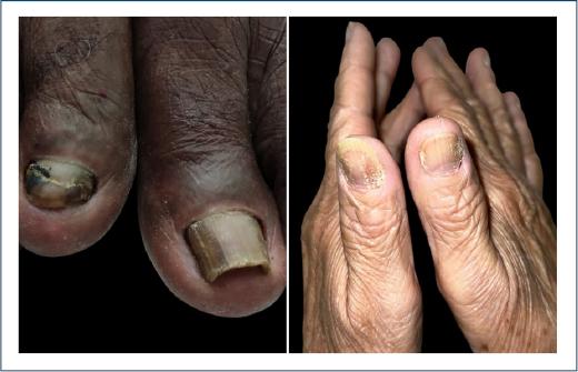
Typical physical examination findings include hyperkeratosis of the nail bed, which often causes varying degrees of nail plate onycholysis26. A white or yellow discoloration of the nail plate is common, as well as subungual debris26. Trauma is a risk factor for onychomycosis, and violaceous/brown/black nail plate discoloration may also be present26. In longstanding or severe cases, there may be extensive onychodystrophy with nail plate thickening, crumbling, ridging, onychocryptosis, and partial or complete nail loss26 (Fig. 4). A dermatophytoma, or fungal abscesses, is a white/yellow or orange/brown longitudinal streak in the nail plate and is quite specific for onychomycosis26.
Dermatoscopic features of onychomycosis include the “ruin appearance,” “longitudinal streaks,” and “spikes” at the proximal margin of the onycholytic area27,28. The “aurora borealis” sign (defined by the combination of multicolored chromonychia with longitudinal streaks, spikes, and onycholysis) demonstrates the highest sensitivity and specificity for this disorder27,28. The irregular, spiked proximal border corresponds to distal-to-proximal invasion of the nail bed’s longitudinal ridges by dermatophytes27,28.
Direct microscopy and fungal culture are the gold-standard methods for diagnosing onychomycosis29. Microscopic examination is performed after the nail is cleaned with 70% isopropyl alcohol, and then subungual debris samples are obtained, typically 8-10 fragments, to improve diagnostic accuracy. In a positive direct examination and fungal culture, hyphae, pseudohyphae, and spores are identified, confirming infection, although the specific organism is not identified in direct microscopy only in culture examination25,29.
Nail biopsy is performed by obtaining a sample from the active site of infection, approximately 4 mm from the free edge30. After paraffin embedding, the material is stained with hematoxylin and eosin and special fungal stains, periodic acid-Schiff and methenamine silver, to visualize fungal structures under microscopy30. However, as with direct microscopic examination, histopathology should always be complemented by culture.
Paronychia
Paronychia is defined as inflammation or infection of the proximal or lateral nail folds31,32. Clinically, it presents with an acute onset of discomfort, tenderness, erythema, and edema. The acute form is characterized by disruption of the nail fold’s protective barrier and pathogen invasion31,32 (Fig. 5). Secondary infections often follow trauma, such as nail-biting or manipulation of ingrown nails, the latter being the most common etiologic factor31.
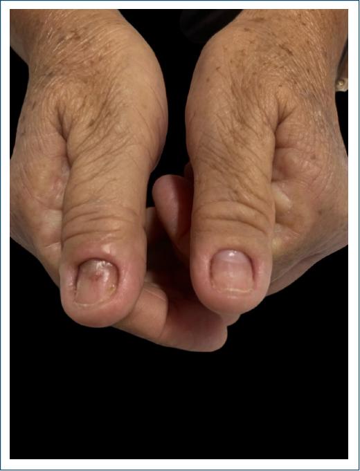
If left untreated, infection may progress to the formation of granulation tissue around the nail fold and abscess development31. An untreated abscess in one fold can extend to involve all nail folds and may even spread into adjacent soft tissues of the affected digit31,32. Staphylococcus aureus is the primary pathogen, although Streptococcus spp., Pseudomonas spp., other Gram-negative bacteria, and Candida albicans can also invade the nail folds31,32.
Diagnosis of paronychia is primarily clinical, based on patient history and physical examination, which helps differentiate it from other nail disorders31-33. For diagnostic confirmation, the digit-pressure test may be employed: when firm pressure over the affected fold produces an area of blanching larger than expected, an underlying abscess is likely present31-33.
Paronychia can be classified as acute or chronic31,33. In the acute form, physical examination may reveal a tender, erythematous, and edematous lateral nail fold, and if an abscess is present, a fluctuant area may be palpable31. In chronic paronychia, the nail fold may be red and swollen, but fluctuation is uncommon; the nail plate may become thickened and discolored, and proximal nail fold retraction, nail dystrophy, and cuticle loss can occur31.
Benign neoplastic nail disorders
Glomus tumor (GT)
GT is a benign vascular hamartoma originating from glomus cells, specialized smooth muscle cells concentrated around dilated vessels and this tumor present as red, purple, or blue lesions beneath the nail plate, classically characterized by the triad of pain, tenderness, and cold sensitivity34,35.
Clinical characteristics of GT typically appear as a small reddish-to-bluish macule under the nail plate or as longitudinal erythronychia with distal notching or fissuring of the nail plate34 (Fig. 6). The predominant symptom is intense nail pain, which may be throbbing or pinpoint in nature; it can occur spontaneously or be provoked by pressure or cold exposure5,34,35.
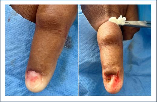
GT are generally small and rarely palpable, rendering clinical examination inadequate for precise localization36. Imaging studies facilitate accurate tumor localization and size assessment, critical factors in selecting the optimal surgical approach36,37. Nail-plate dermatoscopy may reveal vascular structures; however, these can sometimes be subtle or absent36. Therefore, dermatoscopy of the nail bed and matrix before tumor excision is recommended, as it assists both in tumor localization and in visualizing the lesion’s vascular pattern36.
Definitive diagnosis is established by performing a nail bed biopsy with nail plate avulsion. Histopathological examination demonstrates a variable admixture of glomus cells, vascular channels, and smooth muscle38. GT are subclassified into three types: glomangiomas, marked by an abundance of vascular channels; solid GTs, composed predominantly of glomus cells; and glomangiomyomas, which show a predominance of smooth muscle elements38.
Onychopapilloma
Onychopapilloma is a benign neoplasm of the distal matrix and nail bed, typically presenting as a longitudinal band of splinter hemorrhages associated with subungual hyperkeratosis39 (Fig. 7). The most common dermatoscopic finding is longitudinal erythronychia, although melanonychia and a V-shaped distal nail-plate notch may also be observed39.
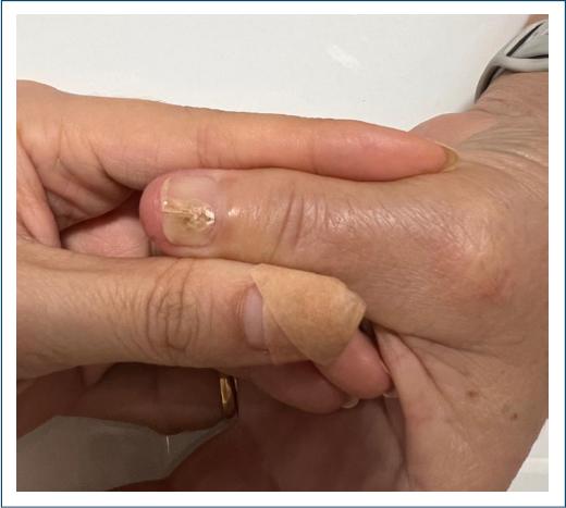
Histopathological examination features include subungual hyperkeratosis with or without focal hemorrhage; in excisional specimens, there is papillomatosis of the nail bed and acanthosis of the nail matrix with layers of subungual parakeratosis and focal parakeratosis39.
Malignant neoplastic nail disorders
Subungual melanoma (SM)
SM is a distinct subtype of cutaneous malignant melanoma that arises from the nail matrix. It is usually a variant of acral lentiginous melanoma, a form of melanoma originating in the palmoplantar region40,41. In two-thirds of cases, SM presents as a longitudinal brown-to-black band on the nail plate, known as longitudinal melanonychia, but it may also be amelanotic and manifest as a red nodule40,41. During its progression, periungual pigmentation may develop, known as Hutchinson’s sign, which, while not pathognomonic, is highly suggestive of melanoma, nail dystrophy may also occur, indicating more advanced disease40. SM is often mistaken for an infection and initial misdiagnosis of SM occurs in 85% of cases41. Dermoscopy assists in distinguishing SM from benign melanocytic pigmented lesions, but biopsy remains the gold standard for diagnosis41.
Dermatoscopy can reveal a gray-brown to black background with longitudinal lines that are irregular in thickness, spacing, and color, as well as fine pigmented granules, features indicative of a melanocytic origin40. The width of the pigmented bands can vary and may progress to total melanonychia40.
Histologic features suggestive of malignancy include lesion asymmetry, infiltrative margins, a markedly increased number of melanocytes in the basal and suprabasal layers with a high propensity to form compact aggregates, presence of cytologic atypia, and dermal inflammation. Malignant melanocytes have large, atypical nuclei with increased mitotic activity. Identification of melanocytes in the nail plate is diagnostic of melanoma42. Invasive SM also exhibits irregularly dispersed dermal nests composed of atypical melanocytes42.
Squamous cell carcinoma (SCC)
SCC is the most common malignant tumor of the nail unit, with the in situ form (Bowen’s disease) occurring more frequently than the invasive variant and usually is associated with the Human Papillomavirus (HPV), mainly type 16, 18, 35, and 5640,43. It typically involves a single digit, most commonly the thumb40,43. The malignancy is usually indolent and painless, affecting the nail bed and periungual regions or both. Lesions are clinically classified into two main categories: periungual type, arising in the epithelium of the nail fold and sulcus and Subungual type, developing in the epithelium of the nail bed40,43.
Dermatoscopy of SCC is characterized by brown linear dots or a clustered glomerular vascular pattern40. Other features include localized subungual hyperkeratosis, erythrochia or leukonychia irregular, lateral detachment (onycholysis), non-parallel longitudinal melanonychia, and splinter hemorrhages40 (Fig. 8).
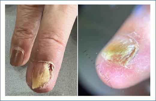
Biopsy with histopathological examination is the gold standard for diagnosing SCC40,43. The histopathological characteristic findings of this tumor include loss of normal epidermal stratification, dyskeratosis, clusters of large cells with hyperchromatic nuclei, atypical mitosis, and, when associated with HPV, perinuclear vacuolization is typically observed40,43.
Traumatic nail disorders
Onychocryptosis
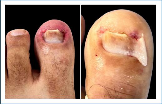
Onychocryptosis, also known as ingrown toenails, is a condition in which the lateral nail fold is penetrated by the nail plate’s edge, causing pain and difficulty ambulating44-47. It is most common in adolescents, young adults, and males44,46. The penetration is often due to nail spicules along the plate’s margin, which elicit an inflammatory response44,46,47. The great toes are most frequently affected44.
The main causes include ill-fitting footwear, improper toenail trimming, abnormalities of the nail apparatus, and excessive perspiration44,47. The condition is unilateral in 80 % of cases and predominantly involves the hallux44. Diagnosis of onychocryptosis is straightforward and classically based on clinical features, without the need for laboratory, radiographic studies, or dermatoscopy44,46,47.
Patients typically present with toe pain that can range from mild discomfort when walking to complete inability to ambulate; depending on lesion progression, there may also be associated swelling, erythema, secondary infection, or seropurulent discharge44-47 (Fig. 9).
Subungual hematoma (SH)
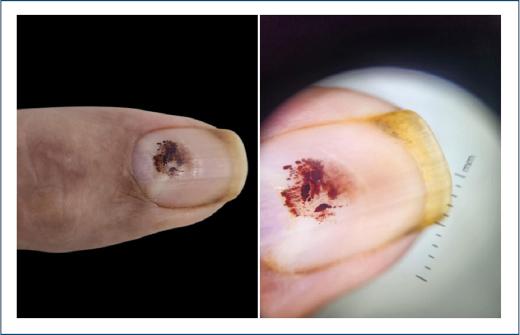
SH is a common nail lesion characterized by the accumulation of blood beneath a fingernail or toenail, typically located between the nail bed and the nail plate48. Trauma is the principal etiology, and the migration of the hematoma distally with nail-plate growth serves as a key diagnostic feature40,48.
Clinically, the hallmark symptom is sudden, throbbing pain following nail injury, resulting from pressure exerted by the subungual blood collection48. SH is the most frequent cause of bluish-red to bluish-black nail pigmentation; unlike melanocytic lesions, it does not form a continuous longitudinal band40,48,49.
Dermatoscopic examination often reveals small, round blood globules at the periphery of the hematoma, distal streaking, and localized leuconychia at the trauma site, accompanied by a bluish-red, bluish-black, or brownish color coloration40,48,49 (Fig. 10).
Conclusion
This study analyzed and discussed the two main types of nail conditions in each classification based on their clinical, etiological features and defined objective criteria to recognize each nail lesion. Diagnostic tools, such as dermoscopy, microscopy, culture, and nail biopsy were described, highlighting their importance and clinical applicability for the early diagnosis of some frequent nail diseases. In summary, by systematizing clinical and histopathological criteria for the diagnosis of nail lesions, this study may contribute to reducing diagnostic errors, guide more precise therapeutic management, and improve the quality of life for patients affected by nail disorders.

