




 Inglês (pdf)
 Artigo em XML
 Referências do artigo
 Tradução automática
 Enviar este artigo por email
 Citado por SciELO
  Similares em
SciELO
Similares em
SciELO 
 Permalink
Introduction
Pseudoxanthoma elasticum (PXE) is a rare genetic disorder with an autosomal recessive mode of genetic transmission affecting multiple organs, such as the skin, eyes, heart, and gastrointestinal system1. PXE, also termed as Gronblad Strandberg syndrome, is a rare disorder due to a mutation in the ABCC6 gene (ATP-binding cassette transporter C6), located in chromosome 16. Prevalence of PXE was found to be 1/25.000 to 1/100.000 inhabitants. It has been found that the prevalence of PXE is 10 times more in women than in men2,3. It encodes an ATP-binding driven anion transporter, seen in the cell membrane of the liver and kidney. PXE is a form of genodermatoses, currently an incurable disease associated with serious complications due to elastic fiber fragmentation and calcification4. Here we report a rare case of PXE with its clinical, histopathological, and ocular findings.
Case report
A 17-years-old female came to the outpatient department with the complaints of itchy, yellowish papules and plaques in the anterior and lateral aspect of her neck for the past 1 year. No relevant personal or family, or any medical history of dermatosis. No history of any consanguineous marriage in the family. On examination, painless, uneven skin-colored reticulated plaques (Fig. 1) without any surrounding inflammatory signs were seen in the anterior aspect and in the nape of the neck, giving a parchment-like skin appearance.
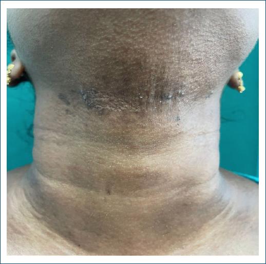
Figure 1 Clinical image of patient showing reticulated plaques over the anterior aspect of the neck.
Complete hemogram and routine blood investigations were found to be normal. The patient was then subjected to a skin biopsy. A punch biopsy was taken from the lesion in the anterior and nape of the neck. Biopsy revealed benign stratified squamous lining with middle and lower thirds of dermis showing fragmented and calcified basophilic elastic fibers amidst collagen bundles. The adjacent area shows dense lymphocytic infiltration and giant cell reaction (Fig. 2).
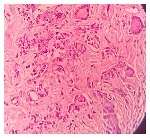
Figure 2 Histopathological image showing fragmented elastic fibers with calcification, lymphocytic infiltration, and giant cell reaction (H&E stain, 40×).
Verhoff Von Gieson stains showed black-colored fragmented elastic fibers in the dermis (Fig. 3).
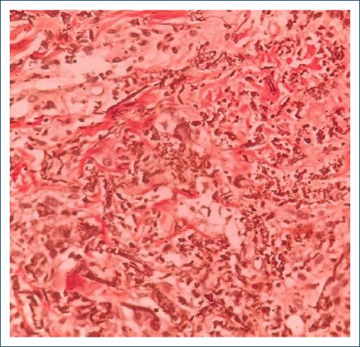
Figure 3 Histopathological image showing fragmented elastic fibers (Verhoeff Von Gieson stain, 40×).
Diagnosed as a case of PXE and the patient was then subjected to ocular examination, which revealed peau d’ orange appearance of retinal blood vessels, which was the earliest retinal manifestation. Following the investigation, the patient was then subjected to cardiovascular evaluation and the results were normal. The patient is currently on follow-up.
Discussion
PXE is a rare inherited genetic disorder, caused due to abnormal mineralization of the connective tissue with elastic fibre degeneration affecting various organs, such as the skin, eyeballs, and cardiovascular system5.
Cutaneous manifestations are seen as yellowish macules or papules, or plaques. PXE can occur at any age and the skin appears lax and wrinkled. The lesions start appearing over the lateral aspects of the neck, followed by skin creases, armpits, popliteal, and inguinal regions. Oral mucosa and genital region can also be affected6,7.
Early ocular involvement starts as peau d’ orange appearance of retinal vessels and progresses to the development of angioid streaks due to lesions over the Bruch membrane. Progressive retinal pigmentation, macular degeneration, choroidal neovascularization, retinal hemorrhage, and scar formation can occur, leading to the most serious complication of complete ocular blindness8.
Various cardiovascular abnormalities occur in a case of PXE, such as bradycardia, hypertension, angina pectoris, atherosclerosis, and cardiac arrest at younger ages. Gastrointestinal manifestations, including melena, hemorrhages, and hematemesis, can be seen. Some patients presented with stroke at younger ages. Major pathogenesis behind these complications was found to be fragmentation of elastic fibers in the lining of blood vessels and in the connective tissue of the pericardium, myocardium, and endocardium of the heart9.
Certain inherited hemolytic disorders, such as beta thalassemia, hereditary spherocytosis, and Sickle cell anemia, can occur in patients with PXE10.
Plomp et al. proposed guidelines for diagnosing PXE which includes major criteria: (a) skin lesions, such as yellow cutaneous plaques and papules, fragmentation, clumping, and calcification of elastic fibers, (b) ophthalmic lesions, such as Peau d’ orange appearance of retina or the presence of angioid streaks, and (c) genetic factors such as mutation of both alleles of ABCC6 gene or a first degree relative affected with PXE. Minor criteria include ophthalmic lesions, such as one angioid streak shorter than 1 disk diameter, one or more comets in the retina, and one or more wing signs in the retina: genetic factors, such as the presence of a mutation of one allele of the ABCC6 gene11.
Major differential diagnosis for PXE includes solar elastosis, PXE-like papillary dermal elastolysis. PXE-like papillary dermal elastolysis occurs in elderly females, where partial or complete loss of elastic fibers were seen but without calcification. Solar elastosis shows irregularly thickened, coarse, disorganized, and tangled elastic fibers12.
Conclusion
This case report deals with a young female with typical clinical and histopathological features of PXE highlighting a need for a multidisciplinary approach for accurate diagnosis of this rare disorder. This helps us to understand the disease better and to discover the newer therapeutic approach to treat the disease and to prevent the complications as early as possible.

