


SciELO - Scientific Electronic Library Online

 
vol.84 número1Múltiplas lesões em arco no pescoço de um adulto: um caso atípico de elastose perfurante serpiginosaAngiossarcomas: caracterização clínica e histopatológica de cinco casos e revisão da literatura índice de autoresíndice de assuntosPesquisa de artigos
Home Pagelista alfabética de periódicos  

Serviços Personalizados

Journal

Artigo

Indicadores

Links relacionados

  • Não possue artigos similaresSimilares em SciELO

Compartilhar


Portuguese Journal of Dermatology and Venereology

versão impressa ISSN 2795-501Xversão On-line ISSN 2795-5001

Port J Dermatol Venereol. vol.84 no.1 Lisboa mar. 2026  Epub 28-Out-2025

https://doi.org/10.24875/pjdv.25000049 

CASE REPORT

Desmoplastic trichoepithelioma on the face mimicking basal cell carcinoma: a case report and literature review

Tricoepitelioma desmoplásico na face simulando carcinoma basocelular: um relato de caso e revisão da literatura

Isabele M. Saldanha1  * 

Airton K. Motizuki2 

Renata M. Oyama-Okajima1 

1Dermatology Service of the Federal University of Pará, João de Barros Barreto University Hospital

2Department of Medicine, Faculty of Medicine of Federal University of Pará. Belém, Pará, Brazil


Abstract

Desmoplastic trichoepithelioma is a rare benign cutaneous neoplasm that differentiates toward germinative cells of the hair follicle. It can be classified as familial multiple trichoepithelioma, solitary, and desmoplastic, the latter being a rare variant with low incidence and clinical features resembling basal cell carcinoma. We report the case of a 54-year-old female patient who presented with a brownish plaque exhibiting areas of dark pigmentation, an irregular outline, a pearly sheen, and surface telangiectasias in the right paranasal region. Complementary studies, including histopathological and immunohistochemical analyses, were performed to establish the definitive diagnosis. Furthermore, this cutaneous lesion displays both clinical and histopathological characteristics that complicate differentiation between benign and malignant tumors, making immunohistochemistry essential to confirm the desmoplastic variant of trichoepithelioma.

Keywords Desmoplastic trichoepithelioma; Differential diagnosis; Basal cell carcinoma

Resumo

O tricoepitelioma desmoplásico é uma rara neoplasia benigna de pele que se diferencia em células germinativas do folículo piloso, e pode ser classificado entre tricoepitelioma múltiplo familial, solitário e desmoplásico, sendo esse último uma variante rara com pouca incidência na população e com similaridades clínicas do carcinoma basocelular. O caso é de uma paciente de 54 anos,que apresentou uma placa acastanhada, com algumas áreas de pigmentos enegrecidos, irregular, com brilho perláceo e telangiectasias em sua superfície na região paranasal direita, sendo realizado exames complementares como o anatopatológico e imunohistoquímico para confirmação completa do diagnóstico. Além disso, essa lesão no tegumento manifesta estruturas tanto clínicas quanto histopatológicas que dificultam na diferenciação de tumores malignos ou benignos, sendo fundamental a realização da imunohistoquímica para confirmar essa variante desmoplásica do tricoepitelioma.

Palavras-chave Tricoepitelioma desmoplásico; Diagnóstico diferencial; Carcinoma basocelular

Introduction

Desmoplastic trichoepithelioma (DT) is a rare benign skin neoplasm of the hair follicle, a variant of trichoblastoma originating from the proliferation of mesenchymal cells1-3. The classification of this cutaneous neoplasm can be divided into three forms: familial multiple trichoepithelioma, solitary, and desmoplastic, with the desmoplastic form characterized by increased fibroplasia in the dermis and this tumor should be considered a differential diagnosis for basal cell carcinoma (BCC)1-3.

In addition, DT shows a predilection for females, particularly young or middle-aged women4,5. Its most common manifestations occur on the facial skin as pink or skin-colored papules and nodules that progressively increase in size, often easily confused with the clinical aspects of BCC4,5. The histopathology of this neoplasm consists of a histological triad of thin cords of basaloid cells, keratin cysts, and desmoplastic stroma, which are important for both histological differential diagnoses and clinical differentiation from BCC3-7.

Case report

A 54-year-old female patient was referred for a dermatological consultation due to a facial lesion that had been present for approximately 2 years, showing progressive growth over time without associated symptoms (Fig. 1).
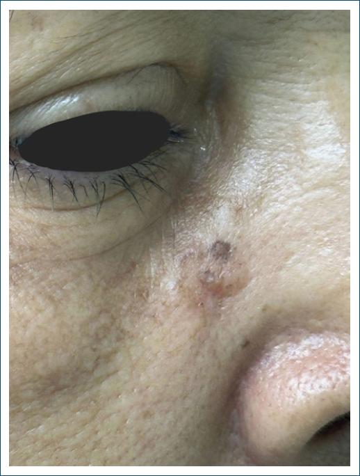
Figure 1 Clinical appearance of the neoplasm. 

On dermatological examination, a brownish plaque with some black-pigmented areas, irregular shape, pearly shine, and telangiectasias on its surface was observed, measuring 1 cm in the right paranasal region (Fig. 2).
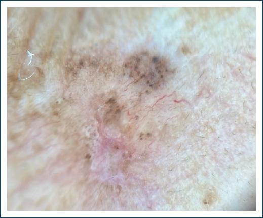
Figure 2 Dermatoscopy of the lesion with irregularly distributed black pigment, tortuous vessels, and mild erythema, measuring 1 cm. 

The patient underwent an incisional biopsy of the lesion, followed by histopathological examination, which showed atrophic epidermis and dermis with diffuse proliferation of small nodular masses and cords of basaloid cells with peripheral palisading, including occasional horn cysts, embedded in desmoplastic and myxoid stroma. Bulb-papilla images suggested follicular differentiation (Fig. 3).
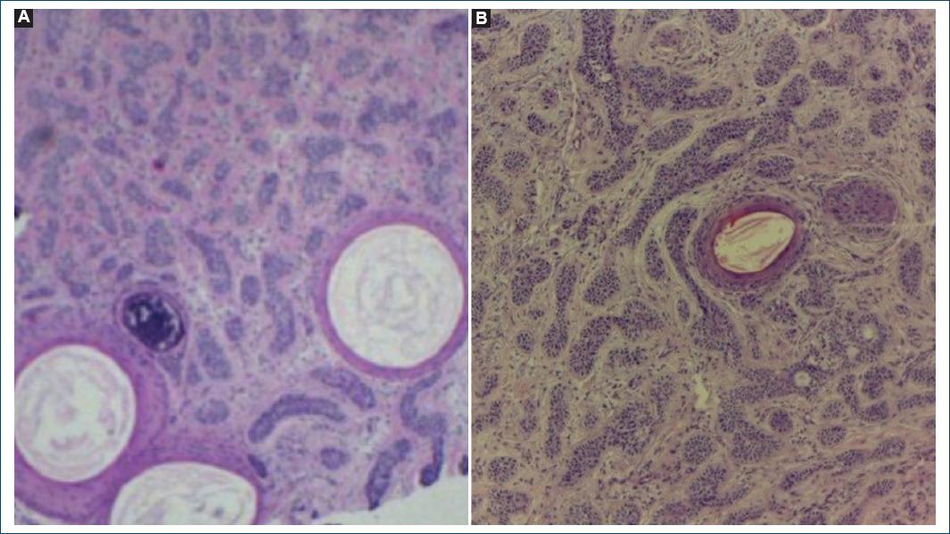
Figure 3 Histopathological findings. A and B: histological sections, horn cysts, embedded in desmoplastic and myxoid stroma (H&E, x40). 

The histopathological examination revealed an intradermal epithelial neoplasm, raising differential diagnoses of DT, micronodular BCC, and microcystic adnexal neoplasm. An additional immunohistochemical study confirmed the diagnosis of DT (Table 1).

Table 1 Results of the immunohistochemical panel 

Antibody Clone Result Observation
Ki-67 (cell proliferation antigen) MIB1 Positive 5%
p63 protein (squamous/transitional epithelia; myoepithelial cells) DAK-p63 Positive -
Epithelial tumor glycoprotein BerEp4 Positive -
Epithelial membrane antigen E29 Negative -
Anti-apoptotic protein BCL-2 124 Negative -

Following the DT diagnosis, clinical follow-up of the patient was chosen.

Discussion

BCC is the most common skin neoplasm among non-melanoma skin cancers. Histopathological classification divides BCC into types such as nodular, superficial, and infiltrative. Despite its rare metastatic potential, BCC significantly impacts patient morbidity1-3. DT is a benign cutaneous tumor that differentiates into germinative hair follicle cells1-3. Representing < 1% of all skin tumors, its clinical and histopathological similarities with BCC components pose a controversy in both dermatologic surgery and clinical settings, as this neoplasm has a rare chance of malignant transformation3-7.

According to Rezze et al. dermoscopy is an essential technique in the clinical approach to skin lesions, playing a key role in identifying malignant lesions such as BCC8. However, dermoscopic findings in DT can mimic BCC structures, such as pearly shine and branching telangiectasias9. While arborizing telangiectasias are common in many cutaneous neoplasms, these vessels are thicker and more branched in nodular BCC, unlike the finer, less branched vessels in DT, which aligns with this patient’s dermoscopic findings9.

Histopathological findings in trichoepithelioma are characterized by basaloid cell proliferation, compact desmoplastic stroma, small cysts lined with keratinized epithelium, and sometimes calcifications. These features overlap with BCC, necessitating immunohistochemical studies for definitive diagnosis, as trichoepithelioma does not express carcinoembryonic antigen10-14.

Studies like Bains et al. used markers CK20, Ki-67, Bcl-2, CD10, and CD34 in immunohistochemistry to evaluate reactivity between the two neoplasms discussed. However, CD34, CD10, and CK20 markers were not used in this case; only Bcl-2 and Ki-67 were applied15. The Ki-67 marker showed 5-10% tumor cell proliferation, consistent with Bains et al.’s findings, while Bcl-2 did not show reactivity, highlighting the need to define an immunohistochemical panel to differentiate this benign tumor from other malignant neoplasms15.

Given this, there is limited scientific evidence on trichoepithelioma and its variants, especially the desmoplastic form, as it is a rare neoplasm with low population incidence. Only one publication exists in Brazilian literature. Furthermore, few studies address specific markers for identifying and differentiating this disease. Thus, continuous monitoring of suspected DT lesions is crucial to correctly rule out malignant cutaneous diagnoses and implement appropriate therapeutic management.

Conclusion

The patient in this case report presents with DT, a rare but benign tumor that clinically and histopathologically resembles BCC. Immunohistochemistry is an essential and necessary test for diagnostic confirmation and for defining the appropriate therapeutic management. In addition, there is limited literature evaluating the desmoplastic form of trichoepithelioma and its histological or immunohistochemical diagnosis. Therefore, careful attention is needed to avoid diagnostic errors and unnecessary treatments for patients diagnosed with this condition.

References

1. Mohammadi AA, Seyed Jafari SM. Trichoepithelioma:a rare but crucial dermatologic issue. World J Plast Surg. 2014;3:142-5. [ Links ]

2. Azulay, RD. Dermatologia. 8th ed. Rio De Janeiro:Guanabara Koogan;2022. [ Links ]

3. Fitzpatrick TB. Tratado De Dermatologia. 7th ed. Rio De Janeiro:­Revinter;2010. [ Links ]

4. Rahman J, Tahir M, Arekemase H, Murtazaliev S, Sonawane S. Desmoplastic trichoepithelioma:histopathologic and immunohistochemical criteria for differentiation of a rare benign hair follicle tumor from other cutaneous adnexal tumors. Cureus. 2020;12:e9703. [ Links ]

5. Jedrych J, Leffell D, McNiff JM. Desmoplastic trichoepithelioma with perineural involvement:a series of seven cases. J Cutan Pathol. 2012;39:317-23. [ Links ]

6. Reiter O, Mimouni I, Dusza S, Halpern AC, Leshem YA, Marghoob AA. Dermoscopic features of basal cell carcinoma and its subtypes:a systematic review. J Am Acad Dermatol. 2021;85:653-64. [ Links ]

7. Simpson W, Garner A, Collin JR. Benign hair-follicle derived tumours in the differential diagnosis of basal-cell carcinoma of the eyelids:a clinicopathological comparison. Br J Ophthalmol. 1989;73:347-53. [ Links ]

8. Rezze GG, SáBC, Neves RI. Dermatoscopia:o método de análise de padrões. An Bras Dermatol. 2006;81:261-8. [ Links ]

9. Pitarch G, Botella-Estrada R. Dermoscopic findings in trichoblastoma. Actas DermoSifiliogr. 2015;106:e45-8. [ Links ]

10. Almeida HL, Boeing M, Parodi RB, Mendonça R. Tricoepitelioma desmoplásico. An Bras Dermatol. 2001;76:4. [ Links ]

11. Heller J, Roche N, Hameed M. Trichoepithelioma of the vulva:report of a case and review of the literature. J Low Genit Tract Dis. 2009;13:186-7. [ Links ]

12. Khan H, Mpyisi L, Azzam MJ, Braudis K. Infiltrative basal cell carcinoma (iBCC) adjacent to a desmoplastic trichoepithelioma (DTE):a case report and literature review of iBCCs and DTEs. Cureus. 2024;16:61893. [ Links ]

13. Brownstein MH, Shapiro L. Desmoplastic trichoepithelioma. Cancer. 1977;40:2979-86. [ Links ]

14. Huet P, Barnéon G, Cribier B. Trichoépithéliome desmoplastique:corrélation dermatopathologie-dermatoscopie. Ann Dermatol Venereol. 2019;146:760-3. [ Links ]

15. Bains A, Vishwajeet V, Lahoria U, Bhardwaj A. Morpheaform basal cell carcinoma masquerading as desmoplastic trichoepithelioma in a young patient. Indian J Dermatol. 2021;66:706. [ Links ]

FundingNone.

Ethical considerations

Protection of human subjects and animals. The authors declare that no experiments on humans or animals were performed for this research.

Confidentiality, informed consent, and ethical approval. The authors have followed their institution’s confidentiality protocols, obtained informed consent from all patients, and secured approval from the Ethics Committee. SAGER guidelines have been followed as applicable to the nature of the study.

Declaration on the use of artificial intelligence (AI). The authors declare that no generative artificial intelligence was used in the writing or creation of the content of this manuscript.

Received: July 28, 2025; Accepted: October 05, 2025

*Correspondence: Isabele M. Saldanha E-mail: isabelemartinsaldanha@gmail.com

Conflicts of interest

None.

Creative Commons License Portuguese Society of Dermatology and Venereology. Published by Permanyer. This is an open access article under the CC BY-NC-ND license