




 Inglês (pdf)
 Artigo em XML
 Referências do artigo
 Tradução automática
 Enviar este artigo por email
 Citado por SciELO
  Similares em
SciELO
Similares em
SciELO 
 Permalink

INTRODUCTION
Antineutrophil cytoplasmic antibody (ANCA) ‑associated vasculitis (AAV) is a rare cause of glomerulonephritis affecting small‑sized blood vessels. Its clinical presentation can be diverse but often presents as rapidly progressive glomerulonephritis (RPGN), sometimes with the need for urgent dialysis. ANCA‑associated AAV can be differentiated into four types, depending on their phenotype: microscopic polyangiitis, granulomatous polyangiitis, eosinophilic granulomatous with polyangiitis and renal‑limited vasculitis.1
Despite recent developments in immunosuppressive treatments and better management of the disease in the acute phase with induction therapy, AAV with renal involvement is still associated with poor renal prognosis and a high risk of end-stage renal disease (ESRD). Furthermore, there is a high risk of immunosuppressant‑associated infectious complications, which are the most common cause of first-year mortality.1-4 This makes it clear that it is fundamental to identify and validate features of prognostic value that could help determine those who are likely to benefit from immunosuppressors and those in which this therapy may cause more harm than good.
Kidney biopsy confirms the diagnosis, details the degree of active inflammation and chronicity of disease, and informs of renal prognosis. Proliferative glomerulonephritis, cellular crescents and fibrinoid necrosis are all features of active disease, which are more likely to respond to immunosuppressants. On the other hand, glomerular sclerosis and tubular atrophy and interstitial fibrosis (IFTA) are determinants of chronicity, indicating poorer renal outcomes.1,2In 2010, Berden et al developed a histologic classification based on glomerular morphology alone, aiming to predict renal outcomes. This classification is divided into four classes: sclerotic (≥50% sclerotic glomeruli), focal (≥50% normal glomeruli), crescentic (≥50% glomeruli with cellular crescents) and mixed (<50% normal, <50% with cellular crescents and <50% sclerotic glomeruli).5 The first validation study correlated the focal class with better renal outcomes, followed by the crescentic class, while the sclerotic class was associated with worst renal outcomes and a higher risk of death at 1‑year after diagnosis.6 Although subsequent studies confirmed these findings in multiple cohorts, results for crescentic and mixed classes were inconsistent. While some studies obtained the same conclusions as the original paper by Brix et al with regards to crescentic class, others found the mixed class as having better renal outcomes.1-3,6-8A lack of precise findings of prognostic value in the proposed score led to the development of a new risk score, the ANCA Renal Risk Score (ARRS). This clinicopathologic score was developed in Germany in 2018, by Brix et al,2 and puts together histological parameters and clinical parameters, attributing points to each as represented in Table 1. Normal glomeruli were defined as those that displayed no signs of vasculitic lesions or glomerulosclerosis. ARRS stratifies patients into three risk categories, based on a score of 0 to 11: low (0 points), medium (2‑7 points) and high risk (>8 points). In the original work, the authors accurately predicted ESRD at 36 months in a training cohort as a 0%, 26% and 68% risk in the low, medium and high‑risk categories, respectively.1 These results were then validated in an independent cohort, with 0%, 27% and 78% risk, respectively. Furthermore, from the risk factors included, only percentage of normal glomeruli (PNG) was able to independently predict renal outcomes, receiving the highest weighting in the score.2,3Other studies have validated this score in cohorts from different countries with similar results, including the United Kingdom, China and Japan,4,6,8except for the high‑risk group that has seen some divergences between studies.1,9
With our work, we aimed to apply the ARRS in a Portuguese cohort and validate it in this population, as well as to assess the difference in overall survival between groups. Moreover, we hoped to compare the predictive value of ARRS against Berden classification in determining the risk of ESRD at 36 months. ESRD was defined as the necessity for renal replacement therapy (RRT) for a minimum of 12 weeks extending through to the last follow‑up.

METHODS
We designed an observational and retrospective study examining AAV in native kidney biopsies evaluated in a Portuguese kidney histomorphology‑dedicated center between 2004 and 2023. Despite all biopsies being evaluated in one center, samples were sent in from various hospitals around the country. The sample was non‑randomized and selected by convenience. Data collected from biopsy reports included PNG and percentage of IFTA. Clinical and demographical data was also collected, including age, sex, estimated glomerular filtration rate (eGFR) at presentation, progression to ESRD and death. From a total of 165 patients, we excluded 3 AAV and anti-glomerular basement membrane disease overlap, 2 due to lack of clinical data, 1 due to insufficient details in the histopathology report and 1 with recurrent disease. In the end, 158 biopsies were considered for this work. Specimens were divided into Berden’s four categories (sclerotic, focal, crescentic or mixed). ARRS was calculated based on eGFR, PNG and IFTA, and patients were classified as being at low, intermediate or high risk of developing ESRD by 36 months. Statistical analysis was performed with SPSS Statistics ® for Apple macOS ® , version 28.0.1.0. Data following a normal distribution is expressed as the mean ± standard deviation, while data following non‑normal distribution is expressed as the median (interquartile range). Survival analysis was executed with Kaplan‑Meier analysis (Log‑rank test). Univariate Cox regression analysis of survival was performed using the three ARRS variables, with results expressed as hazard ratios (HR) with 95% confidence intervals (95% CI). The time‑dependent receiver operating characteristic (ROC) curve and the area under curve (AUC) were used to evaluate the discrimination for both predictive models. A p value of <0.05 was considered statistically significant.
RESULTS
A total of 158 biopsies were obtained. The mean patient age at diagnosis was 67.1 ± 14.1 years and 53.8% (n=85) were male. There was a predominance of MPO‑related disease (77.2%; n= 122) and the most common presentation was RPGN (68.4%; n=108), followed by followed by nephritic syndrome (17.7%; n=28). Almost a third of patients (29.7%; n=47) required urgent hemodialysis, of which only 1 was dialysis ‑free at discharge. Other 7 patients required RRT during their hospital stay, and none recovered kidney function. After release from inpatient care, over time 36.2% (n=38) of those previously dialysis-free developed ESRD with the need of RRT. When using Berden classification, 13.3% of biopsies were classified as focal, 30.1% mixed, 30.8% crescentic and 25.8% sclerotic. Kaplan Meier kidney survival estimates by 36 months revealed a significant worse prognosis in the sclerotic and crescentic classes, with mean time to RRT of 11.4 and 20.8 months, respectively (Log ‑R test: (2) = 12.9, p=0.005), compared to the focal and mixed classes which, in this cohort, were similar (23.0 vs 24.7 months, Log -R test: (2) = 0.1, p=0.78) (Fig. 1).
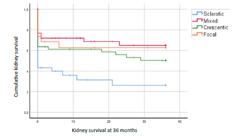
Figure 1. Kaplan‑Meier curves representing kidney survival at 36 months, according to Berden classification.
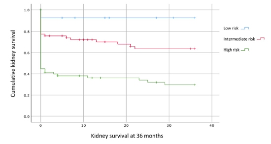
As for the ARRS, the median score in our cohort of patients with AAV was 6.5 ± 3.6, with 10.8% (n=17), 44.3% (n=70) and 44.9% (n=71) of biopsies classified as being in the low, intermediate and high‑risk groups, respectively. Kaplan Meier estimates for renal survival at 36 months revealed an overall mean time to RRT of 20.0 months. This time interval varied by risk category, with patients in the lo ‑risk group experiencing a mean time to dialysis of 33.4 months, in the intermediate group 24.7 months and in the high‑risk group 12.7 months (Log‑R test: (2) = 24.9, p <0.001). Kidney survival by ARRS category at 36 months is represented in Fig. 2 (92.9%, 63.6% and 29.7% in the low, intermediate and high‑risk groups, respectively). For longer follow‑up, Kaplan Meier estimates for renal survival in the ARRS‑stratified classes showed an overall mean time to RRT of 103.6 months.
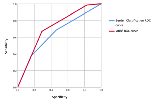
Overall patient survival at 36 months was also analysed, and there was no statistically significant difference be- tween groups: 88.2%, 87.1% and 87.3% in the low, intermediate and high‑risk groups, respectively (Log‑R test: (2) = 0.3, p=0.9). Univariate Cox regression analysis plotted for ESRD at 36 months using the three ARRS score variables identified eGFR at presentation as the strongest predictor of kidney survival (HR: 0.96, 95% CI: 0.93‑0.99, p=0.004), where-as PNG and IFTA did not achieve statistical significance (p=0.189 and p=0.066, respectively). A ROC curve was designed for both classification methods. In the Berden classification (Fig. 3), the model used sclerotic as the highest risk class of progression to ESRD, followed by crescentic, and finally mixed and focal classes together in the lowest risk. ARRS followed the low, medium and high‑risk scores. Both curves were significant with ARRS showing the highest statistical significance (AUC 0.72, p<0.001 vs 0.65, p=0.03).
DISCUSSION
To the best of our knowledge, this is the first study validating ARRS and comparing its performance to Berden classification in a Portuguese cohort. The prevalence of middle‑aged patients and the male gender in our cohort is consistent with known disease epidemiology and aligns with findings from other published cohorts.2,4Consistent with published evidence, in our study the sclerotic class in Berden classification was found to be associated with the worst renal outcomes, with a mean time to RRT of 11.4 months (Fig. 1).6,8,10As previously stated, crescentic and mixed classes have delivered inconsistent renal prognoses in prior investigations.3,6,7In our study, the crescentic sub-group presented worse outcomes than mixed subgroup (which in our cohort was equal to the focal class). This may be due to interobserver variability when examining kidney fragments under light microscopy, which could also explain the discrepancies in available evidence. When applying ARRS, we observed a high prevalence of both intermediate (44.3%; n=70) and high‑risk (44.9%; n=71) groups, with the latter group having a mean time to RRT of just 12.7 months. The high prevalence of both these classes could be explained by the often‑varied clinical presentation of AAV that can delay diagnosis and kidney biopsy, which often already reveals high IFTA scores and low PNG. Another possible explanation is that in some cases, diagnosis is made without the need for a biopsy, which could justify the lower prevalence of low‑risk ARRS samples in our cohort (10,8%; n=17).
In our study, kidney survival at 36 months in the low‑risk group was 92.9%, findings similar to results from Brix et al original cohort and other validation studies.2,4,9,11Regarding the intermediate and high‑risk groups, our results exhibited a lower kidney survival estimate than most studies (63.6% and 29.7%, respectively). Although we were not able to gather this data, this could be due to a higher prevalence of comorbidities in our cohort. Chronic kidney disease prevalence in Portugal is well above European and world average, with high prevalence of hypertension, diabetes and obesity in our population.12 Another possible reason for these differences could be in the dependence of different treatment schemes used from 2004 to 2023, especially with the emergence of rituximab as both an induction and maintenance immunosuppressant. Despite in the original ARRS study authors stating treatment did not influence the scoring system,2 in fact, a study developed in a Scottish cohort concluded that the inclusion of treatment modalities increased ARRS predictive strength.13 Regarding overall patient survival, we found no statistically significant difference between the three groups.
Unlike Brix et al original paper, in our study eGFR emerged as the only independent predictor of renal outcomes (HR: 0.96, 95% CI: 0.93‑0.99, p=0.004). However, we recognize eGFR in the setting of acute kidney disease is difficult to interpret and serum creatinine is a more reliable marker of kidney injury. A recently published article tested a new revised model of ARRS which substituted eGFR for serum creatinine, with similar performances and better discriminative power.9 In our study, ARRS was a statistically better predictor of ESRD at 36 months than Berden classification (Fig. 3). This superiority was also demonstrated in other studies though not always attaining statistical significancy and results are not consistent in available evidence.8,14,15There is a lack of comparative studies between both predictive models and prospective studies with longer follow-up could clarify if one tool is indeed superior to the other. Our study has several caveats regarding its design and sampling. First, we incurred in a selection bias since there was no randomization of the sample and data was collected from a limited time interval (2004 to 2023). Secondly, our data is limited to Portuguese patients, and although there may be foreign ancestry in some, we cannot safely translate our results to other populations. Finally, we do not have information on comorbidities or treatment options used for each patient, which may interfere in renal prognosis.
CONCLUSION
In conclusion, although AAV can be diagnosed without the need for a kidney biopsy, histology is essential to better assess the degree of active inflammation and chronicity of disease and to determine renal prognosis. ARRS performed well in a Portuguese cohort, but replacing eGFR with serum creatinine and the inclusion of treatment modalities could enhance the score’s predicting power. Between the two tested risk scales, ARRS performed better than Berden classification in determining progression to ESRD at 36 months in our cohort. ARRS is a simple tool that may aid clinicians in tailoring immunosuppression depending on risk estimate, favoring its use in patients who are likely to recover some kidney function. This could halt infectious complications and potentially diminish morbidity and mortality in AAV patients. While ARRS has been validated in several small cohorts, its performance has still to be replicated in large‑scale prospective interventional studies to allow for worldwide recommended use.

