




 Inglês (pdf)
 Artigo em XML
 Referências do artigo
 Tradução automática
 Enviar este artigo por email
 Citado por SciELO
  Similares em
SciELO
Similares em
SciELO 
 Permalink

INTRODUCTION
Hyponatremia is the most common electrolyte disorder, affecting up to 35% of hospitalized patients.¹ Accurate assessment of volume status is essential for both the diagnosis and management of hyponatremia.² Since the introduction of the venous excess ultrasound grading system (VExUS)-originally developed to predict diuretic responsiveness in the setting of acute kidney injury following cardiac surgery³-its use has been explored in various clinical contexts, including acute kidney injury⁴ and myocardial infarction.⁵ More recently, ultrasound‑based techniques have been proposed for evaluating volume status in patients with hyponatremia,⁶ with reported application in critically ill patients⁷ and in the assessment of severe hyponatremia in hospitalized individuals.⁸ The objective of this study was to evaluate the usefulness of VExUS in guiding the management of acute hypoosmolar hyponatremia in hospitalized patients, to determine its ability to identify discrepancies between clinical examination and ultrasound‑based assessments, and to assess its potential role in predicting clinical outcomes in this population.
METHODS
We conducted a prospective observational study between November 1, 2023, and August 31, 2024, in which patients were managed according to established clinical guidelines for hyponatremia.⁹ Within the first 24 hours of enrolment, each patient underwent an ultrasound examination assessing at least two of the following: inferior vena cava diameter and collapse, Doppler flow of the hepatic veins, portal vein, and renal veins. The treating physician was blinded to the ultrasound findings and had no access to the sonographer’s assessment. Ultrasound examinations were performed by multiple experienced clinicians.
Population and Sample
Adult patients diagnosed with moderate to severe asymptomatic hypoosmolar hyponatremia (plasma sodium ≤130 mEq/L) were eligible for inclusion. Symptomatic patients were also included once clinically stabilized.
Exclusion criteria comprised patients with pseudohyponatremia, diabetic decompensation, uncontrolled active multiple myeloma, ongoing thiazide therapy, post-urological procedure hyponatremia, known chronic hyponatremia, stage V chronic kidney disease or those receiving dialysis, and individuals under palliative care or approaching end of life. Patients with known decompensated liver cirrhosis, severe tricuspid regurgitation, or pregnancy were also excluded, as these conditions were deemed likely to interfere with ultrasound assessment.
Patients for whom at least two valid ultrasound measurements could not be obtained were likewise excluded.
Main Outcome
The primary study variable was serum sodium concentration, measured at 24, 48, and 96 hours after inclusion. We compared patients who exhibited discrepancies between volume status assessments based on clinical examination and those based on the VExUS grading system. Additionally, we compared the final etiological diagnosis-established after reviewing complementary diagnostic tests-with the VExUS classification.
According to the predefined interpretation criteria, a discrepancy was considered present when the clinical assessment suggested a non-hypervolemic state, but VExUS indicated venous congestion. Conversely, if the clinical evaluation indicated hypervolemia and VExUS did not show venous congestion, this was not regarded as a discrepancy, as not all hypervolemic states are necessarily associated with venous congestion.¹⁰
Secondary Outcomes
Clinical data were collected regarding medication use and symptoms at the time of study inclusion. Physical examination findings included the presence of oedema, inspiratory crackles, ascites, and hepatojugular reflux. An overall clinical assessment of the patient’s volume status was also recorded.
At study entry, all patients underwent testing for urinary osmolality, urinary electrolytes, and venous blood gas analysis. Patients with evidence of pseudohyponatremia on blood gas analysis were excluded. It was recorded whether the hyponatremia was considered antidiuretic hormone (ADH)‑mediated or non‑antidiuretic hormone (non‑ADH)‑mediated based on urinary osmolality, whether the results were influenced by diuretic use, and whether urinary sodium levels were consistent with hypovolemia or hypervolemia.
Within the first 48 hours, laboratory tests were performed including creatinine, estimated glomerular filtration rate (eGFR), serum osmolality, low‑density lipoprotein cholesterol (LDL), high-density lipoprotein cholesterol (HDL), triglycerides, total cholesterol, total proteins, albumin, N‑terminal pro B‑type natriuretic peptide (NT‑proBNP), uric acid, cancer antigen 125 (CA‑125),11,12albumin-to-creatinine ratio, thyroid‑stimulating hormone (TSH), adreno-corticotropic hormone (ACTH), and baseline cortisol. Data were also collected on treatments administered, including any changes in therapy after 24 hours or following the availability of additional test results.
Volume Status Assessment Using VExUS
Based on previous evidence in the setting of cardiac surgery‑related kidney failure3 and heart failure, 13 patients were considered non‑congestive under the following conditions:
Or
Not determined IVC and normal venous Doppler waveforms.
Patients were considered congestive under the following conditions:
All images were saved for subsequent review.
Statistical Analysis
Descriptive and inferential statistical analyses were performed. A normality test (Shapiro‑Wilk) was conducted beforehand, and a non‑parametric test was used when appropriate (Mann‑Whitney, Kruskal‑Wallis). Additionally, the relationship between VExUS findings and analytical surrogates of volume status-namely, fractional excretion of sodium (FENa%), N‑terminal pro B‑type natriuretic peptide (NT‑proBNP), and cancer antigen 125 (CA‑125)-was explored.
Data analysis was carried out using R Studio.
Ethical Considerations
This clinical study has been designed and conducted in strict accordance with the ethical principles set out in the Declaration of Helsinki.14 It has also been reviewed and approved by the Research Ethics Committee of Hospital La Fe in Valencia on October 25th of 2023 with registration number 2023‑880‑1, ensuring compliance with national and international regulations on research involving human subjects.
All participants were duly informed about the study’s objectives, procedures, benefits, and possible risks, and voluntarily provided their informed consent before inclusion in the research.
RESULTS
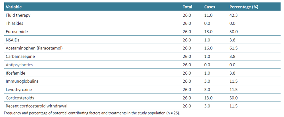
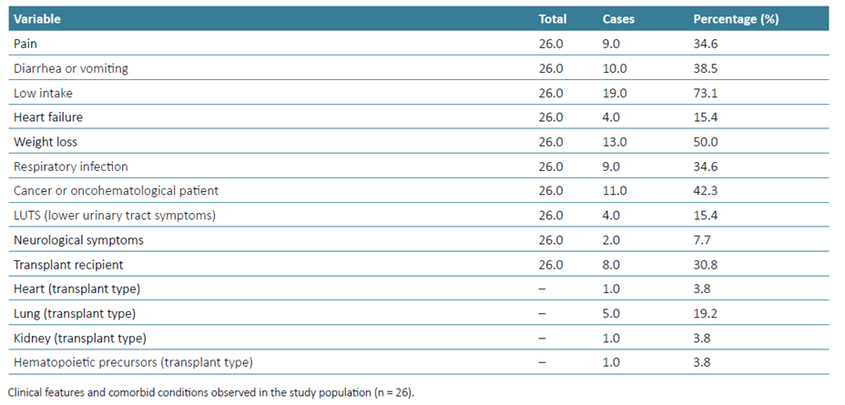
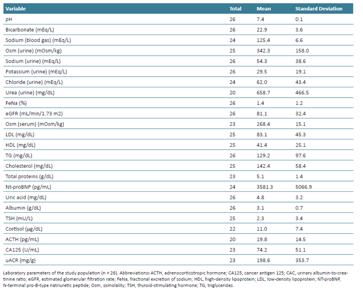
A total of 28 patients were enrolled during the study period. Of these, two were excluded: one for insufficient ultrasound data acquisition and another due to newly diagnosed cirrhosis decompensation. The characteristics of the included patients and the laboratory results obtained are summarized in Tables 1‑4. A total of 27% (n=7) of the patients had a solid organ transplant, and one patient had undergone hematopoietic stem cell transplantation.

Volume Status Assessments
Based on standard clinical assessment, including medical history and physical examination, 34% (n = 9) of patients were classified as euvolemic, 46% (n = 12) as hypervolemic, and 19% (n = 5) as hypovolemic. After incorporating the results of additional diagnostic tests, 11.5% (n = 3) were found to have hyponatremia not mediated by antidiuretic hormone (ADH). Among the ADH‑mediated cases, 30.8% (n = 8) were consistent with the syndrome of inappropriate antidiuretic hormone secretion (SIADH), 15.4% (n = 4) with hypervolemia, 23.1% (n = 6) with hypovolemia, and in 19.2% (n = 5), the classification was deemed inconclu-sive due to ongoing diuretic therapy.
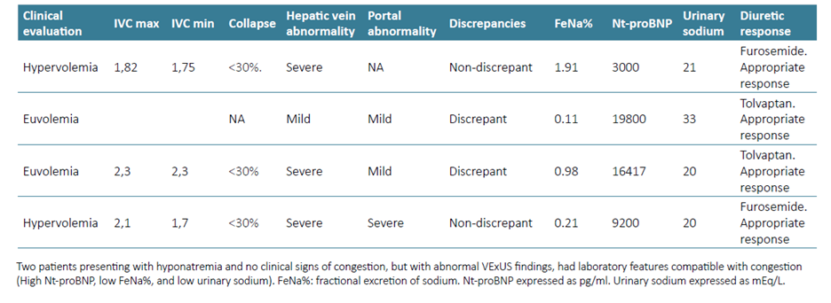
According to the venous excess ultrasound grading system (VExUS), 15.4% (n = 4) of patients were classified as congestive, while 84.6% (n = 22) were classified as non‑congestive. All patients diagnosed with SIADH were categorized as non‑congestive according to VExUS. Discrepancies between clinical and VExUS‑based volume status assessments were identified in 7.7% (n = 2) of patients. No discrepancies were observed between the VExUS‑based assessment and the final volume status classification following integration of complementary laboratory tests. Congestive cases and laboratory findings are summarized in Table 5.
There were no statistically significant differences in serum sodium levels between discrepant and non‑discrepant groups at any of the time points assessed: baseline (0 h), p = 0.409 (n = 26); 24 h, p = 0.884 (n = 26); 48 h, p = 0.598 (n = 24); and 96 h, p = 0.351 (n = 23). The evolution of sodium levels by discrepancy status is illustrated in Fig. 1. The proportion of missing ultrasound measurements by anatomical region was as follows: inferior vena cava, 34.6% (n = 9); hepatic veins, 15.4% (n = 4); portal vein, 3.8% (n = 1); and renal veins, 30.8% (n = 8).
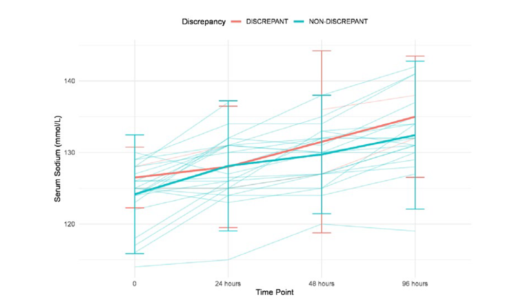
Figure 1 Evolution of serum sodium levels according to the presence or absence of discrepancies between clinical and ultrasound volume assessment. Lines represent individual patient trajectories. Bold lines indicate the group mean, with vertical bars representing ±2 standard deviations. No statistically significant differences were observed at any time point using the Kruskal‑Wallis test (p > 0.1). A bootstrap analysis (1000 samples) confirmed the robustness of these findings.
Comparison Between Volume Status Assessment and Surrogate Laboratory Parameters of Volume
A statistically significant difference was found in NT‑proB-NP levels between the congestive (n = 4) and non‑congestive (n = 20) groups (Wilcoxon, p = 0.003), as shown in Fig. 2.17
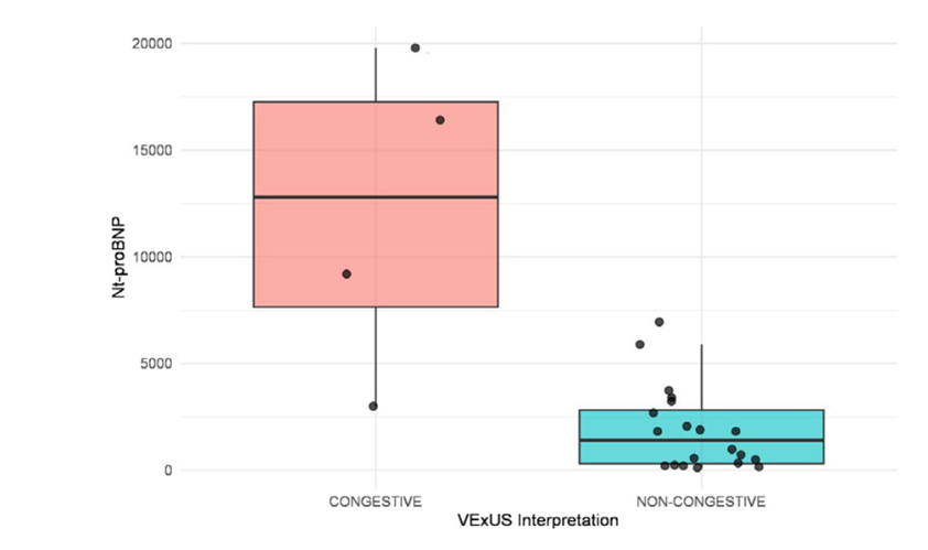
Figure 2 Comparison of biomarker values between patients classified as congestive and non‑congestive according to VExUS interpretation. Each boxplot displays the distribution of biomarker values in the two groups. A significant difference was observed using the Wilcoxon ranksum test (p = 0.003). Median biomarker levels were markedly higher in the congestive group (n = 4) compared to the non‑congestive group (n = 20).
No statistically significant difference was found in CA125 between the congestive (n = 4) and non‑congestive (n = 19) groups (t-test, p = 0.844), indicating comparable biomarker distributions between both categories as defined by VExUS interpretation.
No statistically significant difference was found in FeNa (%) between the congestive (n = 4) and non‑congestive (n = 22) groups (Wilcoxon, p = 0.201).
Treatments
A total of 65.4% (n = 17) of patients were treated with fluid restriction, while 30.8% (n = 8) received fluid therapy. Among those who received diuretics, furosemide was the most used in 26.9% (n = 7) of patients, followed by tolvaptan in 15.4% (n = 4). Two patients (7.7%) received urea19 as the initial treatment. Hypertonic saline was administered as initial therapy in 23.1% (n = 6) of patients, and 19.2% (n = 5) continued to receive it after 24 hours. Two patients (7.7%) experienced early clinical deterioration, both of whom required initiation of 3% saline infusion.9,18In one case, cotrimoxazole was discontinued as a cause of hyponatremia; in another, fluid administration was suspended; and in a third, spironolactone was withdrawn. One patient (3.8%) presented with tacrolimus toxicity requiring dose adjustment, and another (3.8%) had levothyroxine reintroduced after it was inadvertently discontinued at admission.
Patients with discrepancies between clinical and ultra-sound‑based volume assessments were managed with diuretics and evolved favourably. Only four patients (15.4%) required a change in treatment 24 hours after study inclusion, and none of them had shown discrepancies between the clinical assessment and VExUS findings.
Among patients treated with diuretics (n=11), a non‑significant (linear mixed effects model; p = 0,119) tendency towards a faster sodium level improvement was observed in the non‑congestive group, as shown in Fig. 3.
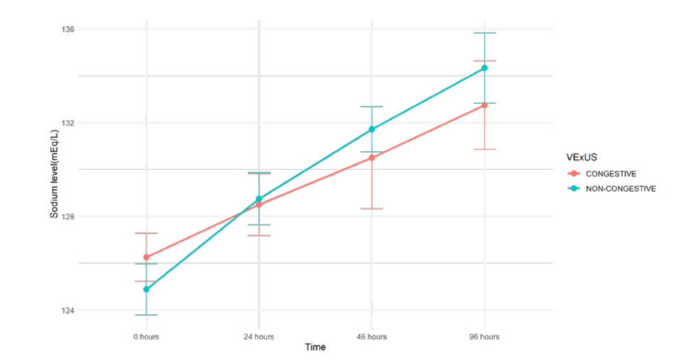
Figure 3 In this mixed‑effects model, serum sodium levels increased over time in the congestive group, with a mean rise of 6.5 mEq/L by 96 hours (p < 0.001). The non‑congestive group showed a slightly greater increase at each time point, the interaction terms between time and group were not statistically significant.
The distribution of attributed aetiologies and the therapeutic approaches selected for hyponatremia are consistent with those documented in large registries; however, the success rate of correction significantly exceeds typical observations, likely a consequence of the controlled study environment.
DISCUSSION
To our knowledge, this is the second study,8 beyond isolated clinical reports, to examine ultrasound measurements for volume status assessment in hospitalized patients with hyponatremia and the first to evaluate the clinical impact of the discrepancies found.
There are significant methodological differences between this study and the one by Rahman et al (2024).8 Rahman et al8 used a global examination, reviewing the presence of pleural effusion or ascites and exclusively assessing the inferior vena cava. The present study adheres to the evaluation proposed by Beaubien‑Souligny et al (2020).3 Furthermore, patients with moderate hyponatremia were not excluded in our study and they comprise most of our patients. Rahman et al (2024) did not exclude patients with tricuspid regurgitation or cirrhosis, a patient group that often presents with hypervolemia and hyponatremia.8 In the present study the number of patients identified as congestive was low (n = 4), suggesting a more limited role for VExUS in the context of hyponatremia, where venous congestion may be less prevalent than in other clinical settings, such as heart failure.20 VExUS alone is insufficient to prove a patient either euvolemic or hypovolemic. Thus, to widen the utility of point‑of‑care ultrasound in the context of hyponatremia, greater attention should be directed toward evaluating lung ultrasound, cardiac ultrasonography and cardiac output in non‑congestive patients with hyponatremia, to help classify the volume status in the future.6,17Discrepancies in volume status classification between clinical assessment and the VExUS system did not predict worse clinical outcomes or a greater need for treatment modifications. The general impact of VExUS in the management of hyponatremia may have been limited by the low prevalence of venous congestion in this population, particularly when patients with severe tricuspid regurgitation and cirrhosis are excluded, and the convergence of therapeutic strategies across different volume status categories.1 For example, diuretics may be used in both syndrome of inappropriate antidiuretic hormone secretion (SIADH) and heart failure, and hypertonic saline is commonly administered regardless of the underlying volume classification.
These findings do not negate the utility of VExUS. After complementary tests were analysed, no discrepancies were observed between the standard diagnostic approach and the VExUS interpretation. This demonstrates that some patients do not exhibit clinical signs of hypervolemia while still presenting with venous congestion in the context of hyponatremia. In fact, the two discrepant cases had initially been considered euvolemic based on clinical evaluation but were classified as congestive by VExUS. This indicates that the technique may be useful in identifying clinically silent venous congestion in hyponatremia, potentially preventing iatrogenic harm in selected cases. This evidence aligns with the results discussed above.8 A significant difference in NT‑proBNP levels was observed between congestive and non‑congestive patients. Interestingly, this association was not replicated with CA‑125, which may indicate that hypervolemic, but non‑congestive states are more prevalent in patients with hyponatremia than in other clinical scenarios such as heart failure, where CA‑125 has shown a stronger correlation with congestion and clinical outcomes.12 A non‑significant tendecy towards a faster improvement of the non‑congestive group treated with diuretics was observed, suggesting a role of the technique in predicting poorer outcomes in congestive patients when compared with other hypervolemic and non‑congestive or SIADH patients. VExUS has proved useful in predicting poorer outcomes in other clinical settings previously, such as heart failure20 and acute kidney injury.4 Ultrasound is safe and non-invasive, and its use may be particularly valuable in specific clinical scenarios, especially when volume status is uncertain, and the administration of intravenous fluids carries potential risk. For instance, in patients with a history of heart failure, ultrasound may provide critical information to guide management. In our cohort, at least two ultrasound parameters were successfully assessed in all but one patient. As a single abnormal finding can often yield clinically relevant insight, the implementation of VExUS3 alongside other clinical evaluations may offer added value, despite incomplete.
Finally, it is important to recognize that VExUS alone, given its limitations in non‑congestive patients, does not obviate the need for comprehensive evaluation as recommended in current clinical guidelines for the diagnosis and management of hyponatremia in most cases.
The study reflects the overall experience of a large hospital over 10 months and is consistent with the daily clinical practice.21 Furthermore, it gathers the largest sample of patients with hyponatremia evaluated with VExUS and includes most of clinical scenarios found when facing hyponatremia.
CONCLUSION
In this study, discrepancies between clinical assessment and VExUS‑based volume classification were identified (n = 2), improving diagnostic accuracy. The number of congestive cases (n = 4) suggests that the general use of VExUS may have a more limited role in hyponatremia than in settings where venous congestion is more prevalent. Nevertheless, ultrasound remains safe and non-invasive, and VExUS proved valuable in detecting clinically silent congestion in selected patients. The evidence presented here suggests that future research should incorporate a broader range of ultrasound assessments, such as cardiac and lung ultrasonography, to evaluate point-of-care ultra-sound in the management of hypoosmolar hyponatremia.
Take-home Messages
The venous excess ultrasound (VExUS) system is a valuable tool for classifying congestive status in patients presenting with hyponatremia.
VExUS plays a potentially critical role in patients where systemic congestion is not clinically apparent, allowing for the timely identification of volume overload and preventing the administration fluid therapy.
VExUS is not suitable as the sole ultrasound tool for clas-sifying the overall volume status of patients once venous congestion has been ruled out. In future research, lung and cardiac ultrasound may add further value in the evaluation of hyponatremia.
Consequently, the broad application of VExUS in hyponatremia management may be of limited utility in a significant number of cases and cannot replace complementary diagnostic workup.

