SciELO - Scientific Electronic Library Online

 
vol.37 issue2Wunderlich Syndrome in a Peritoneal Dialysis Patient with Warfarin Iatrogenic Coagulopathy: Should We Really be so Aggressive on Cardioembolic Events Prevention? author indexsubject indexarticles search
Home Pagealphabetic serial listing  

Services on Demand

Journal

Article

Indicators

Related links

  • Have no similar articlesSimilars in SciELO

Share


Portuguese Journal of Nephrology & Hypertension

Print version ISSN 0872-0169

Port J Nephrol Hypert vol.37 no.2 Lisboa June 2023  Epub June 30, 2023

https://doi.org/10.32932/pjnh.2023.07.243 

CASE REPORT

Nephrotic Syndrome and Waldenstrom’s Macroglobulinemia: A Case Report

1 Internal Medicine Department, Hospital de Vila Franca de Xira, Vila Franca de Xira, Portugal

2 Nephrology Department, Hospital de Vila Franca de Xira, Vila Franca de Xira, Portugal

3 Laboratory of Renal Morphology, Hospital de Curry Cabral, Centro Hospitalar e Universitário de Lisboa Central, Lisboa, Portugal


ABSTRACT

Waldenstrom’s macroglobulinemia (WM) is an IgM associated lymphoplasmacytic lymphoma and kidney disease is a rare complication. We report a case of a 41-year-old male with a history of WM diagnosed 16 years ago, currently with no indication for treatment. The patient presented with nephrotic syndrome and acute kidney injury, hypertension, and multiple lymphadenopathies. A kidney biopsy was performed, showing minimal change disease with lymphomatous infiltration of B-lymphocytes CD20+. Therapy with bortezomib, dexametahasone and rituximab was started and after 6 months of follow-up he presented progressive recovery of renal function and remission of proteinuria.

This case illustrates the importance of screening renal disease in WM patients. A kidney biopsy should be performed in those presenting with otherwise unexplained renal failure and/or nephrotic syndrome.

Keywords: Biopsy; Kidney/pathology; Nephrotic Syndrome; Waldenstrom Macroglobulinemia

INTRODUCTION

Waldenstrom’s macroglobulinemia (WM) is a B-cell lymphoproliferative disorder characterized by bone marrow infiltration by a lymphoplasmacytic lymphoma infiltration of bone marrow and a sérum immunoglobulin M (IgM) paraprotein.1,2 It is a rare lymphoid neoplasia, accounting for 2% of all hematological malignancies.3

WM can manifest with tissue infiltration by malignant cells, deposition of IgM and changes caused by specific properties of the sérum IgM, namely hyperviscosity. Although patients with WM can presente with an extensive range of signs and symptoms, most of them are related to tumoral infiltration (cytopenia and hepatosplenomegaly), monoclonal protein accumulation in the circulation (cryoglobulinemia and serum hyper viscosity syndrome), monoclonal protein accumulation in tissues (amyloidosis) or autoantibody production (neuropathy and hemolytic anemia).4,5

Although several pathogenic mechanisms that can affect multiple organs have been described regarding this disease, kidney involvement in WM is rare.2,6

The most common kidney biopsy findings include AL amyloidosis, cryoglobulinemic glomerulonephritis and direct infiltration of the kidney by atypical lymphoid cells along with large intracapillary aggregation of IgM. These injuries result from the infiltration of malignant cells, an immune mediated reaction to the cells or from the production of an abnormal paraprotein.2,7-9

We describe a case of WM with acute renal injury (AKI) and nephrotic syndrome (NS) due to a minimal change disease.

CASE REPORT

A 41-year-old Caucasian male presented to the hospital with a 3-week history of periorbital edema, slowly progressive bilateral lower extremity edema and weight gain. His past medical history includes MW diagnosis 16 years before. After diagnosis he received 8 cycles of R-CHOP (rituximab, cyclophosphamide, hydroxydaunorubicin hydrochloride, vincristine-oncovin and prednisolone) and after which his hematologist considered the patient to have an indolent form of MW, so the patient started a follow up with annual clinical and laboratorial evaluation. On physical examination the patient had a blood pressure of 146/96 mmHg, a 4+ bilateral lower extremity edema up to thighs, multiple palpable cervical, axillary, and inguinal lymph nodes and hepatosplenomegaly. The remaining clinical examination was unremarkable.

Laboratory results showed hypoalbuminemia (1.8 mg/dL), normal iron metabolism and beta 2 microglobulin was 10.786 mg/dL. Urinalysis showed of protein 400 mg/dL with a protein to creatinine ratio (P/C ratio) of 3.53 g/g. The remaining analytic results are presented in Table 1. Protein electrophoresis revealed a marked hypoalbuminemia and a monoclonal spike in the y region (Fig. 1 and Table 1). Immunoelectrophoresis showed an IgM kappa monoclonal protein (Table 1).

Figure 1 Protein electrophoresis. 

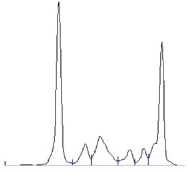
Table 1 Analytical results 

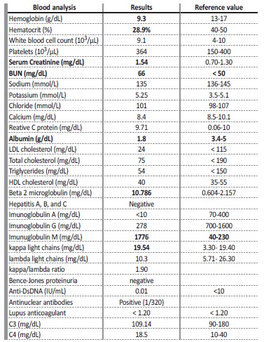
Anti-DsDNA - anti double stranded DNA; BUN - Blood urea nitrogen; HDL - High density lipoprotein

cholesterol; LDL-Low - density lipoprotein

Thorax, abdomen, and pelvis computed tomography revealed hepatosplenomegaly and multiple axillar lymphadenopathies, intra-abdominal and inguinal regions. The kidney ultrasound showed kidneys with preserved form and a slightly enlarged dimensions on the left; an obstructive component was excluded.

As a patient with a known history of WM, he was observed by ophthalmology and neurology and complications related to hyperviscosity were ruled out. The fat abdominal biopsy was negative for amyloidosis.

Kidney biopsy (left kidney) showed an interstitial lymphomatous infiltration (Fig. 2). Glomeruli were unremarkable. Immunohistochemistry showed interstitial staining for CD20 + B cells, (Fig. 3), CD5 - and CD10 -. Immunofluorescence was negative and electron microscopy showed the presence of diffuse effacement of podocytes foot processes and no electron dense deposits were presented. A diagnosis of minimal change disease (MCD) and renal parenchymal infiltration by lymphoplasmacytic lymphoma was made.

Figure 2 Light microscopy: PAS staining - Normal glomeruli, lymphomatous infiltrate (100x). 

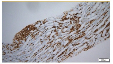
Figure 3 Immunohistochemistry for CD20: Lymphoplasmocytic infiltrate with CD 20 expression (ME 200x) 

The patient was evaluated by hematology and steroids were started during hospitalization. After discharge, he was followed up in a hematology and nephrology outpatient clinic. He started treatment with bortezomib, dexamethasone and rituximab with progressive recovery of renal function (serum creatinine 1.01 mg/dL), and gradual remission of proteinuria after 6- months. (P/C ratio 0.185 g/g; serum albumin 3.8 g/dL).

Table 2 Renal manifestations of WM 

Adapted from: Uppal NN, et al. Kidney diseases associated with Waldenström macroglobulinemia.

Nephrol Dial Transplant. 2019;34:1644-52.2

DISCUSSION AND CONCLUSION

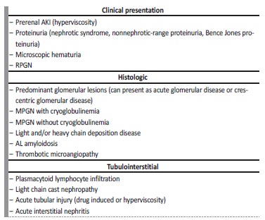
Although infrequent, kidney involvement in Waldenström macroglobulinemia is well documented (Table 2). Renal manifestations are most likely related to abnormal monoclonal protein and/or tissue infiltration by underlying lymphoplasmacytic neoplasm. Only less than 3% of the patient with WM develop end-stage renal disease and NS is present in less than 7% of the patients with WM.2,3,10,11

NS in the presence of lymphoplasmacytic lymphoma with IgM monoclonal gammopathy (WM) can have several causes but immunoglobulin light chain (AL) amyloidosis is the most frequent cause of NS in these patients, and non-amyloid nephrotic syndromes are very rare in the worldwide literature.6,7,12,13

Minimal change disease, a common complication of Hodgkin’s lymphoma, seems to occur very rarely in patients with WM.3,10,13

The pathophysiology of MCD remains poorly understood, however a T-lymphocyte disorder is suggested by the occurrence of the disease in the setting of immune challenge initiated by infectious diseases, drugs, allergies or vaccinations, and also the sensitivity to immunosuppressive agents. In WM, as well as in Hodgkin’s lymphoma patients, a T-cell disorder leading to a reduced CD4/CD8 ratio and an abnormal secretion of lymphokines has been documented and may be associated to the occurrence of MCD.12,14-18

AKI is also an uncommon presentation of MW. The most common causes of AKI in monoclonal gammopathies, such as hypercalcemia, uric acid nephropathy, dehydration, cast nephropathy, drug induced and sepsis, were not present in this case, so we admitted that the severe interstitial lymphocytic infiltrate was the cause of AKI.3

This case illustrates the importance of screening for renal disease in WM patients. Minimal change disease should be considered as a rare, but possible, cause of nephrotic syndrome in WM. A kidney biopsy should be performed in those presenting with otherwise unexplained AKI and/or nephrotic syndrome. Early detection of kidney disease and adequate treatment can preserve renal function while controlling the hematologic disease.19,20

References

1. Swerdlow SH, Campo E, Harris NL, Jaffe ES, Pileri SA, Stein H, et al. WHO Classification of Tumors of Haematopoietic and Lymphoid Tissues. Lyon: IARC Press; 2008 [ Links ]

2. Uppal NN, Monga D, Vernace MA, Mehtabdin K, Shah HH, Bijol V, et al. Kidney diseases associated with Waldenström macroglobulinemia. Nephrol Dial Transplant. 2019;34:1644-52. doi: 10.1093/ndt/gfy320. [ Links ]

3. Salviani C, Guido G, Serriello I, Giannakakis K, Rocca AR. Renal involvement in Waldenström’s macroglobulinemia: case report and review of literature. Ren Fail. 2014;36:114-8. doi: 10.3109/0886022X.2013.832859. [ Links ]

4. Banwait R, Aljawai Y, Cappuccio J, McDiarmid S, Morgan EA, Leblebjian H, et al. Extramedullary Waldenström macroglobulinemia. Am J Hematol. 2015;90:100-4. doi: 10.1002/ajh.23880. [ Links ]

5. Gnemmi V, Leleu X, Provot F, Moulonguet F, Buob D. Cast nephropathy and light-chain deposition disease in Waldenström macroglobulinemia. Am J Kidney Dis. 2012;60:487-91. doi: 10.1053/j.ajkd.2012.01.030. [ Links ]

6. Castro H, Valenzuela R, Ruiz P, Lenz O, Monrroy M. Nephrotic syndrome and kidney failure due to immunocomplex-mediated renal damage in a patient with Waldenström’s Macroglobulinemia: a case report. Cases J. 2008;1:333. doi: 10.1186/1757-1626-1-333. [ Links ]

7. Lee B, Smith RS, Tanphaichitr N, Novak R, Robertson S, Haller NA. Waldenström’s macroglobulinemia and nephrotic syndrome with membranous nephropathy. Scand J Urol Nephrol. 2011;45:473-7. doi: 10.3109/00365599.2011.568954. [ Links ]

8. Chauvet S, Bridoux F, Ecotière L, Javaugue V, Sirac C, Arnulf B, et al. Kidney diseases associated with monoclonal immunoglobulin M-secreting B-cell lymphoproliferative disorders: a case series of 35 patients. Am J Kidney Dis . 2015;66:756-67. doi: 10.1053/j.ajkd.2015.03.035. [ Links ]

9. Audard V, Georges B, Vanhille P, Toly C, Deroure B, Fakhouri F, et al. Renal lesions associated with IgM-secreting monoclonal proliferations: revisiting the disease spectrum. Clin J Am Soc Nephrol. 2008;3:1339-49. doi: 10.2215/CJN.01600408. [ Links ]

10. Grabe DW, Li B, Haqqie SS. A case of nephrotic syndrome with minimal-change disease and Waldenstrom’s macroglobulinemia. J Clin Med Res. 2013;5:481-3. doi: 10.4021/jocmr1387w. [ Links ]

11. Haraguchi S, Tomiyoshi Y, Aoki S, Sakemi T. Nephrotic syndrome due to immunologically mediated hypocomplementic glomerulonephritis in a patient of Waldenström’s macroglobulinemia. Nephron. 2002;92:452-5. doi: 10.1159/000063286. [ Links ]

12. Terrier B, Buzyn A, Hummel A, Deroure B, Bollée G, Jablonski M, et al. Serum monoclonal componente and nephrotic syndrome--it is not always amyloidosis. Diagnosis: WM complicated by retroperitoneal and renal infiltration and associated with a minimal change disease. Nephrol Dial Transplant. 2006;21:3327-9. doi: 10.1093/ndt/gfl467. [ Links ]

13. Swaneveld FH, van Dorp WT, Visser O, de Klerk G. Waldenström’s macroglobulinaemia presenting with nephrotic syndrome. Neth J Med. 2012;70:411-3. [ Links ]

14. Javaugue V, Debiais-Delpech C, Nouvier M, Gand E, Chauvet S, Ecotiere L, et al. Clinicopathological spectrum of renal parenchymal involvement in B-cell lymphoproliferative disorders. Kidney Int. 2019;96:94-103. doi: 10.1016/j.kint.2019.01.027. [ Links ]

15. Mwamunyi MJ, Zhu HY, Zhang C, Yuan YP, Yao LJ. Pseudothrombus deposition accompanied with minimal change nephrotic syndrome and chronic kidney disease in a patient with Waldenström’s macroglobulinemia: A case report. World J Clin Cases. 2019;7:2393-400. doi: 10.12998/wjcc.v7.i16.2393. [ Links ]

16. Vos JM, Gustine J, Rennke HG, Hunter Z, Manning RJ, Dubeau TE, et al. Renal disease related to Waldenström macroglobulinaemia: incidence, pathology and clinical outcomes. Br J Haematol. 2016;175:623-30. doi: 10.1111/bjh.14279. [ Links ]

17. Higgins L, Nasr SH, Said SM, Kapoor P, Dingli D, King RL, et al. Kidney Involvement of Patients with Waldenström Macroglobulinemia and Other IgM-Producing B Cell Lymphoproliferative Disorders. Clin J Am Soc Nephrol . 2018;13:1037-46. doi: 10.2215/CJN.13041117. [ Links ]

18. Lagrue G, Branellec A, Blanc C, Xheneumont S, Beaudoux F, Sobel A, et al. A vascular permeability factor in lymphocyte culture supernants from patients with nephrotic syndrome. II. Pharmacological and physicochemical properties. Biomedicine. 1975; 23: 73-5. [ Links ]

19. San Miguel JF, Caballero MD, Gonzalez M. T-cell subpopulations in patients with monoclonal gammopathies: essential monoclonal gammopathy, multiple myeloma, and Waldenstrom macroglobulinemia. Am J Hematol. 1985; 20: 267-73. [ Links ]

20. Hory B, Saunier F, Wolff R, Saint-Hillier Y, Coulon G, Perol C. Waldenström Macroglobulinemia and Nephrotic Syndrome with Minimal Change Lesion. Nephron . 1987; 45:68-70. doi: 10.1159/000184075 [ Links ]

Received: April 20, 2023; Accepted: June 22, 2023

Corresponding Author: Ana Isabel Oliveira Internal Medicine Department, Hospital de Vila Franca de Xira Estrada Carlos Lima Costa, 2600-009 Vila Franca de Xira, Portugal E-mail: ana.oliveira@hvfx.min-saude.pt

Creative Commons License This is an open-access article distributed under the terms of the Creative Commons Attribution License