SciELO - Scientific Electronic Library Online

 
vol.30 número420 anos de relatórios de primavera - percurso de aprendizagensSíndrome de Dravet − experiência de uma unidade de neuropediatria índice de autoresíndice de materiabúsqueda de artículos
Home Pagelista alfabética de revistas  

Servicios Personalizados

Revista

Articulo

Indicadores

Links relacionados

  • No hay articulos similaresSimilares en SciELO

Compartir


Nascer e Crescer

versión impresa ISSN 0872-0754versión On-line ISSN 2183-9417

Nascer e Crescer vol.30 no.4 Porto dic. 2021  Epub 31-Dic-2021

https://doi.org/10.25753/birthgrowthmj.v30.i4.18998 

Original article

Orbital complications in pediatric acute sinusitis: eight-year experience

Complicações orbitárias de sinusite aguda em pediatria: experiência de oito anos

João Fonseca Neves1 
http://orcid.org/0000-0003-2922-9717

João Filipe Simões2 

Sofia Paiva1 

Felisberto Maricato1 

Luís Filipe Silva1 

1.Department of Otorhinolaryngology, Centro Hospitalar e Universitário de Coimbra. 3004-561 Coimbra, Portugal. jfonsecaneves@gmail.com; sofpaiva@gmail.com; felisbertomaricato@gmail.com; luis.f.silva2006@gmail.com.

2.Faculdade de Medicina da Universidade de Coimbra. 3000-370 Coimbra, Portugal. jofsim@gmail.com.


Abstract

Objectives:

Acute sinusitis accounts for up to 82% of orbital infection cases. Infection spreads very quickly, especially through the ethmoid sinus, and orbital complications may arise even under antibiotic therapy. The aim of this study was to describe an 8-year hospital experience with these children.

Methods:

All cases of acute sinusitis with orbital complications admitted to the Department of Otorhinolaryngology of Centro Hospitalar e Universitário de Coimbra between 2010 and 2017 were retrospectively reviewed.

Results:

Sixty-four patients met the inclusion criteria, with a mean age of 9 ± 4.7 years. Male:female ratio was 1.67:1. Most subjects were admitted in the winter period (57.8%), with 2.9 ± 2.5 days of clinical evolution. The mean Lund Mackay score was 10.6 ± 4.9, with maxillary and ethmoid being the most prevalent involved sinuses (96.4% and 94.6%, respectively), and was inversely correlated with age (p<0.05). Preseptal cellulitis was the most common complication (56.3%). Abscesses were identified in 18.7% of patients, but only four (6.25%) required surgery. Seven cases (10.9%) recurred shortly after hospital discharge and required prolonged antibiotic course. All patients recovered well, without further complications.

Conclusion:

Results showed that orbital complications of sinusitis respond well to high doses of endovenous antibiotherapy and patients tend to recover without local comorbidities. Close monitoring of these patients during the first months after hospital discharge is crucial to prevent early relapse.

Keywords: complications; orbital disease; sinusitis

Resumo

Objetivos:

A sinusite aguda é responsável por até 82% dos casos de infeções orbitárias. As infeções propagam-se facilmente, em particular através das células etmoidais, mesmo em crianças sob antibioterapia. O objetivo deste estudo foi descrever a experiência de oito anos de um centro hospitalar com esta patologia.

Métodos:

Revisão retrospetiva dos casos de sinusite aguda com complicações orbitárias admitidos no Departamento de Otorrinolaringologia do Centro Hospitalar e Universitário de Coimbra entre 2010 e 2017.

Resultados:

Sessenta e quatro doentes foram incluídos no estudo, com uma idade média de 9 ± 4,7 anos e uma proporção de rapazes:raparigas de 1,67:1. A maioria dos doentes foi internada nos meses de inverno (57,8%), com 2,9 ± 2,5 dias de evolução clínica. O score de Lund Mackay foi de 10,6 ± 4,9, com os seios maxilar e etmoidais mais prevalentemente envolvidos (96,4% e 94,6%, respetivamente), e correlacionou-se inversamente com a idade (p <0,05). A celulite pré-septal foi a complicação mais comum (56,3%). Foram identificados abscessos em 18,7% dos doentes, mas apenas quatro (6,25%) necessitaram de tratamento cirúrgico. Sete (10,9%) casos ocorreram logo após a alta hospitalar e exigiram antibioterapia prolongada. Todos os doentes recuperaram bem, sem lesões sequelares.

Conclusão:

Os resultados obtidos demonstram que as complicações orbitárias da sinusite respondem bem à terapêutica médica e os doentes frequentemente recuperam sem comorbilidades locais. O acompanhamento clínico destes doentes durante os primeiros meses após a alta hospitalar é essencial para evitar recidivas precoces.

Palavras-chave: complicações; doença orbital; sinusite

Introduction

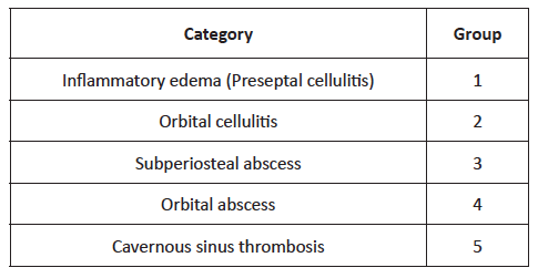
Acute sinusitis is responsible for up to 82% of cases of orbital infection. (1 Before the widespread use of antibiotherapy, 17% of patients affected by orbital cellulitis died from meningitis and 20% suffered permanent visual loss. (2 Orbital complications can occur either directly through a defect in the lamina papyracea or from an septic emboli. (3 Diagnosis is usually established through the combination of clinical examination and radiologic findings. (4 Chandler’s classification (Table 1), published fifty years ago, still represents the most complete and popular classification of orbital infection severity. (5

The best pharmacological modality for these patients remains controversial, and the optimal surgical approach has been debated, especially since the widespread use of endoscopic nasal surgery. (6 The aim of this study was to analyze the outcomes of patients admitted to the Department of Otorhinolaryngology of our institution with orbital complications due to acute sinusitis over an eight-year period.

Table 1 Chandler classification 

Material and methods

A retrospective review of medical records of all children (age <18 years) diagnosed with orbital complications of acute sinusitis admitted to our hospital department between 2010 and 2017 was conducted. Data retrieved included demographics, clinical signs and symptoms, laboratory study, radiologic evidence of orbital inflammation and sinusitis, treatment with intravenous antibiotic, and surgical intervention.

Stata® 15.0 was used for data descriptive and analytical statistics. Associations between dichotomic variables were assessed with chi-squared test (or Fisher’s exact test, when applicable), and continuous measures were compared using a two-sample student t- test. Statistical significance was set at 0.05.

Results

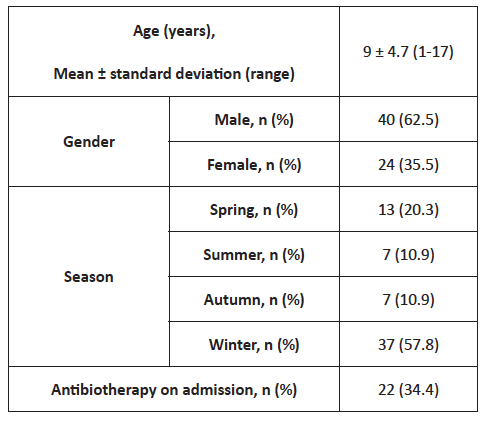
A total of 64 children were admitted to our institution between January 2010 and December 2017, with a mean age of 9 ± 4.7 (range 1-17) years and predominantly (62.5%) male. Most patients were admitted during winter months (57.8%) and 65.6% were not under any medication on admission (Table 2).

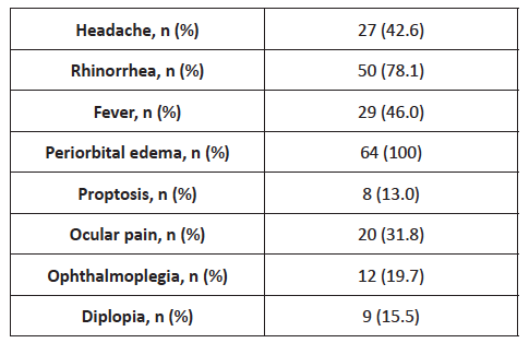
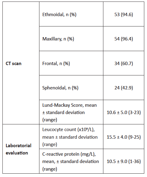
Clinical findings are resumed in Table 3. Most patients were admitted with rhinorrhea (78.1%) and only 46% presented with fever. Patients with rhinorrhea tended to be admitted earlier to the Emergency Department compared with patients without this symptom (2.6 vs. 4 days, respectively; p=0.05). Ocular signs were uncommon, with ocular pain being the most relevant ophthalmologic sign (31.8%), followed by ophthalmoplegia, diplopia, and proptosis. Computed tomography (CT) scan was performed in 65 (87.5%) patients, with ethmoidal and maxillary being the most commonly affected sinuses (95.6% and 96.4%, respectively; Table 4) . Frontal disease was more prevalent in older patients (11.0 ± 4.0 vs. 7.9 ± 4.2; p=0.01). Lund-Mackay score was used for sinusitis radiologic staging, showing a mean value of 10.6 ± 5.0 (range 3-23) and inverse correlation with age (p=0.01). Most (53.8%) patients with proptosis on admission presented abscesses in CT scan (p=0.001). Laboratory findings showed a mild rise in white blood cells (15.5 ± 4.0 x 10⁹/L) and C-reactive protein (10.5 ± 9.0 mg/L; Table 4).

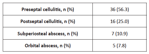
Cellulitis was the most prevalent complication in this patient population (Table 5). Thirty-six patients (56.3%) developed preseptal (Chandler type I) and 16 patients postseptal (Chandler type II) cellulitis. Overall, 12 patients (18.8%) presented subperiosteal (n=7) or orbit (n=5) abscesses. Two patients with orbital abscesses developed intracranial complications: cerebral abscess in one patient and cavernous sinus thrombosis in another patient.

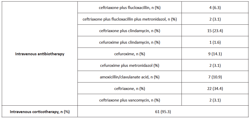
Treatment predominantly included high doses of intravenous ceftriaxone combined with other drugs (flucloxacilin, clyndamicine, or metronidazole; Table 6). Sixty-one (95.3%) patients received concomitant intravenous corticotherapy. Patients exclusively treated with medical therapy had a lower mean age (8.75 ± 4.8 years; p>0.05). Only four patients, with a mean age of 12.8 ± 3.0 years, required surgical drainage, three of whom with external approach and one with combined approach. Two of these patients presented positive culture, one for Fusobacterium necrophorum and the other for Streptococcus constellatus. The mean length of hospital stay was 8.0 ± 5.5 days (range 2-38), and was longer in patients submitted to surgery (11.3 ± 2.9 vs. 7.8 ± 5.6 days in patients undergoing pharmacotherapy, p >0.05) and in older patients (R=0.2546, p= 0.04).

Seven patients (10.9%) recurred shortly after hospital discharge (mean 10.6 ± 7.3 days) due to relapse of orbital signs and were submitted to a longer period of parenteral antibiotherapy, with full recovery. Programmed adenoidectomy was performed in two of these patients (with five and eight years, respectively), and functional endoscopic sinus surgery was performed in a 13-year-old patient to improve nasal breathing.

Table 2 Epidemiological characteristics of the study population 

Table 3 Clinical findings 

Table 4 Imaging and laboratory study 

Table 5 Orbital complications 

Table 6 Medical therapy 

Discussion

Ethmoid sinuses are separated from the orbit by the lamina papyracea, a paper-thin layer that contains many perforations for nerves and blood vessels, as well as some natural dehiscences. (7 Ethmoidal and maxillary sinuses are usually the only cavities present in childhood, and complicated sinus infections mainly result from disease in these sinuses. The orbit is a cone-shaped structure lined by periosteum and surrounded by paranasal sinuses. The orbital septum is a thin connective tissue membrane that separates the superficial portion of the lids (preseptal region) from the deeper orbital structures (postseptal region). Preseptal infection usually only results in cellulitis. However, postseptal infection can evolve from cellulitis to an abscess collection, either intraconal (orbital), extraconal (SPA), or intracranial (directly or through cavernous sinus thrombosis). (8)-(9 Sinus involvement and orbital extension can be difficult to predict based only on clinical examination, and account for the high number of patients (87.5%) evaluated with CT scan in the present study. Patients in this retrospective cohort were slightly older (mean age of 9 ± 4.7 years) compared with other studies, (1), (10), (11), (12), (13 what may explain the high prevalence (60.7%) of frontal sinus involvement found. In agreement with the results obtained, other authors have also reported a strong statistical correlation between ophthalmoplegia and proptosis and presence of orbital abscesses. (1), (2), (7), (12 Gonçalves R et al. investigated a group of 110 children and found that ophthalmoplegia (p <0.001) and proptosis (p <0.001) were significant features for post-septal infections. (7 Although abscess volume was not measured in the present study, bigger abscesses are more prone to surgical drainage, with abscesses larger than 1.250 mm3 likely requiring surgical drainage, according to Todman MS and colleagues. (12), (13), (14), (15

Management of these patients remains a critical issue. Several studies reported a high percentage of patients successfully treated with medical therapy only, (4), (11), (14), (16), (17 in agreement with findings from this study. Parenteral therapy should include broad-spectrum agents, with some authors recommending maintaining it for up to 2-3 weeks. (18 Itzhak Brook recommended an association with a beta-lactam/beta-lactamase inhibitor (e.g., ampicillin-sulbactam, amoxicillin-clavulanate, piperacillin-tazobactam), a carbapenems (e.g. imipenem, meropenem), or a third-generation cephalosporin (e.g. ceftriaxone, cefotaxime) and metronidazole or clindamycin to cover anaerobic bacteria. (19 In this study, patients were preferentially treated with high doses of ceftriaxone, associated with metronidazole or clindamycin in cases of high suspicion of abscess.

Improvement can be assessed by clinical (decrease of ocular edema, erythema, and discomfort) and laboratory progress, with serial CT scans not recommended, for not being a reliable indicator of clinical improvement. (5), (9), (12 In this study, all patients requiring surgery were above nine years of age. However, results in the literature remain inconclusive regarding age as a potential risk factor for surgical therapy. The odds ratio for requiring surgical treatment increase by 1.5 with each year of age above 5 (p=0.004, 95% CI 1.33-1.89), according to a study by Ryan J et al. (14), (17 However, another study reported that age was not a predictor of surgical intervention, (13 in agreement with results from the present study. Sciarretta et al. proposed that surgical treatment should be guided by Chandler system score (Table 1), as stage I and stage II are usually best managed with medical therapy and other stages usually require surgical approach to clear the purulent collection. (1 Regular evaluation of these patients is critical, as failure to improve after the first 48 hours likely reflects treatment failure and need for surgical intervention. (2

Conclusion

This eight-year experience indicates that early diagnosis and prompt institution of appropriate intravenous antibiotic therapy in hospitalized children with orbital complications of acute sinusitis can lead to favorable clinical outcomes without surgical intervention in most children. All patients should be closely monitored with serial ophthalmologic examination, and any deterioration should lead to timely drainage.

References

1. Sciarretta V, Demattè M, Farneti P, Fornaciari M, Corsini I, Piccin O, et al. Management of orbital cellulitis and subperiosteal orbital abscess in pediatric patients: A ten-year review. Int J Pediatr Otorhinolaryngol. 2017;96:72-76. https://doi.org/10.1016/j.ijporl.2017.02.031. [ Links ]

2. Bedwell JR, Choi SS. Medical versus surgical management of pediatric orbital subperiosteal abscesses. Laryngoscope. 2013;123(10):2337-2338. https://doi.org/10.1002/lary.24014. [ Links ]

3. Eviatar E, Sandbank J, Kleid S, Gavriel H. The role of osteitis of the lamina papyracea in the formation of subperiosteal orbital abscess in young children. Int J Pediatr Otorhinolaryngol. 2014;78(12):2267-2270. https://doi.org/10.1016/j.ijporl.2014.10.031. [ Links ]

4. Rubin F, Pierrot S, Lebreton M, Contencin P, Couloigner V. Drainage of subperiosteal orbital abscesses complicating pediatric ethmoiditis: comparison between external and transnasal approaches. Int J Pediatr Otorhinolaryngol. 2013;77(5):796-802. https://doi.org/10.1016/j.ijporl.2013.02.014 [ Links ]

5. Chandler JR, Langenbrunner DJ, Stevens ER, The pathogenesis of orbital complications in acute sinusitis. Laryngoscope. 1970;80:1414-1428. https://doi.org/10.1288/00005537-197009000-00007. [ Links ]

6. Tanna N, Preciado DA, Clary MS, Choi SS. Surgical treatment of subperiosteal orbital abscess. Arch Otolaryngol Head Neck Surg. 2008;134(7):764-767. https://doi.org/10.1001/archotol.134.7.764. [ Links ]

7. Gonçalves R, Menezes C, Machado R, Ribeiro I, Lemos JA. Periorbital cellulitis in children: Analysis of outcome of intravenous antibiotic therapy. Orbit. 2016;35(4):175-180. https://doi.org/10.1080/01676830.2016.1176205. [ Links ]

8. Soon VT. Pediatric subperiosteal orbital abscess secondary to acute sinusitis: a 5-year review. Am J Otolaryngol. 2011;32(1):62-68. https://doi.org/10.1016/j.amjoto.2009.10.002 [ Links ]

9. Emmett Hurley P, Harris GJ. Subperiosteal abscess of the orbit: duration of intravenous antibiotic therapy in nonsurgical cases. Ophthalmic Plast Reconstr Surg. 2012;28(1):22-26. https://doi.org/10.1097/IOP.0b013e31822ddddc. [ Links ]

10. Stokken J, Gupta A, Krakovitz P, Anne S. Rhinosinusitis in children: a comparison of patients requiring surgery for acute complications versus chronic disease. Am J Otolaryngol. 2014;35(5):641-646. https://doi.org/10.1016/j.amjoto.2014.05.008. [ Links ]

11. Todman MS, Enzer YR. Medical management versus surgical intervention of pediatric orbital cellulitis: the importance of subperiosteal abscess volume as a new criterion. Ophthalmic Plast Reconstr Surg. 2011;27(4):255-259. https://doi.org/10.1097/IOP.0b013e3182082b17 [ Links ]

12. Gavriel H, Yeheskeli E, Aviram E, Yehoshua L, Eviatar E. Dimension of subperiosteal orbital abscess as an indication for surgical management in children. Otolaryngol Head Neck Surg. 2011 Nov;145(5):823-7. https://doi.org/10.1177/0194599811416559. Epub 2011 Jul 21. PMID: 21778515. [ Links ]

13. Nation J, Lopez A, Grover N, Carvalho D, Vinocur D, Jiang W. Management of Large-Volume Subperiosteal Abscesses of the Orbit: Medical vs Surgical Outcomes. Otolaryngol Head Neck Surg. 2017;157(5):891-897. https://doi.org/10.1177/0194599817728490. [ Links ]

14. Ryan JT, Preciado DA, Bauman N, Pena M, Bose S, Zalzal GH, et al. Management of pediatric orbital cellulitis in patients with radiographic findings of subperiosteal abscess. Otolaryngol Head Neck Surg. 2009;140(6):907-911. https://doi.org/10.1016/j.otohns.2009.02.014. [ Links ]

15. Tabarino F, Elmaleh-Bergès M, Quesnel S, Lorrot M, Van Den Abbeele T, Teissier N. Subperiosteal orbital abscess: volumetric criteria for surgical drainage. Int J Pediatr Otorhinolaryngol. 2015;79(2):131-135. https://doi.org/10.1016/j.ijporl.2014.11.021. [ Links ]

16. Taubenslag KJ, Chelnis JG, Mawn LA. Management of frontal sinusitis-associated subperiosteal abscess in children less than 9 years of age. J AAPOS. 2016;20(6):527-531.e1. https://doi.org/10.1016/j.jaapos.2016.08.007. [ Links ]

17. Quintanilla-Dieck L, Chinnadurai S, Goudy SL, Virgin FW. Characteristics of superior orbital subperiosteal abscesses in children. Laryngoscope. 2017;127(3):735-740. https://doi.org/10.1002/lary.26082. [ Links ]

18. Ketenci I, Unlü Y, Vural A, Dogan H, Sahin MI, Tuncer E. Approaches to subperiosteal orbital abscesses. Eur Arch Otorhinolaryngol. 2013;270(4):1317-1327. https://doi.org/10.1007/s00405-012-2198-x. [ Links ]

19. Brook I. Microbiology and choice of antimicrobial therapy for acute sinusitis complicated by subperiosteal abscess in children. Int J Pediatr Otorhinolaryngol. 2016;84:21-26. https://doi.org/10.1016/j.ijporl.2016.02.022. [ Links ]

Received: December 06, 2019; Accepted: February 05, 2021

Correspondence to João Fonseca Neves Department of Otorhinolaryngology Centro Hospitalar e Universitário de Coimbra Praceta Professor Mota Pinto, 3004-561 Coimbra Email: jfonsecaneves@gmail.com

Creative Commons License This is an open-access article distributed under the terms of the Creative Commons Attribution License