


 English (pdf)
 Article in xml format
 Article references
 Send this article by e-mail
 Cited by SciELO
  Similars in
SciELO
Similars in
SciELO 
 Permalink
A previously healthy nine-month-old boy presented to the Pediatric Emergency Department with a two-week history of disseminated rash. The rash started in the diaper area and later extended to the trunk, limbs and face. No improvement was reported over the previous week despite treatment with emollients and topical clotrimazole. The mother reported discomfort and mild pruritus. There was a family history of psoriasis in the brother and maternal grandmother. Physical examination revealed good general condition; multiple small and erythematous papules on the face, trunk, and limbs (Figures 1a, 1c, 1d); erythematous papules on the upper eyelids (Figure 1b); erythematous and scaly plaques on the antecubital fold of the left arm (Figure 1d); large and erythematous plaques with well-demarcated margins in the diaper area covered with emollient (Figure 2).
What is your diagnosis?
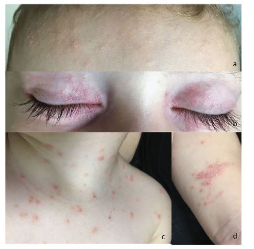
Figures 1a−d Physical examination showing multiple small, erythematous papules on the face (a), trunk (c), and extremities (d); erythematous papules on the upper eyelids (b); and erythematous and scaly plaques on the antecubital fold of the left arm (d).
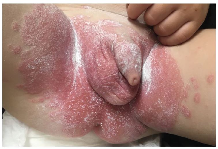
Figure 2 Physical examination showing large, erythematous plaques with well-demarcated margins in the diaper area covered with emollient.
Discussion
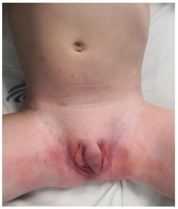
The next day, the child was observed in the Dermatology Department where the diagnosis was confirmed. Topical therapy was started with bath oil, emollient (twice daily), and diaper ointment with zinc (twice daily), followed by petrolatum healing ointment. On the face, prednisolone ophthalmic ointment was used on the eyelids (once daily at bedtime for one week, then two to three times a week), and tacrolimus (0.03%) was used on the remaining lesions (twice daily). For body lesions, a betamethasone with calcipotriol ointment was used at night, with subsequent weaning. This ointment mixed with petrolatum was applied to the diaper area once daily on a similar weaning schedule, along with clotrimazole, which was maintained until complete resolution of the lesions. The child was reevaluated after one month, with almost complete resolution of the lesions (Figure 3).
Psoriasis is a chronic inflammatory disorder primarily involving the skin.¹ It affects 2.0-3.5% of the world population, with increasing incidence and prevalence over the past few decades.²,³ Clinical onset can occur at any age, from the neonatal period to adulthood, with one third of patients manifesting the disease in pediatric age.¹,² Onset at this age correlates with a worse prognosis and also with a positive family history, typically in a first-degree relative, which is usually helpful for the child’s diagnosis.²,⁴-⁶
The pathogenesis of the disease is multifactorial: complex genetic mechanisms, inadequate immune response, and environmental factors play an important role in the unpredictable development and exacerbation of the condition.1-3,5 Some risk factors have been consistently identified as disease triggers, including infections, skin trauma (Koebner phenomenon), drugs, tobacco exposure, and emotional stress.¹,³,⁴ The Koebner phenomenon refers to the emergence of psoriatic lesions at sites of injury, such as scratches, scars, burns, or preexisting conditions (such as seborrheic or atopic dermatitis).³,⁴ Although typical, it is not pathognomonic of psoriasis.³
The most common manifestation of psoriasis is plaque psoriasis, which is defined by well-demarcated erythematous plaques covered with silvery-white scales that are bilaterally and symmetrically distributed on the scalp, face, and extensor surfaces of the limbs.²,³ However, differences in the morphology and distribution of psoriatic lesions allow the classification of psoriasis into several recognized clinical subtypes, such as guttate, pustular, erythrodermic, inverse, napkin, or nail psoriasis. In children, the clinical presentation can be very heterogeneous, and guttate, inverse, or napkin psoriasis are more common than in adults. In fact, guttate psoriasis is the second most common form of psoriasis in children and adolescents, and 40% of cases progress to the classic type.2,³ This form of psoriasis is characterized by an abrupt onset of multiple, small (<1 cm), and drop-like (guttate) scaly papules with a symmetric distribution on the trunk, face, and proximal extremities.²-⁴ Its onset often occurs a few weeks after group A β-hemolytic streptococcal pharyngitis or perianal infection.²-⁵ Another important subtype is inverse psoriasis, which affects flexural and intertriginous areas such as the neck, axillae, groin, and genital or perianal areas.²,⁵ In infants, it typically affects the napkin area and is called napkin psoriasis.² This variant is a common presentation under two years of age and typically resolves with the acquisition of sphincter control (probably reflecting the importance of the Koebner phenomenon induced by urine and stool).²-⁴ Napkin psoriasis is characterized by well-defined, bright, erythematous plaques without typical scales and involves the inguinal folds (unlike candidiasis).²,⁵,⁶ In cases of doubtful diagnosis, the most valuable finding is the recognition of psoriatic lesions elsewhere on the body, which is common.²,³,⁶ Sometimes napkin psoriasis rapidly progresses to a widespread form called psoriatic napkin rash with dissemination.² Facial psoriasis and periorbital involvement with subtle lesions are more common in children than in adults, and differentiation from eczema is more challenging when only this area is affected.²,³ Nevertheless, facial psoriasis typically presents as a psoriatic napkin rash with dissemination, which helps in the diagnosis.² Pruritus is more common in children than in adults.³,⁴,⁷
The diagnosis of psoriasis is mostly clinical.²-⁴ Guttate and plaque psoriasis must be distinguished from viral exanthems and other papulosquamous conditions such as pityriasis rosea or pityriasis lichenoides chronicus.³,⁴ Napkin psoriasis must be differentiated from candidiasis, irritant or seborrheic dermatitis, perianal streptococcal disease, and allergic contact dermatitis.²,⁴ However, it is not uncommon for diaper candidiasis to be superimposed on psoriatic lesions, and the diagnostic clue is the appearance of satellite red and slightly scaly papules on the skin surrounding the plaques.
Patient and parent education is essential to understand the unpredictable course of psoriasis and the importance of the Koebner phenomenon to reduce skin trauma and the risk of exacerbations.³,⁴ Treatment selection depends on age, type of psoriasis, location, severity, previous treatments, and cost.⁴,⁷ Therapeutic options include topical agents, phototherapy, systemic immunosuppressants, and biologic response modifiers.⁴,⁷ First-line treatment is topical therapy, which includes emollients, corticosteroids, vitamin D analogs (calcipotriol or calcitriol), and calcineurin inhibitors (tacrolimus or pimecrolimus). Topical agents are safe, available, easy to use, and less expensive.³,⁴,⁷ Emollients reduce scaling, dryness, and pruritus, thereby enhancing the effect of the remaining topical treatment.²,³,⁷ Topical corticosteroids are usually the first choice, either alone or in combination with other agents such as vitamin D analogues.³,⁷ In some cases, phototherapy and/or systemic therapy may be required for moderate to severe generalized psoriasis or when the disease is unresponsive to topical agents.²,⁴
Authorship
Bárbara Leal - Conceptualization; Administration; Resources; Writing - original draft; Writing - review & editing
Daniela Ribeiro - Resources; Writing - review & editing
Catarina Matos - Conceptualization; Validation; Writing - review & editing
Sónia Coelho - Supervision; Validation; Writing - review & editing

