Imaging cases
Ear pain − Not always otitis
Otalgia − Nem sempre otite
1. Department of Pediatrics, Unidade 2, Unidade Local de Saúde de Gaia/Espinho. 4434-502 Vila Nova de Gaia, Portugal. joanaipires@gmail.com; inesgbv@mail.com; m.luis.tome@gmail.com; raquelguedes@hotmail.com; teresa.pena.fernandes@gmail.com
2. Department of Otolaryngology, Unidade 1, Unidade Local de Saúde de Gaia/Espinho. 4400-129 Vila Nova de Gaia, Portugal. leandro.ribeiro@ulsge.min-saude.pt
Abstract
Acute auricular perichondritis is an infection of the external ear that can lead to potentially serious complications if not promptly diagnosed and treated. The authors present the case of a five-year-old boy who was diagnosed with acute perichondritis after presenting to the Pediatric Emergency Department with acute ear redness, swelling, and pain. The diagnosis of acute perichondritis was established based on the classic physical examination findings of erythema and edema with sparing of the earlobe.
Keywords: auricle, cartilage; ear; perichondritis; pinna
Resumo
A pericondrite auricular é uma infeção do ouvido externo que pode causar complicações graves se não for diagnosticada e tratada prontamente. É apresentado o caso de um rapaz de cinco anos de idade diagnosticado com pericondrite aguda após recorrer ao Serviço de Urgência Pediátrico com queixas de rubor, edema e dor no pavilhão auricular direito. O diagnóstico de pericondrite foi estabelecido com base no exame físico clássico: eritema e edema com preservação do lóbulo.
Palavras-chave: aurícula; cartilagem; ouvido; pavilhão auricular; pericondrite
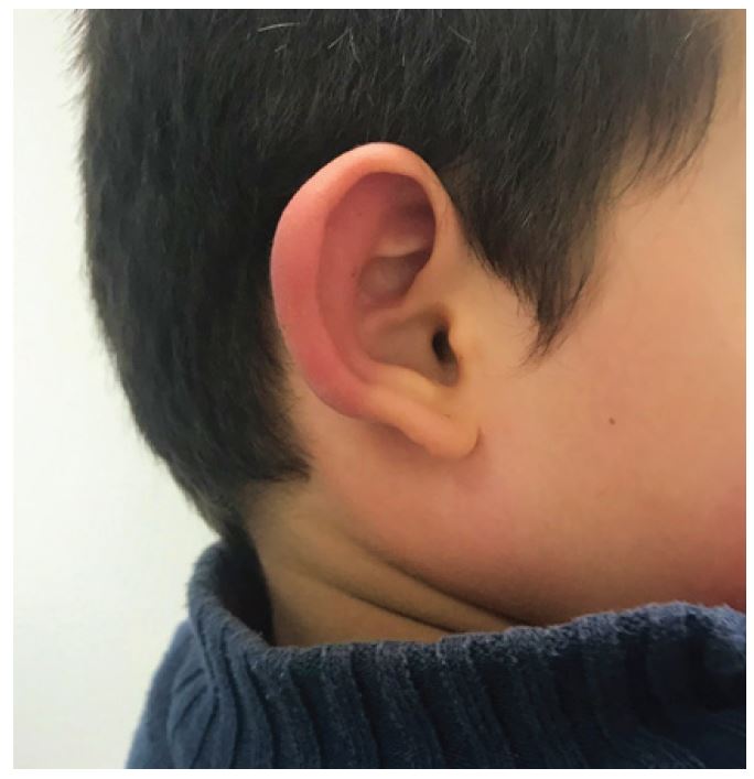
A five-year-old male child was admitted to the Pediatric Emergency Department with pain, redness, and edema of the right ear pinna since the previous day. There was no history of trauma, insect bite, or instrumentation of the affected ear, and no previous medical or surgical history. Physical examination revealed pain, heat, erythema, and swelling of the cartilaginous portion of the auricle with preservation of the earlobe (Figure 1). The mastoid area showed no signs of inflammation. The ear canal was clear with no evidence of external otitis, and the tympanic membrane was normal. The remainder of the examination was unremarkable.
What is your diagnosis?
Diagnosis
Acute auricular perichondritis
Discussion
Auricular perichondritis is inflammation of the cartilaginous portion of the ear, classically sparing the fatty earlobe.1-4 Perichondritis usually results from piercing, insect bite, trauma, or surgical procedures, although this child had no obvious predisposing factors.1,2,5 Common pathogens include P. aeruginosa, S. aureus, E. coli, and Proteus species.2,4 The diagnosis of perichondritis is clinical and based on the classic physical examination revealing erythema and edema of the cartilaginous portion of the auricle with preservation of the earlobe.4 This key finding is very important and helps to differentiate perichondritis from other causes of otalgia.2,4 Auricular perichondritis must be diagnosed and treated promptly to avoid complications. Antibiotic therapy is the mainstay of treatment and should be targeted to cover skin flora and pseudomonas.3-5 Surgical incision and drainage may be considered if an abscess is present to prevent long-term deformities (e.g., cauliflower ear).1-5 This patient was treated conservatively with oral antibiotic (cefuroxime) and topical corticosteroid (mometasone), with marked improvement.
Authorship
Joana F. Pires - Conceptualization; Project administration; Writing - original draft
Inês Bileu Ventura -Conceptualization; Writing - review
Maria Luís Tomé -Conceptualization; Writing - review
Teresa Pena - Writing - review & editing; Visualization
Raquel Guedes - Writing - review & editing; Visualization
Leandro Ribeiro - Writing - review & editing; Visualization
References
1. Lucerna A, Espinosa J. Acute atraumatic pinna (auricular) perichondritis. World Journal of Emergency Medicine. 2018;9(2):152-3.
[ Links ]
2. Rivera-Morales MD, Rodríguez-Belén JL, Vera A, Ganti L. Perichondritis: not all ear pain is otitis. Cureus. 2020;12(10):e11141.
[ Links ]
3. Usoro A, Ehmann MR. Acute Auricular Perichondritis With an Effusion. Clinical practice and cases in emergency medicine. 2019;3(4):453-4.
[ Links ]
4. Bress E, Cohn JE. Perichondritis: inspect the lobule. International Journal of Emergency Medicine. 2020;13(1):1-2.
[ Links ]
5. Davidi E, Paz A, Duchman H, Luntz M, Potasman I. Perichondritis of the auricle: analysis of 114 cases. The Israel Medical Association journal : IMAJ. 2011;13(1):21-4.
[ Links ]


 Inglês (pdf)
 Artigo em XML
 Referências do artigo
 Enviar este artigo por email
 Citado por SciELO
  Similares em
SciELO
Similares em
SciELO 
 Permalink